Transcription
Spine9 mmISSN 2231-0746Volume 10 / Issue 1 / January-June 2020Annals of Maxillofacial Surgery Volume 10 Annals of Maxillofacial SurgeryIssue 1Official Publication of The Indian Academy of Maxillofacial Surgery Estd : 1996January-June 2020 Pages ***-***
Original Article ‑ Prospective StudyEffects of Corticobasal Implant Protrusion inside theNasal and Maxillary SinusFadia Awadalkreem, Abdelnasir Gafar Ahmad1, Stefan Ihde2, Motaz Osman3Department of Oral Rehabilitation, Prosthodontic Division, Faculty of Dentistry, University of Khartoum, 1Department of Oral and Maxillofacial Surgery, Facultyof Dentistry, International University of Africa, 3Department of Implantology, Khartoum Teaching Dental Hospital, Federal Ministry of Health, Khartoum, Sudan,2Department of Evidence and Research, International Implant Foundation, Munich, GermanyAbstractBackground: Implant protrusion into the nasal and maxillary sinuses presents a challenge in cases of severely resorbed maxillae.Aim: The aim of this study was to evaluate the clinical and radiographic effects of BECES implant penetration depth into the nasal andmaxillary sinuses. Setting and Design: This was an observational study conducted in a tertiary institution. Materials and Methods:Forty‑nine BECES implants were inserted into the maxilla of patients who presented with severely resorbed ridges but no history ofsinusitis. Forty‑five implants protruded into the sinus cavities. Patients were examined clinically and radiographically at 1 week and 3, 6,12, and 18 months after insertion. Maxillary sinus health, survival and success rates, and peri‑implant health were assessed using the plaqueindex (PI), calculus index, modified gingival index (MGI), and probing pocket depth (PPD). Statistical Analysis: Wilcoxon signed‑ranktest and Mann–Whitney test were used in this study. Results: Four (8.16%) of the 45 implants that penetrated the cavities reached the sinusfloor without disrupting the membrane; the penetration depth was 4 mm in 20 implants (44.44%) and 4 mm in 25 (55.56%). No patientshowed clinical or radiographic signs of sinusitis during the observation period. There were significant differences in the PI, MGI, and PPDvalues between baseline and the 18‑month follow‑up with no association with the penetration depth. All implants showed radiographicallydirect bone‑to‑implant contact. Where the implant tip barely reached the sinus floor, the membrane healed uneventfully while when deeplypenetrating the sinus, the membrane healed around the implant but did not cover the tip. All prostheses and implants survived during theobservation period. Conclusion: Penetration depth of polished implants with cortical engagement into the maxillary sinus or the floor ofthe nose does not negatively affect implant survival, the success rate of the treatment, nor peri‑implant soft‑tissue health. It also does notprovoke the development of sinusitis.Keywords: BECES implant, cortical engagement of implants, corticobasal implant, maxillary sinus, nasal sinusIntroductionMany methods have been described for the management ofcomplete and partial edentulism. Recently, implant treatmenthas become the gold standard for replacing missing teeth ifadequate bone is present.[1] Cases of severe alveolar ridgeresorption remain a challenge in dental implantology.[1‑3] Theposterior maxilla often presents with limited bone height andpoor bone quality, which jeopardizes the primary stability ofthe implant. A further problem is the complication of maxillarysinus “pneumatization.”[1,3‑6]Conventional maxillofacial therapy has devised methods toincrease the bone quantity in cases of severe bone loss, suchAccess this article onlineQuick Response Code:Website:www.amsjournal.comDOI:10.4103/ams.ams 42 20114as sinus lifting and sinus augmentation procedures with bonegrafts harvested from the hip, tibia, ribs, or chin followedby insertion of the implant.[1,3] This procedure was firstdocumented by Boyne and James in 1980[7] and subsequentlyAddress for correspondence: Dr. Fadia Awadalkreem,Department of Oral Rehabilitation, Prosthodontic Division, P.O. Box 102,Faculty of Dentistry, University of Khartoum, Army Rd., Khartoum, Sudan.E‑mail: fadiadent@hotmail.comSubmitted: 11‑02‑2020Accepted: 29-02-2020Revised: 22-02-2020Published: 08-06-2020This is an open access journal, and articles are distributed under the terms of theCreative Commons Attribution‑NonCommercial‑ShareAlike 4.0 License, whichallows others to remix, tweak, and build upon the work non‑commercially, as longas appropriate credit is given and the new creations are licensed under the identicalterms.For reprints contact: WKHLRPMedknow reprints@wolterskluwer.comHow to cite this article: Awadalkreem F, Ahmad AG, Ihde S, Osman M.Effects of corticobasal implant protrusion inside the nasal and maxillarysinus. Ann Maxillofac Surg 2020;10:114-21. 2020 Annals of Maxillofacial Surgery Published by Wolters Kluwer ‑ Medknow
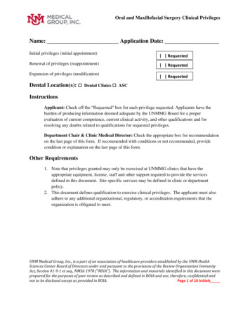
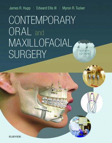
Awadalkreem, et al.: Protrusion of corticobasal implants into sinus cavitiesby Tatum.[8] Summers[9] introduced the sinus floor elevationtechnique using an osteotome with a flapless crestal approach,possibly accompanied by simultaneous placement of animplant.Although a bone augmentation procedure can increase the boneheight and width, it has some disadvantages, including theneed for two independent invasive surgical procedures, whichnot only prolongs the treatment significantly but also resultsin additional costs and risks as well as patient discomfort.[10,11]Numerous postoperative complications of sinus liftingprocedures have been reported, including wound dehiscence,acute/chronic sinusitis, mucocele formation, swelling,discharge of graft material into the sinus, and graft infectionresulting in complete loss of the graft and continuing morbidityat the donor site.[10‑15]Furthermore, sinus lifting procedures have been associatedin many studies with the risk of perforation of the sinusmembrane. The literature reports a 44% incidence of sinusmembrane perforation during implant osteotomies and/orimplant placement.[15] Consequently, implants protrude intothe nasal or maxillary sinuses. Reports on the effects of thisprotrusion on the health of the nasal and maxillary sinuses aremixed. Several groups have found no adverse effects[1,3,4,16-25]while others have reported mild adverse effects.[3,24]An electronic and manual literature search by Ragucci et al.[3]reported after penetration of implants through the floor of themaxillary sinus a clinical complication rate of 14.8%, amongwhich epistaxis being the most common side effect. Theyalso found a radiographic complication rate of 3.4%, the mostcommon being thickening of the Schneiderian membrane,with no significant difference according to the level of implantpenetration. However, they documented a survival rate ofabout 95.6%, with no significant effect of level of implantpenetration. They classified the implant penetration depthsinto two groups ( 4 mm and 4 mm), and the results for bothgroups were the same.In the past few years, several authors have recommendedthe use of Corticobasal implants to achieve bicorticalanchorage (e.g., crest of the ridge and maxillary sinus floor)and to ensure implant stability in patients with severe ridgeresorption or compromised bone support.[26‑36] Consequently,implants may have exposed to the nasal and maxillarysinuses [Figure 1] with possible challenging effects. Althoughmany studies have reported that Corticobasal (BECES )implants have very favorable outcomes with limited andmanageable complications,[27‑29,34‑36] there is still some doubtconcerning the effect of their penetration into the nasal andmaxillary sinuses. In 2018, Lazarov[37] published a prospectivecohort study in which patients treated with Strategic Implant technology who underwent placement of 375 BCS implantsin 105 maxillary sinuses were followed for 3–4.5 years. Themaxillary sinuses were assessed preoperatively on panoramicviews and clinical examination. Patients with symptoms ofFigure 1: A clinical photograph showing an implant that is potentiallyprotruding into the nasal cavity using sinoscopyacute sinusitis were not treated until their symptoms haddisappeared. Postoperative control panoramic views weretaken and clinical examinations were performed in all patientsat 3 years and 4.5 years. Only one sinus showed radiographicand clinical symptoms of a sinusitis. Lazarov concluded thatpolished implants placed in such a way to intentionally orunintentionally penetrate the maxillary sinus do not cause anysinus reaction, and they can remain stable for many years.Furthermore, all symptoms of sinusitis were transient andresolved completely after various treatments even though theimplants were left in place. Little information is availableconcerning the peri‑implant health of the mucosa aroundpenetration areas in the maxillary and nasal sinuses.The aim of the present study was to evaluate the effect ofprotrusion of a BECES implant inside the nasal and maxillarysinuses and to investigate the effect of penetration depth onperi‑implant health and the implant survival rate.Materials and MethodsCharacteristics of the study sampleThe study was approved by the ethics committees of thehospital and the Ministry of Health, Khartoum, Sudan. Allpatients who underwent Corticobasal implant treatmentbetween 2015 and 2017 and presented with severe alveolarridge resorption with possible protrusion of an implant insidethe nasal or sinus cavity were asked to participate in the study.All patients provided informed consent for participation in thestudy and publication of the results after receiving a detailedexplanation of the objectives of the study, the protocol, andthe possible complications.The study inclusion criteria were age 18–80 years and completeor partial edentulism. All patients had severe ridge resorptionand requested implant treatment. The patients were questionedabout their history and symptoms of maxillary sinusitis,[38]including nasal bleeding, congestion, or obstruction, nasalsecretion, and pain or tenderness in the infraorbital region.Annals of Maxillofacial Surgery Volume 10 Issue 1 January-June 2020115
Awadalkreem, et al.: Protrusion of corticobasal implants into sinus cavitiesPatients with a history of sinusitis or bisphosphonate therapywere excluded.Surgical and prosthetic proceduresPreoperative cone‑beam computed tomography (CBCT)scans (Planmeca ProMax; Planmeca, Budapest, Hungary)were obtained for all patients. The scans were obtained using astandardized technique with the same scanner. All images wereanalyzed under standardized conditions. The implants wereplaced under local infiltration anesthesia with 2% lidocaineand epinephrine 1:100,000 after application of Betadine 5% atthe implant sites. The implant osteotomy was performed usinga drill with copious irrigation, followed by cortical penetration,administration of 2 ml of Betadine 5%, and placement ofthe implant. All implants (BECES Brand, ManufacturerSimpladent GmbH, CH‑8737, Gommiswald, Switzerland)were inserted using a standard one‑stage surgical procedure.The length and width of the implant were selectedaccording to the amount of available bone. High primarystability was achieved and tested using the reverse‑torquetechnique at 35 Ncm. Amoxicillin and clavulanatepotassium (Megamox, 1 mg; Hikma Pharmaceuticals,Amman, Jordan), diclofenac potassium (Rapidus, 50 mg;Tabuk Pharmaceutical Manufacturing Co., Tabuk, Kingdomof Saudi Arabia), and xylometazoline (Otrivine, adult nasaldrops; GlaxoSmithKline, Brentford, UK) were prescribedfor all patients. The penetration depth of the implant wasmeasured on CBCT scans obtained postoperatively using themachine’s software.Impression copings were secured to the head of the implant,and an impression was taken using monophase vinylpolysiloxane (Ivoclar Vivadent AG, Schaan, Liechtenstein).The metal framework was constructed on the following day,and a heavy body silicone jaw relation was taken. This metalframework was used to provide implant splinting, to reduceload per unite area, and to ensure better force distributionthat minimizes the possible causes of implant overloading.Either acrylic or composite veneer material was applied tothe framework depending on the extent of loss of soft andhard tissues. One day later (within 72 h), the final prosthesiswas inserted and cemented using Fuji cement (Fuji I LutingCement, GC Corporation, Tokyo, Japan). Patients werescheduled for follow‑up clinical and radiographic examinationsat 1 week and 3, 6, 12, and 18 months.Follow‑up evaluationSinusesAny signs and symptoms of sinusitis such as mucopurulentdrainage, nasal obstruction, facial pain/pressure/fullness,headaches, and an impaired sense of smell were noted.[38]Implant success and survival ratesIn line with the literature, implant survival wasdefined as the presence of the implant in the mouth atthe time of examination. [39] The implant success ratewas assessed using the modified James–Misch implantquality of health scale[39] (I, success [optimum health]; II,116survival [satisfactory health]; III, survival [compromisedhealth]; and IV, failure [clinical or absolute failure]), theAlbrektsson[40] criteria for implant success, which includesa bone loss of 1.5 mm in the 1st year and 0.2 mm yearlythereafter, and the presence of suppuration and peri‑implantinfection, continuous pain, mobility, or persistent peri‑implantradiolucency.[40] The sample was categorized into two groups,i.e., those with an implant penetration depth 4 mm and thosewith a penetration depth 4 mm measured on CBCT.[3]Clinical examinationPeri‑implant soft tissueThe plaque index (PI) is a three‑point scale devised by Mombelliet al.[41] (0, plaque detected; 1, plaque detected by running aprobe across the implant; 2, plaque visible to the naked eye; and3, abundance of plaque). The modified gingival index (MGI)was determined according to the modified Loe and Silnessindex[42] (0, absence of inflammation [normal peri‑implantmucosa]; 1, mild inflammation [slight change in color] andslight edema; 2, moderate inflammation [redness, edema,and glazing]; and 3, severe inflammation [marked rednessand edema] and ulceration). The calculus index (CI) wasreported based on its presence or absence of calculus aroundthe implants both labially and lingually (1, presence and0, absence). The CI was reported as a yes/no variable. Theprobing pocket depth (PPD) was measured in millimeters usinga short‑shank probe with gentle pressure from the mucosalmargin to the bottom of the pocket in both a labial/buccal andpalatal direction.[42] If complete peri‑implant soft‑tissue healingaround the implants had been achieved, the pocket depth wasreported as 0 mm; introduction of the probe was prohibitedgiven that “consensus on probing around basal implants”[43]does not recommend probing as a routine step. However, thesedata should be assessed and reported for comparison withother implant studies. Patients were asked about their level ofsatisfaction, any complaints, and whether or not they wouldchoose the same treatment modality again.The implant penetration depth and the mean PI, MGI, and PPDscores were recorded at five follow‑up visits. The mean wasthen categorized as: 0 0, 0.2–0.4 1, 0.6–1.0 2, and 2 3for calculation of the associations between penetration depthand the index scores and PPD using contingency coefficientand Chi‑square tests.ProsthesesThe prostheses were inspected for decementation, lip support,extent of visible smile, mobility, unnatural wear of the opposingdentition, and fracture of the veneer material.Radiographic evaluationRadiographic assessment was performed using CBCT todetect implant loss and/or fracture, to evaluate bone‑to‑implantcontact, and to detect any changes in the nasal and maxillarysinuses at 1 week and 6, 12, and 18 months. The intraraterreliability was determined using repeated measurements atvarying time intervals spanning several days and was 0.95, 1,1, and 0.90 for the PI, CI, MGI, and PPD, respectively.Annals of Maxillofacial Surgery Volume 10 Issue 1 January-June 2020
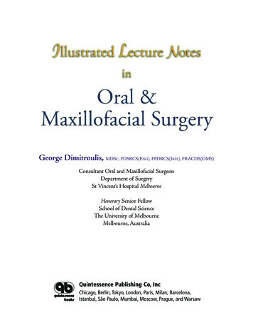
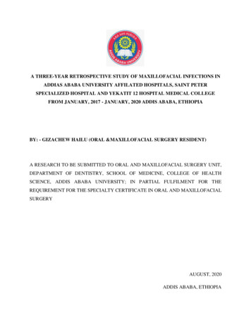
Awadalkreem, et al.: Protrusion of corticobasal implants into sinus cavitiesStatistical analysisThe Wilcoxon signed‑rank test was used to compare the PI,modified MGI, and PPD between baseline and the 18‑monthfollow‑up visit. All data recording and statistical analyseswere performed using the Statistical Package for the SocialSciences (SPSS) software (SPSS version 22; IBM Corp.,New York, NY, USA). P 0.05 was considered statisticallysignificant. The 95% confidence interval was also calculated.ResultsSample distributionForty‑nine implants were found to have penetrated into thenasal and maxillary sinus cavities. The mean patient age was63.6 14.22 years. The immediate (baseline) postoperativeradiographs revealed that 4 (8.16%) of the 45 implants that hadpenetrated the nasal and sinus cavities had reached the sinusfloor without disrupting the membrane; the penetration depthwas 4 mm for 20 implants (44.44%) and 4 mm in 25 (55.56%).[3]Nasal and maxillary sinusesClinical findingsNone of the patients showed any signs or symptoms of rhinitisor sinusitis during follow‑up. All patients maintained good oralhygiene without pain or implant‑related exudates.Radiographic findingsComparison of the radiographs obtained at 6, 12, and18 months with those obtained at baseline did not reveal anyinflammatory reactions at or around the implants, signs ofosteolytic reactions, polyps or granulation around the implantsinside the sinuses, lost implants, or implant fractures. Allimplants showed newly regenerated bone surrounding thethreads, indicating an increase in the bone‑to‑implant contactdespite the different penetration depths [Figures 2 and 3]. Forthose implants that merely penetrated or disrupted the sinusmembrane, it was found that the membrane had healed andtended to cover the implant tips [Figure 2a and b]. When theimplant penetrated the maxillary sinus deeply, there was stillan increase in bone contact around the implant; in these cases,the membrane healed around the tip of the implant but did notcover it [Figure 3a and b].aImplant health and survival rateAccording to the implant health assessment scale,[39] all patientspresented with optimum health at the 18‑month follow‑up,with no pain or tenderness on function, no (zero) mobility,not more than 2 mm of radiographic bone loss from baseline,and no history of exudates. Therefore, the implant survivaland success rates were both 100%.[39]Peri‑implant soft tissueThere was a statistically significant difference in the PI scorebetween baseline and the 18‑month follow‑up [P 0.038,Wilcoxon signed‑rank test; Table 1]. Despite the reportedincrease in the PI readings in some patients, the score rangedbetween 0 and 2 [Table 1]. The CI did not change over timein any case [mean 0.00 0.00; Table 1]. There was also asignificant decrease in the MGI score (P 0.025, Wilcoxonsigned‑rank test). The mean was 0.22 (range, 0–1). Therewas a significant decrease in the PPD (P 0.003, Wilcoxonsigned‑rank test) with a range of readings from 0 mm to 3 mm.Clinical examination revealed that the soft tissue healed veryrapidly even in sites where implants were inserted into freshextraction sockets and a complete soft‑tissue collar presentaround the vertical shaft of the implants.Association between implant penetration depth andplaque index, modified gingival index, and probing pocketdepth valuesThere was no significant difference in the PI, MGI, or PPDvalue according to implant penetration depth [P 0.066,P 0.283, and P 0.550, respectively, Mann–Whitney test;Table 2]. Moreover, there was no association between implantpenetration depth ( 4 mm or 4 mm) and the PI, MGI, andPPD values [P 0.116, P 0.262, and P 0.480, respectively;Tables 3‑5]. There was an apparent increase in the peri‑implantbone level exhibited around the vertical shafts and horizontalplates in all implants [Figures 2 and 3].abFigure 2: Cone‑beam computed tomography scans showing animplant that just barely made contact with the Schneiderian membrane.(a) Immediately after implant insertion. (b) At the 18‑month follow‑up visit.There are no inflammatory reactions at or around the implant, no signs ofan osteolytic reaction, no polyps, and no granulation around the implantinside the sinuses. The implant shows an increase in bone–implant contactbFigure 3: A cone‑beam computed tomography scan showing an implantthat penetrated the Schneiderian membrane and beyond, deep intothe maxillary sinus. (a) Immediately after implant insertion. (b) At the18‑month follow‑up visit. There are no inflammatory reactions at oraround the implant, no signs of an osteolytic reaction, no polyps, and nogranulation around the implant inside the sinuses. The implant shows anincrease in bone–implant contactAnnals of Maxillofacial Surgery Volume 10 Issue 1 January-June 2020117
Awadalkreem, et al.: Protrusion of corticobasal implants into sinus cavitiesPatient satisfactionAll patients were satisfied with their treatment, reported animprovement in their quality of life, and said they would choosethe same treatment again.DiscussionResorption of the residual alveolar ridge following a toothextraction, as well as pneumatization of the maxillaryTable 1: Comparison of the plaque index, modifiedgingival index, and probing pocket depth values betweenbaseline and the 18‑month follow‑up visitPeri‑implantsoft tissueMean SDBaselineP18 monthsPI0.000 0.0000.18 0.5350.038CI0.000 0.0000.000 0.000‑MGI0.110 0.3180.090 0.2880.025PPD0.580 0.8180.200 0.5050.003Nonparametric Wilcoxon signed‑rank test. SD: Standard deviation,PI: Plaque index, CI: Calculus index, MGI: Modified gingival index,PPD: Probing pocket depthTable 2: Comparison of the plaque index, modifiedgingival index, and probing pocket depth values betweenbaseline and the 18‑month follow‑up visit according toimplant penetration depthPeri‑implantsoft tissuePenetration depth (mean SD) 4 mmP 4 mmPI0.040 0.1150.130 0.2080.066CI0.000 0.0000.000 0.000‑MGI0.040 0.1000.170 0.1170.283PPD0.256 0.4600.240 0.2490.550The Mann‑Whitney test was used. SD: Standard deviation, PI: Plaqueindex, CI: Calculus index, MGI: Modified gingival index, PPD: Probingpocket depthTable 3: Association between implant penetration depthand plaque index valuePIPenetration depthTotal 4 mm 4 .62044.4451000CountPercentage of total0.2‑0.4CountPercentage of total0.6‑1.0CountPercentage of totalTotalCountPercentage of totalPI: Plaque index118P0.116sinus, can compromise the placement of a dental implant inthe maxillary posterior region. Several researchers[26,32,33] haverecommended anchorage of bicortical crestal bone and thesinus floor implant to improve implant stability in patients withlimited bone quality. Consequently, implants may protrude intothe maxillary or nasal sinuses, resulting in perforation of themaxillary sinus membrane.Although some studies have reported an incidence of sinusitisassociated with implant penetration inside the maxillary sinus,other studies have reported no change in the maxillary sinus,and there are a number of reports of epistaxis as commonclinical complications and thickening of the Schneiderianmembrane as the main radiographic complication. Therefore,there was a need for a clinical study with a longer observationperiod to investigate these controversial results.According to the literature,[1,44] sinusitis of dental originaccounts for about 10%–12% of all cases of maxillary sinusitisand is associated with many etiological factors, including dentalinfections, such as periapical granuloma, periapical abscess,periodontal infection, inflammatory cyst, oroantral fistula,a foreign body introduced inside the sinus (such as fillingmaterial, root fragment, broken instrument, and largeodontogenic cyst),[45‑47] and a migrated dental implant.[5,48-51] Inthe present study, although some of the implants investigatedshowed penetration of 4 mm, there was no clinical orradiographic evidence of adverse effects in the nasal ormaxillary sinuses, such as nasal decongestion, sinusitis, polypformation, or mucocele. This finding is in accordance withprevious reports[1,4,16‑23] that revealed no association betweenimplant protrusion into the maxillary sinus per se with theincidance of maxillary sinusitis. This observation could beattributed to the smooth surface of the implant and the smallinsertion tip that limits the interruption of the blood supply,resulting in the rapid healing of peri‑implant soft tissue.[27,28,32]Petruson[49] reported normal mucosal conditions with no signsof increased secretion or infection around implants in whichthe tip penetrated the nasal or maxillary sinus even thoughthe surface of the implants used in their study was rough.[49]Variable implant penetration levels were noted in this study; inthe case of minor penetration, the membrane healed and coveredthe tip of the implant. This is confirmed the observations ofZhong et al.,[4] who reported that a surgically disrupted membranearound the apical portion of an implant healed and coveredthe tips of the implants if the protruding depth was 2 mm.Jung et al.[17,18] found that the sinus mucosa covered implantspenetrating the sinus floor by 2 mm in mongrel dogs. Computedtomography scans showed that implant protrusion of 4 mm inthe maxillary sinus caused thickening of the sinus mucosa aroundthe implants; however, these sinuses remained asymptomatic.In contrast, Nooh[24] reported that perforation of the membranewith dental implants may be associated with minor manageablecomplications, such as epistaxis and sinusitis.[24]The high implant survival rate in this study was in accordancewith that in the study by Ragucci et al.,[3] who reported survivalAnnals of Maxillofacial Surgery Volume 10 Issue 1 January-June 2020
Awadalkreem, et al.: Protrusion of corticobasal implants into sinus cavitiesTable 4: Association between implant penetration depthand modified gingival indexMGIDepth of penetration 4 mmTotalP 4 mm00.262Count21Percentage of total46.70.2‑0.4Count4Percentage of total8.9TotalCount25Percentage of total55.6MGI: Modified gingival index1431.13577.8613.31022.22044.445100Table 5: Association between implant penetration depthand probing pocket depthPPDPenetration depth 4 mmTotalP 4 mm00.480CountPercentage of total0.2‑0.4CountPercentage of total0.6‑1.0CountPercentage of totalTotalCountPercentage of totalPPD: Probing pocket .82555.62044.445100rates of 99.5% and 98.5% with implants that have a penetrationdepth of 4 mm and 4 mm, respectively, with no significantdifference between the two depths. Nooh[24] mentioned anoverall survival rate of about 98.4% after 1 year of follow‑up.Seven patients showed mild epistaxis during the immediatepostoperative period, and there was one case of sinusitis.The mean PI reported in this study was in line with thatreported by Mombelli et al.,[52] who rated the amount of plaqueaccumulation around the surface of the submucosal aspect ofthe implant‑abutment on a scale from 0 to 3. The use of acrylicresin as a veneering material to compensate for the severeloss of soft and hard tissues may have caused the increasedplaque accumulation on the restorations. This finding confirmsthe observations of other investigators[53‑55] who reported thatthe micropores and structural inconsistencies of the acrylicsurface supported adhesion of dental plaque and microorganisms.The excellent results for the MGI are credited to the smoothpolished surface that allows rapid attachment of soft tissuearound the implants. The polished vertical implant slotprevents adherence of plaque and apical transmission of thebacterial load inside the basal aspect of the implant, far awayfrom the site of bacterial infection (the oral cavity). Ourobservations confirmed the results of Ihde et al.,[31,33] Lazarov,[27]Ahmad et al.,[28] and Singh et al.,[29] who recommended thisimplant design for use at compromised implant sites, such asthe maxillary sinus, the floor of the nose (where retrogradeperi‑implantitis could occur), or extraction sockets. Theseresults are similar to the experimental and clinical resultsin maxillofacial traumatology and orthognathic surgery, inwhich penetration of smooth bone screws into the sinus andthe nasal aspect of the floor of the nose did not result in anycomplications, infection, or bone loss.[56,57]The increased bone‑to‑implant contact found in this studyis consistent with the finding of Khairnar and Gaur,[20] whoreported increased bone formation and excellent primarystability after indirect elevation of the nasal membrane withsmooth polished implant. Zhong et al.[4] found high removaltorques and intimate bone‑to‑implant contact in bicorticalimplants in rabbits. Furthermore, Kim et al. reported a 100%implant survival rate with a significant increase in bone aroundthe implants when the initial residual bone is 5 mm in height.All patients in this study presented with severe ridgeresorption that precluded rehabilitation with conventionalimplant‑supported prostheses unless preceded by bone‑graftingprocedures. However, any removable prostheses made forsuch patients may be associated with compromised retentionand stability due to the poor status of the denture‑bearingarea. Rehabilitation of these patients with fixed basalimplant‑supported prostheses greatly improved theirsatisfaction. This result is in line with other studies conductedwith conventional dental implants.[58‑61]ConclusionBased on the clinical and radiographic evaluation of thepatients in this study (within the limitations of a small samplesize and short follow‑up period), we can conclude thatprotrusion of a BECES implant into the nasal or maxillarysinuses does not compromise the success or survival rate ofthe implant nor the health of the sinuses. B
concerning the peri‑implant health of the mucosa around penetration areas in the maxillary and nasal sinuses. The aim of the present study was to evaluate the effect of protrusion of a BECES implant inside the nasal and maxillary sinuses and to investigate the effect of penetration depth on peri‑implant health and the implant survival rate.