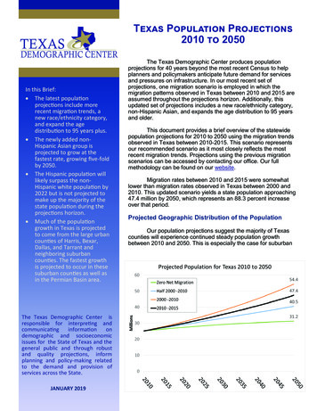
Transcription
Bosire et al. AIDS Research and Therapy 2013, RESEARCHOpen AccessPopulation specific reference ranges of CD3, CD4and CD8 lymphocyte subsets among healthyKenyansErick M Bosire1*, Anthony K Nyamache2, Michael M Gicheru1, Samoel A Khamadi3, Raphael W Lihana3and Vincent Okoth3AbstractBackground: The enumeration of absolute CD4 counts is of primary importance for many medical conditionsespecially HIV infection where therapeutic initiation depends on the count. These ranges tend to vary acrosspopulations. However, these ranges have not been comprehensively established in the Kenyan population.Therefore, this study aimed at establishing the reference ranges for the CD4 and CD8 T-lymphocytes in normalhealthy individuals in Kenya.Methods: A total of 315 individuals of the ages between 16 and 60 years old, in 5 different regions of the country,were recruited into the study. They were screened for diseases that potentially cause lymphocyte homeostasisperturbation. CD4/CD8 Counts were performed by use of a FACSCalibur flow cytometer (Becton-Dickinson, NJ)equipped with automated acquisition and analysis software. Results were analysed according to age, sex and region.Results: Results were presented as means and ranges (in parenthesis) generated non parametrically as 2.5 and 97.5percentiles as follows; In general population; CD3 1655 (614-2685 cells/μL ), CD4 920 (343-1493 cells/μL), and CD8 646(187-1139 cells/μL), while according to sex, females; CD3 1787 (697-2841 cells/μL), CD4 1010 (422-1572 cells/μL), CD8659 (187-1180 cells/μL); males; CD3 1610 (581-2641 cells/μL), CD4 889(320-1459 cells/μL) and CD8 644 (185-1140 cells/μL).The general reference ranges for CD4/CD8 ratios were as follows; general population 1.57(0.50-2.74), males 1.51(0.49-2.64)and females 1.69(0.55-2.95).Conclusion: The lymphocyte reference ranges for the Kenyan population are fairly comparable to those established inother African populations. The ranges also differ appreciably from those established in Germany, Italy and Switzerland.Furthermore, the study reported significant differences in the ranges of different population clusters within Kenya, as wellus between males and females.Keywords: CD4/CD8 count, Lymphocyte subsets, HIV-1, Flow cytometry, Reference rangesIntroductionLymphocytes co-ordinate the immune system’s responseand play a central role in cell mediated immunity [1].Lymphocyte subset may include; Helper T cells (CD4T-cells), Cytotoxic T cells (CTLs or CD8 T-cells), MemoryT cells and Regulatory T cells (Treg cells). Upon encounterwith antigens, CD4 T cells become activated and proliferaterapidly secreting cytokines that sends signals and maintain* Correspondence: bosireerick@yahoo.com1Department of Zoological Science, Kenyatta University, P.O. Box 43844-0100,Nairobi, KenyaFull list of author information is available at the end of the articleactive immune response. On the other hand, the CD8T-cells destroy virally infected cells and tumor cells, andremain inactivated when there is no foreign antigen. Inhuman immunodeficiency virus-1 (HIV-1) infected individuals, lymphocytes (specifically CD4 T-cells) are the viralprime targets. Therefore in these individuals, a CD4 countprovides a picture of immune system competence, withhigher CD4 counts typically signifying healthier immunesystems [1,2]. In such application, flow cytometry is usedto provide absolute counts, percentages and or ratiosof these lymphocyte subsets. Besides HIV management,flow cytometric analysis of peripheral blood lymphocyte 2013 Bosire et al.; licensee BioMed Central Ltd. This is an open access article distributed under the terms of the CreativeCommons Attribution License (http://creativecommons.org/licenses/by/2.0), which permits unrestricted use, distribution, andreproduction in any medium, provided the original work is properly cited.
Bosire et al. AIDS Research and Therapy 2013, phenotypes has proven a useful aid in managing a widerange of medical conditions, including autoimmunity,immunodeficiency, infection, malignancy and transplantation [3-5]. Essential to the effective application of thisapproach is availability of accurate reference values againstwhich results can be meaningfully compared.Reference ranges that are currently used in Kenya arederived from data obtained from Caucasians who are notAfrican Kenyans. In addition, these ranges do not includeage and sex which are very important factors that influence lymphocyte counts [6]. Furthermore, several factorshave been associated with these differences in lymphocytecounts. These may include; demographic and geneticfactors, current exposure to infectious diseases and behavioral factors on CD3, CD4 and CD8 in HIV-negativepopulations [1,2,6]. Averagely, healthy African and Asianpopulations have been shown to have lower CD4 lymphocyte counts than their western European and Caucasiancounterparts [2,7,8]. In addition, women tend to havehigher CD4 levels than men with comparable demographic and behavioral patterns [8,9]. However, there isstill limited data from specific countries to confirmthese differences.The pattern of lymphocyte generation has been shownto affect the levels of circulating lymphocytes in males,females and individuals of different ages. The pattern ofT lymphocyte generation in aging has been associatedwith dynamic changes in thymic and extrathymic functions along with sequential developmental steps from cellsto mature cells [10]. Increased absolute numbers of peripheral blood CD4 cells in females compared to maleshave been reported [11]. This is perhaps due to androgenswhich accelerate thymocytes apoptosis and subsequentlyinfluence T cell repertoire with males tending to haveless CD4 cells than females [12,13]. Sex and age relatedvariations in the lymphocyte subsets contribute to ageand sex related diseases including autoimmune disordersin females and leukemia or lymphoma in males and elderly patients [14,15]. Other factors which have receivedlittle attention but also affect the normal lymphocytelevels include; dietary patterns, body mass index andsmoking habits [16-18]. The influence of these manyfactors point out to the fact that the reference rangesof one population might not be accurately used as areference range for another. This might give an inaccurateinterpretation of the immune status of the individuals.Several efforts to establish reference ranges have indicated differences in lymphocyte levels between Africanand Western populations [2,3,9,19]. In one communityin Kenya, general clinical ranges have been shown to significantly differ from the ranges from other parts of thecontinent [20]. These ranges are also appreciably different from those reported in a neighboring country [21].The two studies in Kenya had indicated a possibility ofPage 2 of 7having remarkably different ranges of lymphocyte subsetswithin the Kenyan population [20,22]. This necessitatesthe need for a comprehensive reference range establishment using subjects from different regions of the country.In this study we report comprehensive reference rangesfor CD3, CD4 and CD8 cutting across representative regions of the country. The study provides ranges specificfor males, females and for different ages.Materials and methodsStudy subjects and samplesA total of 315 adults (222 men and 85 female) were recruited into the study after giving consent. This studywas ethically approved by Kenya National ethical Reviewcommittee before execution. The individuals who wereincluded in the study were healthy adults between 16 and55 (categorized as 16-19, 21-30 and 31-55) years of agepresenting for routine blood donation at the regional centers. These regional blood transfusion centers included;Mombasa, Nakuru, Kisumu, Embu, and Nairobi. A structured questionnaire was used to collect information onthe history and general health status of the donors (SeeAdditional file 1). Donors who confirmed to have a historyof conditions that cause immune perturbations wereexcluded from the study. Further, the samples werescreened for HIV, Hepatitis B virus (HBV), Hepatitis Cvirus (HCV), malaria, and syphilis after blood collection,and samples that tested positive were excluded from thestudy. Blood samples were collected by venipunctureinto EDTA vacutainer tubes and analyzed immediatelyafter collection.Screening for malaria, syphilis, HIV, HCV and HBVEnzyme linked immunoassays were used in screening forthese conditions. Commercially available kits containingpurified antigens were used. The kits included: ICE*Syphilis, Murex anti-HCV (version 4.0), HepanostikaHBsAg Ultra kit, Murex HIV-1.2.0 (Abbot) and malariaantigen detection kit (Creative diagnostics).Sample collection and analysisThe lymphocyte subsets were analyzed on a FACSCaliburflow cytometer (Becton Dickinson ImmunocytometrySystems, San Jose, California) with three monoclonalantibodies (BD Tritest CD4-FITC/CD8-PE/CD3-PerCPreagent) according to the manufacturer’s instructions.The FACSCaliber was calibrated and reconfirmed dailyusing the BD Calibrite beads and BD-FACSComp software, version 2.0 for setting the photomultiplier tubevoltages, setting the fluorescence compensation and checking instrument sensitivity before use. A control sampleof commercially available whole blood was run daily tooptimize the instrument settings. The CD45 versusthe side scatter (SSC) dot plot was visually inspected
Bosire et al. AIDS Research and Therapy 2013, Page 3 of 7Table 1 The means and medians of the lymphocyte subsets in the study populationT lymphocyte subsetCD3CD4CD8RatiosAbsolute count*% Absolute count*% Absolute count*% 360.50-2.74*Expressed as cells/μL of blood. Expressed as a percentage of total lymphocytes.to make sure they appear as a bright compact cluster,with low SSC. Analysis did not proceed if populationswere diffuse and if there was little or no separationbetween clusters [15].These reference ranges were also compared across thesampled geographical regions, by age as well as sex.Data analysisThe CD3, CD4 and CD8 counts and ranges for the generalstudy population were determined. The means, median,percentages and ranges of these lymphocytes, as well asthe CD4/CD8 ratios were determined (Table 1).Non parametric reference range determination was employed by estimating the 2.5 and 97.5 percentile [23].The significance of differences between the mean absolute counts and percentages of the Lymphocyte subsetswere estimated using the Analysis of Variance (ANOVA)and T-test. Multiple comparisons were done using Tukey’stest. Correlation analysis was applied in determining therelationship between lymphocyte subset levels and age.ResultsAmong the 315 individuals between the ages 16-60 years,61.7% (222) were men and 38.4% (85) were female. Theirblood samples were analyzed for CD3, CD4 and CD8lymphocyte subset for establishment of reference ranges.General distribution of the T-lymphocytes for the studypopulationLymphocyte subsets in the different Kenyan regionsMean absolute counts of the different regions were compared using ANOVA. Significantly different means werefurther subjected to Tukey’s test to show differencesbetween each of the regions. The difference in the meanCD3 of Nairobi and Rift valley regions was minimal compared to the difference in the CD3 mean of the LakeVictoria basin region. The Lake Victoria region had thelowest CD3 mean compared to all other regions. Thesedifferences in the CD3 absolute count means wereTable 2 Means, medians and ranges of the lymphocyte subsets for the different geographical regions in KenyaRegionLymphocyte subsetCD3 countCD3%CD4 countCD4%CD8 countCD8%NairobiMean SD1841 486c69 6a1046 283c39 6a717 260c26 627-48266-116516-35Mean SD1728 642b64 7a949 209b37 7b650 329ab24 021-50213-108311-34Mean SD1876 883d67 8b1029 559ab37 7c752 421d27 23-47217-131513-39Mean SD1195 449abcd66 7c690 263abc38 6d454 231acd25 26-47135-79112-36Mean SD1617 574a66 7d881 333a36 7a675 289a28 22-47210-117916-37EasternRift valleyLake VictoriaCoastThe absolute counts of CD4, CD3 and CD8 are presented as cells/μL of blood.Means followed by the same letters within a column are not significantly different from each other according to Tukey’s test.
Bosire et al. AIDS Research and Therapy 2013, Page 4 of 7significant. General reference ranges were also determinedas mentioned earlier (P 0.05; Table 2).T-lymphocyte subset for males and femalesFemales had a significantly higher CD3 mean absolutecounts (P 0.05) compared to men. There were significant differences in CD4 counts for males and females.However, there were no significant differences in themean CD8 absolute counts. T lymphocyte subset reference ranges for males and females separately were alsodetermined at 2.5 and 97.5 percentiles (Table 3).Table 3 Means, medians and ranges of the lymphocytesubsets for males and femalesLymphocyte subsetSexFemalep-valueT-lymphocyte subset for the different age groupsThe means and medians of the lymphocyte subsets werealso determined for the different age groups. There wereno significant differences in the mean absolute countsin the age groups that were considered in this study(Table 4).Comparison of the ranges of the current study with thoseestablished by other studiesThe reference ranges established in this study showed variations with the ones used in MultiSET (BD FACScaliber)as well as other studies in different populations. Thestudies compared below included normal (HIV negative)Table 4 The lymphocyte subset levels in the different agegroupsMaleSubsetCD3 Absolute count*CD3 Absolute N55196620.024Mean6567670.180Median6567670.261CD8 Absolute count*CD8 Absolute 0.498CD8% 10.256Mean2426270.138Median232526.0.090CD4 Absolute count**CD4 Absolute 54CD4% 01Mean3837380.222Median3637370.184CD4/CD8 ratioCD4/CD8 4 0.001Mean1.701.541.54 0.0010.030Median1.631.451.510.135**Expressed as cells/μL of blood.Expressed as a percentage of total lymphocytes. P valueN*Mean31-55CD3% MeanMean20-300.039CD3% MeanAge group16-19*Expressed as cells/μL of blood.Expressed as a percentage of total lymphocytes.ANOVA was used to test the differences in means of the lymphocytes in theage groups.
Bosire et al. AIDS Research and Therapy 2013, Page 5 of 7populations in their study populations. Most of the studieswere regional, that is, having been conducted in specificcountries or in some specific regions within the countries(Table 5).DiscussionThe reference values of the main circulating lymphocytesubsets have been established by many studies throughout the world and have shown some variability according to geographical locations and methodology. Resultsin Table 5 show comparative values of CD3, CD4 andCD8 subsets levels from other countries, showing variations based on geographical region and platform methodsused.In this study, the mean CD3 count was found to begenerally higher compared to those reported in Sweden(1075) but lower in CD3% mean [3]. In comparison withother African countries the lymphocyte counts and percentages were found to be fairly comparable in some casesbut in others slightly different. In addition, Kenya likemany African countries (Tanzania, Ghana, Senegal andEthiopia) had lower lymphocyte subset levels comparedto those reported in Sweden [3,25,26].Considering the two important lymphocyte subsets CD4and CD8, there were striking variations with the otherreported ranges within and outside Africa. The meanCD8 value for this study was similar to those obtainedin other African countries but slightly higher than thosereported in Tanzania and Senegal [8,25]. The CD4 absolute counts were significantly higher than those reportedin Ethiopia and Senegal [17] but lower compared to thosereported in Kampala Uganda [21]; Table 5. However, theranges cannot be conclusively compared since the studiesdiffer in experimental set up. For example the study inUganda included patients who visited an AIDS information center in Kampala, whereas our study was amongblood donors across the country. In comparison to othercountries outside Africa, the CD4 absolute counts werehigher compared to Asian populations [27]. It can bespeculated that, these differences in the lymphocytecounts are due to the many factors already mentioned,including genetic predisposition [1-3]. Many of thesefactors including the genetic influence have not beencomprehensively confirmed.In this study, females had higher absolute counts forCD3 and CD4 lymphocytes compared to males. This wasprobably due to biological factors (Table 4). It has beenspeculated that gender and age-related variations withinthe immune system parameters may contribute to thepathogenesis of several gender and age-related diseasessuch as autoimmune disorders in female patients [17,19,24].Further, it has been shown that there are gender differencesin the generation of CD8 cells during HIV-1 infection,due to increased immune activation compared to men[28]. However, a comparison of mean CD8 absolute countsbetween males and females, revealed no significant differences. These findings were consistent with previous studies[2,3,19,24].The study also compared the differences in absolutescounts across regions. There were significant differencesacross regions with Lake Victoria region, consistentlyrecording lower lymphocyte counts compared to otherregions. The CD8 absolute count of Nairobi were appreciably similar with ones reported by Embree [19] in theircontrols of a study carried out in Nairobi. These observations show that, there is need to understand the impact ofgenetic makeup and altitude on the levels of T lymphocytes. The CD4 median absolute counts for the Rift valley(810 cells/μL) were similar to the ones reported in oneof the studies in Kericho which is located within thevalley ([20] Table 2).Analysis of the lymphocyte subsets in the different agegroups revealed no significant differences. A more comprehensive study design might be required to give a clearpicture of the lymphocyte changes with advancing age. Itis however well known that the pattern of T lymphocytegeneration with age originates from dynamic changes inthymic as well as extrathymic functions, along withTable 5 Comparison of the reference ranges developed in this study with reference ranges of other 00-2460780-2240540-1790CD3% 140-820CD8%13.3894-36.392513-41NANANANANACD4/CD8 .0 NANot available.*Expressed as cells/μL of blood. Expressed as a percentage of total lymphocytes.Kenya-Current study, MultiSET-BD Biosciences, Tanzania [19], Ghana [2], Italy [24], Germany [25], Switzerland [3].
Bosire et al. AIDS Research and Therapy 2013, sequential developmental steps from stem cell to ultimately mature cells [29].ConclusionOur study has established ranges and reveals that lymphocyte subsets among the Kenyan population are differentfrom those of other populations in the other populationssuch as Germany, Italy and Switzerland. However, theranges are fairly comparable to those reported in someAfrican countries. The ranges are also slightly differentfrom those that are being used currently. This studyhas established reference ranges for the lymphocytesubsets among healthy Kenyan individuals of the agesbetween 16-60 years using FACSCalibur flow cytometer(Becton Dickinson Immunocytometry Systems, San Jose,California). The study has further shown the possibility of having varied lymphocyte levels among Kenyansubpopulations, due to many unconfirmed factors. Therefore, the current ranges should be more comprehensivelydetermined and further revised, in order to have accuratecomparisons during flow cytometry.Additional filePage 6 of 73.4.5.6.7.8.9.10.11.12.Additional file 1: Questionnaire for sample collection.AbbreviationsHIV: Human immunodefieciency virus; CTL: Cytotoxic T lymphocytes;SSC: Side scatter; ANOVA: Analysis of variance.Competing interestsThe authors declare that they have no competing interests.13.14.15.Authors’ contributionsEB designed the study, collected samples, analysed them and drafted themanuscript. MMG helped in data analysis and interpretation and reviewedthe manuscript. SAK conceptualized the idea, helped in designingexperiment, analysis of the samples, interpretation of data and review of themanuscript. AKN helped in drafting and critically revising the manuscript. RLhelped in critically reviewing the manuscript. VO helped in critical review ofthe paper. All authors read and approved the final manuscript.AcknowledgementsThe authors wish to thank individuals who participated in the study, the staffof all the Regional Blood Transfusion centers for their participation and theDepartment of Zoological Sciences, Kenyatta University for financial support.16.17.18.19.20.Author details1Department of Zoological Science, Kenyatta University, P.O. Box 43844-0100,Nairobi, Kenya. 2Department of Plant and Microbial Sciences, KenyattaUniversity, P. O. Box 43844-0100, Nairobi, Kenya. 3Kenya Medical ResearchInstitute, Centre for Viral Research, P.O Box 54840-00200, Nairobi, Kenya.Received: 15 May 2013 Accepted: 5 November 2013Published: 7 November 2013References1. Ahmadi K, Hall M, Norman P, Vaughan R, Snieder H, Spector T, Lanchbury J:Genetic determinism in the relationship between human CD4 and CD8T lymphocyte populations. Genes Immunol 2001, 2:38–387.2. Ampofo W, Torpey K, Mukadi Y, Koram K, Nolan K, Amenyah R, Kaitoo E,Antwi P, Ofori-adjei D, Lamptey P: Normal CD4T lymphocyte levels in HIV21.22.23.24.seronegative individuals in the Manya/Yilo Krobo communities in theEastern Region of Ghana. Viral Immunol 2006, 19:260–266.Bisset L, Lung T, Kaelin M, Ludwig E, Dubs R: Reference values forperipheral blood lymphocyte phenotypes applicable to the healthyadult population in Switzerland. Eur J Haematol 2004, 72:203–212.Illoh O: Current applications of flow cytometry in the diagnosis ofprimary immunodeficiency diseases. Arch Pathol Lab Med 2004, 128:23–31.Davis W, Hamilton M: Uses of flow cytometry to characterizeimmunodeficiency syndromes in Camelids. Small Ruminant Res 2006,61(2):187–193.Ullrich K, Koenigsmann M, Mohren M, Franke A: Lymphocyte subsets’reference ranges in an age-and gender-balanced population of 100healthy adults—a monocentric German study. Clin Immunol 2005,116:192–197.Howard R, Fasano C, Frey L, Miller C: Reference intervals of CD3, CD4, CD8,CD4/CD8, and absolute CD4 values in Asian and non-Asian populations.Cytometry 1996, 26:231–232.Mandy F, Nicholson J, McDougal J: Revised guidelines for performingsingle-platform absolute CD4 T-Cell determinations with CD45 gatingfor persons infected with human immunodeficiency virus. MMWR 2003,52:1–13.Klose N, Coulibaly B, Tebit D, Nauwelaers F, Spengler H, Kynast-Wolf G,Kouyaté B, Kräusslich H, Böhler T: Immunohematological reference valuesfor healthy adults in Burkina Faso. Clin Vaccine Immunol 2007, 14:782–784.Lahita R: Sex hormones and systemic lupus erythematosus. Rheum DisClin North Am 2000, 26:951–968.Vajpayee M, Kaushik S, Sreenivas V, Wig N, Seth P: CDC staging based onabsolute CD4 count and CD4 percentage in an HIV-1-infected Indianpopulation: treatment implications. Clin Exp Immunol 2005, 141:485–490.Kalayjian R, Landay A, Pollard R, Taub D, Gross B, Francis I, Sevin A, Pu M,Spritzler J, Chernoff M, Namkung A, Fox L, Martinez A, Waterman K, Fiscus S,Sha B, Johnson D, Slater S, Rousseau F, Lederman M: Age related immunedysfunction in health and in human immunodefieciency virus (HIV)disease: association of age and HIV infection with naive CD8 celldepletion, reduced expression of CD28 on CD8 cells and reduced thymicvolumes. J Infect Dis 2003, 187:1924–1933.Olsen N, Kovacs W: Effects of androgens on T and B lymphocytedevelopment. Immunol Res 2001, 23:281–288.Clerici M, Butto S, Lukwiya M: Immune activation in Africa is environmentallydriven and is associated with upregulation of CCR5: Italian–Ugandan AIDSproject. AIDS 2000, 14:2083–2092.Menard D, Joelle M, Bem M, Kassa E, Gresenguet G, Talarmin A:Immunohematological reference ranges for adults from the centralAfrican republic. Clin Diagn Lab Immunol 2003, 10:443–445.Schaberge T, Theilacker C, Nitschke O, Lode H: Lymphocyte subsets inperipheral blood and smoking habits. Lung 1997, 175:387–394.Mair C, Hawes S, Agne H, Sow P, N’dove I, Manhart L, Fu P, Gottlieb G,Kiviat N: Factors associated with CD4 lymphocyte counts in HIV-negativeSenegalese individuals. Clin Exp Immunol 2007, 151:432–440.Feldman G, Minkoff H, Schneider F: Association of cigarette smoking withHIV prognosis among women in the HAART era: a report from thewomen’s interagency HIV study. Am J Pub Health 2006, 96:1060–1065.Ngowi B, Mfinanga S, Bruun J, Morkve O: Immunohaematologicalreference values in human immunodeficiency virus-negative adolescentand adults in rural northern Tanzania. BMC Infect Dis 2009, 9:1–7.Kibaya R, Bautista C, Sawe F, Schaffer D, Sateren W, Scott P, Michael N,Robb M, Birx D, Souza M: Reference ranges for the clinical laboratoryderived from a rural population in Kericho Kenya. PloS ONE 2008,3(10):e3327. Doi:10.1371/journal.pone.0003327.Tugume S, Piwowar E, Lutalo T, Mugyenyi P, Grant R, Mangeni F, Pattishall K,Katongole-Mbidde E: Hematological reference ranges among healthyUgandans. Clin Diagn Lab Immunol 1995, 2:233–235.Embree J, Bwayo J, Nagelkerke N, Njenga S, Nyange P, Ndinya-Achola J,Pamba H, Plummer F: Lymphocyte subsets in human immunodeficiencyvirus type 1-infected and uninfected children in Nairobi. Pediatr Infect DisJ 2001, 20:397–403.NCCLS: How to define and determine reference intervals in the clinicallaboratory; approved guideline, Volume 20. 2nd edition. Wayne, PA: NationalCommittee for Clinical Laboratory Standards; 2000:13. C28-A2.Santagostino A, Garbaccio G, Pistorio A, Bolis V, Camisasca G, Pagliaro P,Girotto M: An Italian national multicenter study for the definition of
Bosire et al. AIDS Research and Therapy 2013, 25.26.27.28.29.Page 7 of 7reference ranges for normal values of peripheral blood lymphocytesubsets in healthy adults. Haematologica 1999, 84:499–504.Koetz K, Bryl E, Spickschen K, O’Fallon W, Goronzy J, Weyand C: T-cellhomeostasis in patients with rheumatoid arthritis. Proc Natl Acad Sci USA2000, 97:9203–9208.Nicholson J, Stein D, Mui T, Mack R, Hubbard M, Denny T: Evaluation of amethod for counting absolute numbers of cells with a flow cytometer.Clin Diagn Lab Immunol 1997, 4:309–313.Chng W, Guat B, Tan S, Kuperan P: Establishment of adult peripheralblood lymphocyte subset reference range for an Asian population bysingle-platform flow cytometry: influence of age, sex, and race andcomparison with other published studies. Clin Diagn Lab Immunol 2004,11:168–173.Meier A, Chang J, Chan E, Pollard R, Sidhu H, Kulkarni S, Wen T, Lindsay R,Orellana L, Mildvan D, Bazner S, Streeck H, Alter G, Lifson J, Carrington M,Bosch R, Robbins G, Altfeld M: Sex differences in the Toll-like receptormediated response of plasmocytoid dendritic cells to HIV-1. Nat Med2009, 15:955–959.Globerson A: Thymocytopoiesis in aging: the bone marrow–thymus axis.Arch Gerontol Geriatr 1997, 24:141–155.doi:10.1186/1742-6405-10-24Cite this article as: Bosire et al.: Population specific reference ranges ofCD3, CD4 and CD8 lymphocyte subsets among healthy Kenyans. AIDSResearch and Therapy 2013 10:24.Submit your next manuscript to BioMed Centraland take full advantage of: Convenient online submission Thorough peer review No space constraints or color figure charges Immediate publication on acceptance Inclusion in PubMed, CAS, Scopus and Google Scholar Research which is freely available for redistributionSubmit your manuscript atwww.biomedcentral.com/submit
perturbation. CD4/CD8 Counts were performed by use of a FACSCalibur flow cytometer (Becton-Dickinson, NJ) equipped with automated acquisition and analysis software. Results were analysed according to age, sex and region. Results: Results were presented as means and ranges (in parenthesis) generated non parametrically as 2.5 and 97.5