Transcription
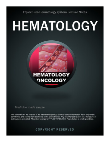
Ftplectures Hematology system Lecture NotesHEMATOLOGYMedicine made simpleThis content is for the sole use of the intended recipient(s) and may contain information that is proprietary,confidential, and exempt from disclosure under applicable law. Any unauthorized review, use, disclosure, ordistribution is prohibited. All content belongs to FTPLECTURES, LLC. Reproduction is strictly prohibited.COPYRIGHT RESERVED
Ftplectures Clinical MedicineCopyright 2014Adeleke Adesina, DOClinical Medicine 2012 ftplectures LLC1133 Broadway Suite 706,New York, NY, 10010The field of Medicine is an ever-changing profession and as new evidence based studiesare conducted, new knowledge is discovered. Ftplectures has made tremendous effort todeliver accurate information as per standard teaching of medical information at the timeof this publication. However, there are still possibilities of human error or changes inmedical sciences contained herein. Therefore, ftplectures is not responsible for anyinaccuracies or omissions noted in this publication. Readers are encouraged to confirmthe information contained herein with other sources.ALL RIGHTS RESERVED. This book contains material protected under Internationaland Federal Copyright Laws and Treaties. Any unauthorized reprint or use of thismaterial is prohibited. No part of this book may be reproduced or transmitted in anyform or by any means, electronic or mechanical, including photocopying, recording, orby any information storage and retrieval system without express written permission fromftplectures.
Anemia part-1 The red blood cellStructure- ‐- ‐- ‐- ‐- ‐- ‐- ‐- ‐- ‐- ‐- ‐- ‐- ‐- ‐BiconcaveMicroscopic- 6 to 8 µmVolume- 90 fentolitreDiameter – 2 µmNo nucleusSmallFew organellesHaemoglobin – can carry 270millionmolecules inside a single RBCCytoskeleton- spectrinCell membrane is bilayered with cholesterol and phospholipidsPhosphotydylserine (PS)RBC is red because of haemoglobin heme (porphrin ring Fe2 ) globin (2αglobin 2βglobin 4 polypeptide chains)Histidine holds the Fe molecule in the centre so that O2 can bind to the Fe.Mainly the RBC is red due to iron in heme group.Functions- ‐- ‐- ‐Carry oxygen but do not use itUndergo anaerobic respiration- use glucose to form pyruvate.Pyruvate forms lactate and releases 2ATP which is used by the Na /K ATPasepump to maintain electroneutrality.Production- ‐- ‐- ‐- ‐- ‐They are made in bone marrow of large bones like femur, radius or ulna.Stem cells – haematocytoblast - proerythroblast (committed stem cell) – erythroblast– normoblast – the nucleus is ejected to form – reticulocyte – a red blood cell/erythrocyte formed finallyErythropoesis- it is the formation of RBC from stem cellsErythropoietin- produced from kidneys has the enhancing effect of erythropoesisTestosterone has enhancing effect on erythropoietin.Lifecycle- ‐- ‐- ‐- ‐- ‐- ‐It has a life span of 120 days.An erythrocyte completes a round in circulatory system within every 20 secondsAfter 120 days the RBS gets weakened plasma membrane.Spleen, liver and bone marrow - area where RBCs are destroyed.Reticuloendothileal system- consists of macrophagesPhosphatidyl serine is in the inner core membrane of RBC. When they are exposedand perceived by macrophages, they are destroyed.
- ‐- ‐The process of destruction is called phagocytosis.The RBC is destroyed to formHeme-it releases Fe3 which binds to transferrinRest of the RBC- biliverdin– bilirubin (b vreductase)Albumin binds to bilirubin and takes it back toliver- ‐- ‐4 polypeptide chainsThrough urine and faeces, RBC is eliminated.The red blood cell can undergo hemolysis by itself. Hemolysed haemoglobin formshaptoglobin.
Anemia part 2- Heme synthesisHeme is made inside the liver and bone marrow.Glycine Succinyl CoAMitochondria ALA Synthetase amino lavulinic acid ( ALA) needs Vit. B6 (pyridoxal phospahate) ALA dehydrasePorphobilinogen (PBG) CytoplasmUroporphyrinogen III Protoporphyringen/ protohemeFe2 FerroketolaseHeme- ‐- ‐Heme has a negative effect on ALA synthase to regulate the pathwayGlucose has a negative effect alsoPorphyrias1. Acute intermittent porphyria –- ‐ Uroporphrinogen I synthetase deficiency- ‐ UPSI converts porphybilinogen to uroporphyringen IIILabs- ‐ High levels of PBG- ‐ High levels of ALASymptoms- ‐ 5 P’s- ‐ Painful abdomen- ‐ Neuropathy- ‐ Psychological problems- ‐ Paranoid, depressed- ‐ Portwine urine which turns pink in the presence of oxygen- ‐ Barbiturates, hypoxia and alcohol have the inhibitory effect on the pathway- ‐ Do not give barbiturates because they worsen abdominal pain by activating cytochromeP450 system found in liver. This system causes more and more of the functioning of theheme synthesis pathway resulting in an increase in PBG levels.2. Porphyria Cutenea Torda- ‐ Most common- ‐ Deficiency of uroporphyrinogen decarboxylase which converts UPSIII to coreporphyrinogen which gets converted to heme.
- ‐- ‐- ‐SymptomsSensitive to light- very very photosensitivieInflammation and blistering of the skinTreatmentGive beta carotene.Vit. B6 deficiency- ‐- ‐Causes sideroblastic anemiaRinged sideroblast seen in peripheral smear.Lead poisoning- ‐- ‐Lead inhibits ALA dehydrase and ferroketolaseMicrocytic sideroblastic anemiaSymptoms- ‐- ‐- ‐- ‐- ‐- ‐- ‐HeadachesNauseaMemory lossLead lines in the gumsLead deposition in abdomen and epiphyses of boneHigh levels of ALA in urineAbdominal pain, diarrhoea
AnemiaDefinitionIt is the decrease in hematocrit/haemoglobin concentration.Hematocrit is the volume % of RBC in bloodstream. It is also called as PCV or packed cell volume.Compensation of anemia1.2.3.4.Heart rate increasesCardiac output increasesStroke volume increasesIncreased extraction of oxygen to tissuesThe Hb dissociation graph shifts to the right.2,3 Diphosphoglycerate decreases the affinity of Hb to oxygen so the curve shifts again to right.On doing CBC, if Hb 7g/dl, 2 units of PRBCs are given.Those who are old or cardiopulmonary problems PRBCs must be given when Hb is below 10g/dl.Clinical features1.2.3.4.5.Pallor in Eyes- pale conjunctivaFatigue, weakness, poor concentrationNausea, vague abdominal discomfortHypotensive (80/40) and tachycardiacJaundice – yellow eyes yellow skin- haemolytic anemia – due to excess of bilirubin spilledin the blood streamDiagnosis1. CBC- complete blood count- check Hb level and hematocrit (H/H)Hb *3 hematocrit1 unit of PRBCs Hb increases by 1Hematocrit increases by 3Side note- pseudo anemia (dilutional anemia)- when 0.9% normal saline is given, Hbconcentration gets diluted so gives the impression of anemia.2. Reticulocyte index – to check if erythropoesis is going on. It can bea. Greater than 2%- making a lot of reticulocytes- indicates excess destruction or loss ofbloodb. Less than 2%- no production of RBCs from the bone marrow3. Blood smear for mean corpuscular volume (MCV)Average volume of RBC 90Fl
Approach to anemiaH/H Retic count%Greater than 2%Blood lossTraumaGI bleedLess than 2%Check for MCVhaemolytic anemia lactate dehydrogenase LDH haptoglobin bilirubinFind sourceMCV 70- microcytic anemiaIron studies80-99- normocytic anemiaAplastic anemia 100 macrocytic anemiaVitamin B12 dficiencyThalassemias ( Alpha andBeta)Anemia of chronic diseaseIron deficiencyLead poisoningSideroblastic anemiaBone marrow fibrosisFolateTumourAnemia of chronic diseaseRenal failureLiver diseaseIn case of emergency, resuscitation is done.
Iron deficiency anemiaDefinition- It is the most common type of microcytic anemia- MCV 70- ferrochetolase brings fe for conversion of protoporhyrin to heme.Causes- ‐- ‐- ‐- ‐- ‐- ‐Chronic blood loss by farMenstrual blood loss by farAny male of age 40 years with iron deficiency anemia secondary to GI bleed, rule out coloncancer.Inadequate supplementation of iron in diet.- like infants growing in breast milk and toddlers(6monts to 3 years)Adolescent femalesPregnant womenClinical features1.2.3.4.5.Fatigue/tiredPallor- conjunctiva and handsDyspnoea on exertionOrthostatic hypotension or lightheadednessHypotensive and tachycardia in acute GI bleed.Diagnosis1. Ferritin level- Ferritin is a form of iron storage.- level is low2. TIBC – total iron binding capacity- level is high3. Transferrin levels- It transfers iron from ferritin to bone marrow for erythropoesis- level behigh4. Iron levels- level is low5. Peripheral smear- microcytic, hypochromic RBC6. Stool Guaic test- check your finger inserted in stool with stool Guaic, if it turns blue, itindicates blood loss.Treatment1. Ferrous sulphate- oral supplements- ADR- nausea, constipation and dyspepsia2. Parentral IV Iron3. Packed Red Blood Cells- hemodynamic instability in case of high heart rate and low BP.
Sideroblastic anemia- ‐- ‐- ‐It occurs due to Vit B6/ pyridoxal phosphate deficiencyThe peripheral smear shows ringed sideroblasts.Vit B6 is important for the action of ALA synthetase for the formation of delta ALA which ifnot formed causes non-formation of heme thereby causing anemia.
ThalassemiaDefinitionIt is a disorder where the globin component of haemoglobin is affected. There is ainadequateproduction of alpha or beta globin chain.TypesBeta thalassemiaBeta globins are missingMore in Indians, Mediterranean’s, MiddleEastern peopleAlpha thalassemiaAlpha component is missingBeta tetramers are formedSeverity depends on the number of globin chainsmissing.Beta thalassemiaMajorHomozygous beta chainmutationAlso called Cooley’s anemiaMore common inMediterranean peopleSymptoms- ‐ Microcytic andhypochromicanemia- ‐ MCV 70 Fl- ‐ Massivesplenomegaly- ‐ Bone marrowexpansion to makemore RBC, andbone looks rugged- ‐ Growth retardation- ‐ Failure to thrive- ‐ Death within firstyear if left untreatedDiagnosis- ‐ HaemoglobinelectrophoresisElevated level ofHbF (fetal)- ‐ Peripheral bloodsmear- microcyticand hypochromicTreatmentBlood transfusion- PRBCshave to be givenDue to lots of transfusion,there may be iron overloadcausing hemochromatosis-MinorHeterozygous beta chainmutationIntermediaIt affects the 2 genes thatcode for the beta globinchainsIt is the most common type ofthalassemiaSymptoms- ‐ Asymptomatic- ‐ MCV 70Fl- ‐ Mild microcytic andhypochromic anemiaSymptoms- ‐ Intermediatetype of anemiaTreatmentNo treatment requiredTreatmentNo treatment required
Deferoxamine ( ironchelating agent)Alpha thalassemiaFor 1 alpha sub-unit 2 genes are required. So in total 4 genes needed for 2 alpha chains.1. Silent carrier- (α-/αα)- If only 1 gene is affected, no treatment is required. NormalHb/Haematocrit2. Alpha thalassemia minor- (α-/α-)- two gene mutations are missing- ‐ Mild Microcytic and hypochromic anemia- ‐ Common in African American patients- ‐ No treatment required3. Haemoglobin H disease- ‐ --/α- ‐ Severe haemolytic anemia- ‐ Splenomegaly- ‐ Diagnosis by Hb electrophoresis- we find Hb H.- ‐ Treatmenti.Blood transfusionii.Splenectomy4. Mutations in all 4 genes- ‐ --/-- ‐ They develop Hydros fetalis- ‐ They do not survive
Anemia of Chronic Disease-microcytic hypochromic anemiaDefinitionA patient is anemic while he has a chronic disease like tuberculosis.Causes- ‐- ‐- ‐- ‐- ‐- ‐TuberculosisLupusLung abscess (parapneumonic effusion- pneumonia)Cancer-Hodgkin’s disease, lung cancer and breast cancerInflammatory pathologies- Rheumatoid arthritis, lupus , Sjogren’s syndromeTraumaChronic infection- releases inflammatory cytokines- inhibits erythropoesisLab- ‐- ‐- ‐- ‐Ferritin- high levelTIBC- low levelSerum Fe – low levelSerum transferrin- low levelDuring chronic disease, the body does not like releasing iron and keeps it away from bacteriaTreatmentTreatment of underlying cause is done.
Aplastic anemiaIt is normocytic normochromic anemia. Bone marrow fails to do its function of production of RBCs,WBCs and platelets. It is called pancytopenia (anemia, thrombocytopenia and dication- Gold, chloramphenicol, sulphonamidesViruses-Parvo viruses, HepB and C and Epstein Barr virus, CMV, HZV and HIVChemicals – insecticideSymptoms1.2.3.4.Anemia-Fatigue. Tired, DyspnoeaThrombocytopenia- easy bruising, petechiaeNeutropenia- more predisposed to infectionAcute leukemiaDiagnosisBone marrow biopsy- hypocellular marrow, absence of progenitor cells.(haematopoetic cell lines)Treatment1. Bone marrow transplant2. PRBCs- blood transfusion3. Immunosuppressive drugs
Vitamin B12 deficiencyMacrocytic anemia1. Odd chain fatty acids/ cholesterol- propionyl CoA (in presence of PCA carboxylase)methylmelonyl coA ( in presence of MMA mutase which is activated by Vitamin B12)succinyl CoA- myelin synthesis and Kreb’s cycle2. N5-methylTHF in presence of methinine synthase (MS) gets converted to THF (active formof folate). B12 gets converted to Methyl B12.3. In the presence of methinne synthase homocysteine gets converted to methinine. Me-B12gives methyl group to homocysteine4. dUMP is converted to dTMP then to thymine5. THF is converted to 5 10 methylene THF which catalyses the reaction 4 and gets converted toDHF.6. DHF is converted to THF in the presence of dihydrofolate reductase.7. Thymine is converted to DNAWhat does Vit B12 deficiency cause?1.2.3.4.Increased level of methyl malonyl coANeuropathy due to non-formation of myelin sheathHomocysteine level is highCells do not proceed in their cell cycle.Liver stores Vit B12 for a period of 3 yearsSource- meat and fishCauses for VitB12 deficiency1. Perinicious anemia- antibodies are produced against parietal cells which produce IF that isresponsible for Vit B12 deficiency.2. Gastrectomy3. Strict vegetarian/ poor nutrition4. Diseases affecting terminal ileum like Crohn’s disease5. Terminal ileum resection6. Fish tapeworm (Diphylobothrium Latum) or bacterial overgrowth - Impaired absorption ofVit. B12Clinical features1. Anemia- Megaloblastic anemia, macrocytosis because cells stuck in G2 phase of mitosis.There is impaired DNA synthesis.2. Neuropathy- nerves of spinal cord affected.3. Loss of vibration and position sense4. Ata
Ftplectures Hematology system Lecture Notes HEMATOLOGY Medicine made simple This content is for the sole use of the intended recipient(s) and may contain information that is proprietary, confidential, and exempt from disclosure under applicable law. Any unauthorized review, use, disclosure, or distribution is prohibited. All content belongs to .
