Transcription
Chinese MedicineBioMed CentralOpen AccessResearchEffects of ginsenosides Re and Rg3 on intracellular redox state andcell proliferation in C6 glioma cellsWai Yee Ng and Mildred S Yang*Address: Department of Biology, Hong Kong Baptist University, Hong Kong SAR, PR ChinaEmail: Wai Yee Ng - 06458815@hkbu.edu.hk; Mildred S Yang* - msyang@hkbu.edu.hk* Corresponding authorPublished: 11 July 2008Chinese Medicine 2008, 3:8doi:10.1186/1749-8546-3-8Received: 26 February 2008Accepted: 11 July 2008This article is available from: http://www.cmjournal.org/content/3/1/8 2008 Ng and Yang; licensee BioMed Central Ltd.This is an Open Access article distributed under the terms of the Creative Commons Attribution License (http://creativecommons.org/licenses/by/2.0),which permits unrestricted use, distribution, and reproduction in any medium, provided the original work is properly cited.AbstractBackground: Cellular redox state is important to cell growth and death. The growth of tumorcells may be modulated by intracellular reduced glutathione/oxidized glutathione (GSH/GSSG). Thepresent study aims to investigate the effects of ginsenosides Re and Rg3 on cellular redox state andcell proliferation in C6 glioma cells.Methods: Cultured C6 glioma cells were exposed to various concentrations of either Rg3 or Refor 24 hours. Cell growth and death were measured by the BrdU incorporation assay and the m bromide (MTT) assay respectively. Cellularredox state was determined by free radical production using flow cytometry and GSH/GSSG usingspectrofluorometry.Results: At a sub-lethal concentration, Re suppressed cell proliferation with a significant decreasein BrdU incorporation. Re did not increase reactive oxygen species (ROS) production butincreased GSH/GSSG via increased activity of gamma glutamylcystenyl synthase (γ-GCS). Incontrast, Rg3 increased free radical production and reduced GSH/GSSG. The effects of Rg3 wereprobably due to increased activity of glutathione peroxidase (GPx).Conclusion: Re and Rg3 alter cellular redox state of C6 glioma cells in opposite directions.Changes in cellular redox state induced by Re and Rg3 are correlated with the proliferation ratesof C6 glioma cells.BackgroundCellular redox state can be monitored by intracellularthiol levels, among which the ratio of reduced glutathione(GSH) and oxidized glutathione (GSSG) is the most useful [1,2]. In the presence of free radicals, mainly H2O2,GSH acts as electron donor and is oxidized into GSSG byglutathione peroxidase (GPx). The GSSG generated is laterconverted back to GSH by glutathione reductase (GR) inwhich reduced nicotinamide adenine dinucleotide phosphate (NADPH) is the hydrogen donor. In living tissues,the level of glutathione is ranged 5–10 mM. In culturedcells, the level of intracellular glutathione is several foldshigher than the level of total adenosine nucleotides [3].Under stable conditions, cells maintain a constant ratio ofGSH/GSSG [1,4,5]. Excessive reactive oxygen species(ROS) decreases the GSH/GSSG ratio [6]. The GSH/GSSGratio is increased by the addition of N-acetylcysteine(NAC, a cysteine analogue) [7] or by improved enzymesfor glutathione synthesis [8]. Antioxidants or moleculesthat increase the GSH/GSSG ratio suppress cell prolifera-Page 1 of 8(page number not for citation purposes)
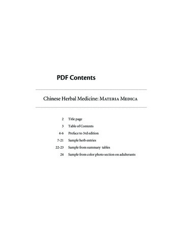
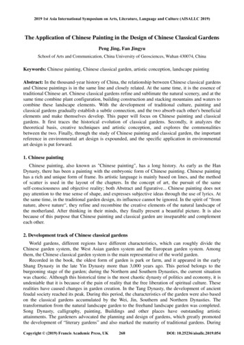
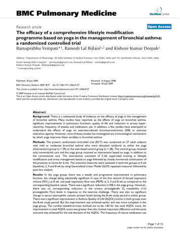
Chinese Medicine 2008, 3:8http://www.cmjournal.org/content/3/1/8tion [9-14], while oxidative stress increases cell proliferation and leads to cell death [2,15-19]. Cellular redox statealso affects cellular signaling pathways, gene expressionand enzymes associated with cell cycle progression[2,6,11,12,15,16,18,19]. Conour et al. identified 92 candidate proteins involved in cell cycle progression that areredox sensitive [20]. Growth and metastasis of tumor cellsmay also be regulated by intracellular redox state[2,17,21,22].Ginseng (Panax ginseng, Renshen) is a popular Chineseherbal medicine known for its wide spectrum of pharmacological actions [23]. Ginseng is composed of a series ofsaponins known as ginsenosides which are derivatives oftriterpene dammarane. There are two categories of ginsenosides, namely protopanaxadiol (PPD, e.g. Ra, Rb, Rc,Rd, Rg3, Rh2) and protopanaxatriol (PPT, e.g. Re, Rf, Rg1,Rg2, Rh1). Each gensenoside is characteristic in its sugarmoiety position on dammarane. Some ginsenosides modulate cardiovascular and neurological functions [24],whereas others act as antioxidants [25-29] to prevent cancer [30-32], protect against chemically induced tissuesdamage [33-36] and delay ageing [25]. Ginsenoside Rehas anti-oxidative abilities apart from its immunomodulatory, antihyperlipidemic and neuroprotective activities.In cardiomyocytes, pretreatment with Re significantlyattenuates H2O2 induced free radical production and protect cell death [37]. Rg3 acts as a prooxidant to accelerate2,2'-azobis (2-amidinopropane) hydrochloride (AAPH)induced haemolysis in human erythrocytes [26]. Rg3 isanti-angiogenic and anticancer through inducing apoptosis [30,38].Ginsenosides are active components in ginseng which hasanticancer properties [23,32,39-41]. The present studyinvestigates how Re and Rg3 suppress the growth of C6glioma cells. The C6 glioma cell is a model for studying aform of malignant brain tumor glioblastoma multiform[42]. The study also investigates the mechanisms throughwhich ginsenosides Re and Rg3 modulate the levels of oxidized and reduced glutathione, and ROS productionMethodsMaterialsCell culture media and all reagents for biochemical assayswere purchased from Sigma Chemical (MO, USA). Perchloric acid (PCA) and potassium hydroxide were purchased from Fisher Scientific (NJ, USA). Antibiotics werepurchased from Life Technologies (NY, USA). Fetalbovine serum (FBS) and 6-carboxy-2,7-dichlorodihydrofluorescein diacetate (H2DCFDA) were purchasedfrom Invitrogen (Scotland, UK) and Molecular Probes(NJ, USA) respectively.Ginsenosides Re and Rg3 (purity 99%) were obtainedfrom International laboratory (USA). The identities ofginsenosdies were confirmed by high-performance liquidchromatography (HPLC) and electrospray ionization –mass spectrometry (ESI-MS). The Rg3, a 20(S) isoform,and the Re were 784 Daltons and 946 Daltons of molecular weight respectively (Figure 1). In this study, the concentration of ginsenosides was based on nominal weight.The ginsenosides were dissolved in dimethyl sulfoxide(DMSO), filtered (0.2 μm membrane) and added to theculture media to achieve various final concentrations.Buthionine-(S, R)-sulfoximine (BSO), an inhibitor ofgamma glutamylcystenyl synthase (γ-GCS), was dissolvedin phosphate buffered saline (PBS) and sterilized througha 0.2 μm membrane. Cultured C6 glioma cells (ATCC noCCL-107) were purchased from the American Type Culture Collection (USA).20(S)-Rg3MW 784 DaltonsReMW 946 DaltonsFigureChemical1 structures of ginsenosides Re and Rg3 (adapted from chemBlink http://www.chemblink.com/)Chemical structures of ginsenosides Re and Rg3 (adapted from chemBlink http://www.chemblink.com/).Page 2 of 8(page number not for citation purposes)
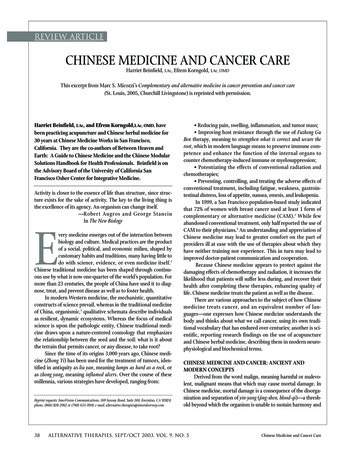
Chinese Medicine 2008, 3:8Cell cultureThe cells were maintained in the F-12 medium supplemented with 10% FBS (tested for lipopolysaccharide) and0.5% antibiotics (amphotericin B and penicillin-streptomycin) in a humidifier under 95% air and 5% CO2 at37 C.Treatment of cellsCells were sub-cultured into 6-well plates until confluence. The incubation media were then removed andreplaced with those containing the required concentrationof Rg3 or Re. The concentration of DMSO in each well waskept at 0.5%, which did not affect intracellular glutathione level and GSH/GSSG. Cells were treated with 100 μMBSO for 24 hours for the study of cell response in theabsence of GSH.Analysis of cell viabilityThe viability of C6 glioma cells treated with Re and Rg3was determined by the m bromide (MTT) assay. Briefly, cellswere cultured in 96-well plates and treated with variousconcentrations (0–800 μg/ml) of Re or Rg3. After incubation, cells were washed twice with PBS. A medium (100μl) containing 500 μg/ml MTT was added to each well.The medium was removed after 3 hours of incubation andDMSO (100 μl) was added to dissolve the formazan crystals. Light absorbance at 595 nm was measured with amicroplate reader (Tecan Infinite F200, Germany). Cellviability was the optical density ratio of a treated cultureover an untreated control.Cell proliferation assayThe effects of either Re or Rg3 on cell proliferation weredetermined by the BrdU cell proliferation kit (Calbiochem, Germany). Cells were cultured in 96-well plates.Upon confluence, they were incubated with either Re orRg3 of various concentrations for 24 hours. BrdU wasadded to the medium 20 hours before the end of incubation. The amount of BrdU incorporated into cells wasdetermined by binding BrdU to mouse anti-BrdU antibody conjugated with horseradish peroxidase. Tetramethylbenzidine, the substrate of horseradish peroxidase,was later added. The color product of the enzymatic reaction was measured in a microplate reader (Tecan InfiniteF200, Germany) at 450–540 nm.Analysis of intracellular glutathione metabolismIntracellular glutathione metabolism is reflected by theinterplay of GSH, GSSG, GPx, GR and γ-GCS. Cells wereincubated for 24 hours before intracellular metaboliteswere extracted by 0.3 M PCA containing 1 mM ethylenediamine-tetraacetic acid (EDTA) with a previouslydescribed method [43]. The levels of GSH and GSSG weredetermined with spectrofluorometry [3]. For , cells were washed twice with pre-warmed PBS andextracted with a protein extraction kit (EMB Biosciences,Calbiochem, USA) with a previously described method[8].Analysis of ROS production by flow cytometryIntracellular production of ROS was evaluated withH2DCFDA which is converted to a fluorescence productdichlorodihydrofluoroscine (DCF) when treated withH2O2. Cells were treated for 24 hours with either 100 μg/ml of BSO or 200 μg/ml of Re. Thirty minutes before theend of experiment, H2DCFDA was added (final concentration 10 μM). The cells were then trypsinized, centrifuged, re-suspended in PBS and passed through theFACScan flow cytometer (Becton Dickinson, U.S.A.). Fluorescence intensities of DCF were measured by excitationwavelength of 488 nm and emission wavelength of 620nm. In each sample, approximately 10000 cells were analyzed by the CellQuest software (Becton Dickinson,U.S.A.).Data analysisCellular redox state was determined by the ratio of GSH/GSSG. Each experiment was repeated 3–8 times. Themeans and standard deviations were calculated. The differences between samples were evaluated by the one wayanalysis of variance (ANOVA), followed by tests for differences between groups by a post-hoc method (Dunnett C)without an assumption of equal variance between groups(SPSS version 14). The differences with P 0.05 were considered statistically significant.ResultsSelection of concentrations of Re and Rg3 for the study ofcell proliferationA 24 and 48 hour dose-response study was conducted.Cells were treated with various concentrations (0–800 μg/ml) of Re and Rg3. There was a significant reduction (P 0.01) in cell viability upon treatment with 100 μg/ml ofRg3 for 24 hours (Figure 2). At 48 hours, a significant difference (P 0.01) in cell viability was observed when Rg3concentration was 50 μg/ml. In contrast, there was no significant change in cell viability after treatment with Re for24 hours. At high concentrations (400 and 800 μg/ml),cell viability decreased 20% after 48 hours.A decrease in cell proliferation was also demonstrated bydecreased BrdU incorporation. While cell viability did notdecrease (Figure 2a) after treatment with low concentrations of Re (50–200 μg/ml), BrdU incorporation (Figure3) decreased 20% (P 0.01). In contrast, Rg3 did notdecrease BrdU incorporation at concentrations below 200μg/ml at which cell viability significantly decreased (P 0.05).Page 3 of 8(page number not for citation purposes)
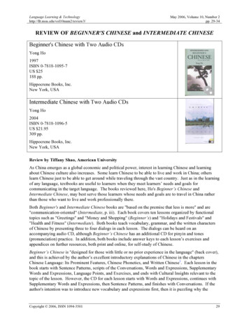
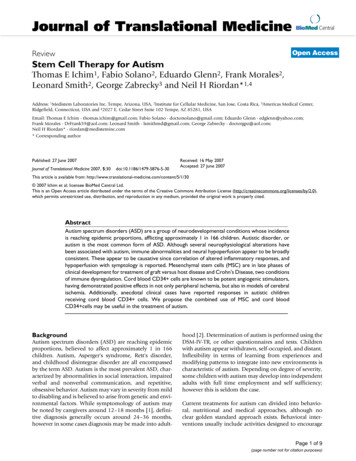
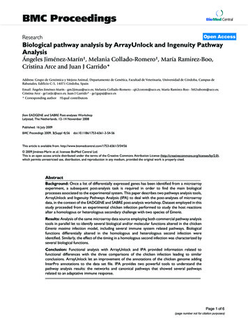
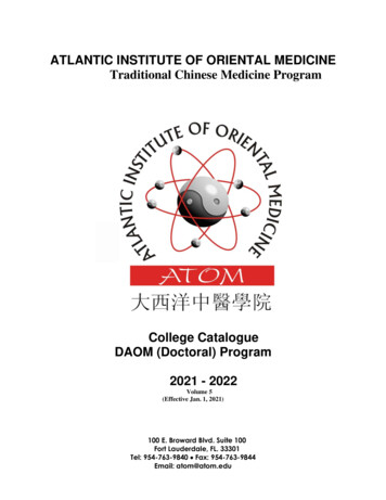
Chinese Medicine 2008, BrdUiousconcentrationsincorporation3ofassayRe inandC6Rg3gliomafor 24cellshourstreated with varBrdU incorporation assay in C6 glioma cells treatedwith various concentrations of Re and Rg3 for 24hours. Each value represents the mean and standard deviation of 8 replicates. *: statistically significant (P 0.05). **:statistically significant (P 0.01).activity of γ-GCS, which is the rate limiting enzyme forGSH synthesis (Figure 8). Rg3 significantly increased (P 0.05) GPx without changing GR (Figure 7b) or γ-GCS(Figure 8).FigureChangesfor24 and2in 48cellhoursviability of cells treated with (a) Re or (b) Rg3Changes in cell viability of cells treated with (a) Re or(b) Rg3 for 24 and 48 hours. Each value represents themean and standard deviation of 8 replicates. **: statisticallysignificant (P 0.01)Glutathione metabolismThe resting level of GSH is three folds higher than that ofGSSG. The GSH/GSSG ratio ranges from 2 (in tumor cells)to 40 (in normal cells) [44]. The GSH/GSSG ratio may bealtered by either toxicants [3] or antioxidants [45]. Retreatment increased GSH but not GSSG (Figure 4a), whileRg3 increased GSSG (Figure 4b) but not GSH. Rg3 treatment decreased the GSH/GSSG ratio, while Re treatmentincreased it (Figure 5). A decrease in the GSH/GSSG ratioafter treatment with Rg3 indicated oxidative stress. ROSproduction was increased after treatment with Rg3 (Figure6). Rg3, which decreased the GSH/GSSG ratio at 200 μg/ml, also significantly increased ROS generation (Figure 6).The results show that Rg3 and Re treatments changed theintracellular GSH/GSSG ratio in opposite directions.Re treatment did not significantly change the activity ofeither GPx or GR (Figure 7a) but increased (P 0.05) theSuppreession of γ-GCS activity by BSO reverses Re inducedchangesTo further confirm that Re acts by changing γ-GCS activity,treatment of cells with BSO, an inhibitor of γ-GCS whichcould significantly lowered (P 0.01) the GSH/GSSG ratio(Figure 9) and increased (P 0.05) cellular ROS production (Figure 6), was able to reverse the Re induced changein both GSH/GSSG (Figure 9) and BrdU incorporation(Figure 10).DiscussionThe present study showed that both Re and Rg3 affectedthe survival of C6 glioma cells. While Re suppressed thegrowth of C6 glioma cells at low concentrations, Rg3caused death of C6 glioma cells via oxidative stress. Furthermore, we demonstrated that the action of Re was correlated with an increase in the intracellular GSH level,thereby raising the GSH/GSSG ratio. BSO (an inhibitor ofγ-GCS) treatment reversed the GSH/GSSG rise induced byRe (Figure 9) and decreased BrdU incorporation in thepresence of Re (Figure 10).γ-GCS is the rate limiting enzyme in glutathione synthesis.The enzyme and its gene expression may be modulated bya number of factors. For example, an increase in the γ-GCSactivity may be stimulated by oxidative metabolites. Shi etPage 4 of 8(page number not for citation purposes)
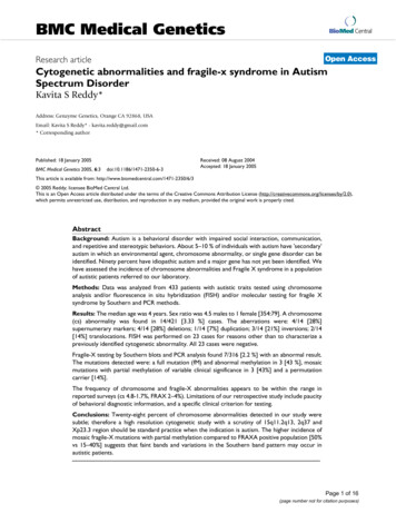
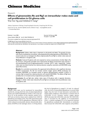
Chinese Medicine 2008, ngewithRe 5inandtheRg3GSH/GSSHfor 24 hoursratio of C6 glioma cells treatedChange in the GSH/GSSH ratio of C6 glioma cellstreated with Re and Rg3 for 24 hours. Each value represents the mean and standard deviation of 4 replicates. *: statistically significant (P 0.05). **: statistically significant (P 0.01).tion [47]. Such feedback activation may be an underlyingcause of cellular adaptation to oxidative stress. Therefore,similar adaptive response may take place in C6 gliomacells following Re treatment.FigureChanges(a)Re or4in(b)theRg3GSHfor and24 hoursGSSG levels upon treatment withChanges in the GSH and GSSG levels upon treatment with (a) Re or (b) Rg3 for 24 hours. Each valuerepresents the mean and standard deviation of 4 replicates. *:statistically significant (P 0.05). **: statistically significant (P 0.01).al. showed that the heavy subunit mRNA level and subsequent enzyme activity increased in response to 2,3dimethoxy-1,4-napthoquinone (DMNQ) through transcriptional activation of γ-GCS [46]. Our previous studiesshowed that the production of quinone metabolite ofbenzo [a]pyrene activated γ-GCS [8]. However, no suchquinone metabolites were detected in the metabolism ofRe in biological tissues. Furthermore, the present studyshowed that Re treatment suppressed free radical production (Figure 6), which did not support the free radical production hypothesis in γ-GCS activation. Another studyshowed that PC12 cells' tolerance of 7-hydroxycholesteroland 15-deoxy-12,13- prostaglandin J2 was achieved viaup-regulation of cellular glutathione by γ-GCS r BSOgliomalevel(100indicatedcellsμM)treatedforby the24withhoursfluoReChanges in the intracellular ROS level indicated bythe fluorescence intensity (FLH-1) in C6 glioma cellstreated with Re (200 μg/ml), Rg3 (200 μg/ml) or BSO(100 μM) for 24 hours. The cytogram represents threeindependent experiments. The x-axis and y-axis denote fluorescence intensity and cell count respectively.Page 5 of 8(page number not for citation purposes)
Chinese Medicine 2008, 3:8http://www.cmjournal.org/content/3/1/8Figure withChangestreated7in the(a)GRRe and GPx(b) Rg3activitiesfor 24ofhoursC6 glioma cellsChanges in the GR and GPx activities of C6 gliomacells treated with (a) Re and (b) Rg3 for 24 hours.Each value represents the mean and standard deviation of 4replicates. *: statistcally significant (P 0.05).FigureChangesReand Rg38in thefor γ-GCS24 hoursactivity of C6 glioma cells treated withChanges in the γ-GCS activity of C6 glioma cellstreated with Re and Rg3 for 24 hours. Each value represents the mean and standard deviation of 4 replicates. *: statistically significant (P 0.05).A number of factors must be considered before using Reto treat brain tumor. Firstly, Re suppresses cell proliferation at a low rate. At a low concentration of 50 μg/ml, Rewas effective in suppressing cell proliferation (Figure 3).However, there was only a 10% decrease in BrdU uptakeafter 24 hours (Figure 2a), and only a 20% decrease in cellviability after 48 hours. Secondly, Re is metabolized byintestinal flora via deglycosylation and fatty acid esterification [48], which may limit the application of Re givenper oral. Finally, it is not clear whether normal astrocytesrespond similarly to Re. In current anticancer strategies,both normal and tumor cells are affected. Menon et al.demonstrated that exposure of both nonmalignanthuman breast epithelial (MCF-10A) and breast cancercells (MCF-7 and MDA-MB-231) to NAC decreased theoxidation of a prooxidant-sensitive dye in MCF-10A cells,but not in MCF-7 and MDA-MB-231 cells, suggesting thatmalignant and non-malignant cells may have differentresponses to oxidative stress [17]. We will further investigate whether C6 glioma cells and normal astrocytesrespond differently to Re treatment.ConclusionRe suppresses the growth and proliferation of C6 gliomacells and increases the cellular GSH/GSSG ratio byenhancing the γ-GCS activity, suppressing ROS generationand reducing cell proliferation rate. Rg3 causes cell deathvia oxidative stress. While both Re and Rg3 eliminate thegrowth and proliferation of C6 glioma cells, they modulate cellular redox state in opposite directions. Yue et al.suggest that ginseng or selected ginsenosides modulatecellular function via specific intracellular receptors in away that may reflect the Ying/Yang actions of ginseng[49]. The opposite actions in modulating cellular redoxPage 6 of 8(page number not for citation purposes)
Chinese Medicine 2008, e; DMSO: dimethyl sulfoxide; EDTA:ethylenediamine-tetraacetic acid; ESI-MS: electrosprayionization – mass spectrometry; Fetal bovine serum(FBS); GPx: glutathione peroxidase; GR: glutathionereductase; GSH: reduced glutathione; GSSG: oxidized glutathione; H2DCFDA: 6-carboxy-2,7-dichlorodihydrofluorescein diacetate; HPLC: high-performance liquidchromatography; MTT: m bromide; NAC: N-acetylcysteine;NADPH: nicotinamide adenine dinucleotide phosphate; PBS: phosphate buffered saline; Perchloric acid (PCA);PPD: protopanaxadiol; PPT: protopanaxatriol; -ROS: reactive oxygen speciesCompeting interestsThe authors declare that they have no competing interests.FigureChangesReand/or9inBSOthe GSHfor 24andhoursGSSG levels upon treatment withChanges in the GSH and GSSG levels upon treatment with Re and/or BSO for 24 hours. Each value represents the mean and standard deviation of 4 replicates.Symbols indicate that the treatment groups are significantlydifferent (P 0.05) from each other.Authors' contributionsWN is an MPhil student who carried out the experimentsand helped draft the manuscript. MY supervised the studyand drafted the manuscript. Both authors read andapproved the final version of the manuscript.Acknowledgementsstate by Re and Rg3 may be another example of the Ying/Yang actions of ginseng.Abbreviationsγ-GCS: gamma glutamylcystenyl synthase; AAPH: 2,2'azobis (2-amidinopropane) hydrochloride; BSO: buthionine-(S, R)-sulfoximine; DMNQ: 2,3-dimethoxy-1,4-nap-The authors wish to thank Prof ZH Jiang, School of Chinese Medicine, HongKong Baptist University for the HPLC and ESI-MS analyses to confirm theginsenosides Re and Rg3 used in this study. This study was supported bythe Hong Kong Baptist University Faculty Research corporation10for 24 hoursassay in C6 glioma cells treated with ReBrdU incorporation assay in C6 glioma cells treatedwith Re and BSO for 24 hours. Each value represents themean and standard deviation of 4 replicates. Symbols indicatethat the treatment groups are significantly different (P 0.05)from each other.9.10.11.Jones DP: Redox potential of GSH/GSSG couple: assay andbiological significance. Methods Enzymol 2002, 348:93-112.Jonas CR, Ziegler TR, Gu LH, Jones DP: Extracellular thiol/disulfide redox state affects proliferation rate in a humancolon carcinoma (Caco2) cell line. Free Radic Biol Med 2002,33:1499-1506.Yang MS, Yu LC, Gupta RC: Analysis of changes in energy andredox states in HepG2 hepatoma and C6 glioma cells uponexposure to cadmium. Toxicology 2004, 201:105-113.Andersson A, Lindgren A, Arnadottir M, Prytz H, Hultberg B: Thiolsas a measure of plasma redox status in healthy subjects andin patients with renal or liver failure. Clin Chem 1999,45:1084-1086.Hultberg B, Andersson A, Isaksson A: Thiol and redox reactiveagents exert different effects on glutathione metabolism inHeLa cell cultures. Clin Chim Acta 1999, 283:21-32.Menon SG, Goswami PC: A redox cycle within the cell cycle:ring in the old with the new. Oncogene 2007, 26:1101-1109.De Flora S, Izzotti A, D'Agostini F, Balansky RM: Mechanisms of Nacetylcysteine in the prevention of DNA damage and cancer,with special reference to smoking-related end-points. Carcinogenesis 2001, 22:999-1013.Lin T, Yang MS: Benzo[a]pyrene-induced elevation of GSHlevel protects against oxidative stress and enhances xenobiotic detoxification in human HepG2 cells. Toxicology 2007,235:1-10.Fisher JC, Kling DE, Kinane TB, Schnitzer JJ: Oxidation-reduction(redox) controls fetal hypoplastic lung growth. J Surg Res 2002,106:287-291.Hoffman A, Spetner LM, Burke M: Cessation of cell proliferationby adjustment of cell redox potential. J Theor Biol 2001,211:403-407.Kim KY, Rhim T, Choi I, Kim SS: N-acetylcysteine induces cellcycle arrest in hepatic stellate cells through its reducingactivity. J Biol Chem 2001, 276:40591-40598.Page 7 of 8(page number not for citation purposes)
Chinese Medicine 2008, .28.29.30.31.32.33.34.Menon SG, Sarsour EH, Spitz DR, Higashikubo R, Sturm M, Zhang H,Goswami PC: Redox regulation of the G1 to S phase transitionin the mouse embryo fibroblast cell cycle. Cancer Res 2003,63:2109-2117.Nkabyo YS, Ziegler TR, Gu LH, Watson WH, Jones DP: Glutathione and thioredoxin redox during differentiation in humancolon epithelial (Caco-2) cells. Am J Physiol Gastrointest Liver Physiol 2002, 283:G1352-G1359.Sekharam M, Trotti A, Cunnick JM, Wu J: Suppression of fibroblast cell cycle progression in G1 phase by N-acetylcysteine.Toxicol Appl Pharmacol 1998, 149:210-216.Burdon RH, Gill V, Rice-Evans C: Oxidative stress and tumourcell proliferation. Free Radic Res Commun 1990, 11:65-76.Kusano C, Takao S, Noma H, Yoh H, Aikou T, Okumura H, AkiyamaS, Kawamura M, Makino M, Baba M: N-acetyl cysteine inhibits cellcycle progression in pancreatic carcinoma cells. Hum Cell2000, 13:213-220.Menon SG, Coleman MC, Walsh SA, Spitz DR, Goswami PC: Differential susceptibility of nonmalignant human breast epithelialcells and breast cancer cells to thiol antioxidant-inducedG(1)-delay. Antioxid Redox Signal 2005, 7:711-718.Burdon RH, Rice-Evans C: Free radicals and the regulation ofmammalian cell proliferation. Free Radic Res Commun 1989,6:345-358.Burdon RH, Gill V, Rice-Evans C: Cell proliferation and oxidativestress. Free Radic Res Commun 1989, 7:149-159.Conour JE, Graham WV, Gaskins HR: A combined in vitro/bioinformatic investigation of redox regulatory mechanisms governing cell cycle progression. Physiol Genomics 2004, 18:196-205.Futakuchi M, Ogawa K, Tamano S, Takahashi S, Shirai T: Suppression of metastasis by nuclear factor kappaB inhibitors in anin vivo lung metastasis model of chemically induced hepatocellular carcinoma. Cancer Sci 2004, 95:18-24.Martin V, Herrera F, Carrera-Gonzalez P, Garcia-Santos G, Antolin I,Rodriguez-Blanco J, Rodriguez C: Intracellular signaling pathways involved in the cell growth inhibition of glioma cells bymelatonin. Cancer Res 2006, 66:1081-1088.Attele AS, Wu JA, Yuan CS: Ginseng pharmacology: multipleconstituents and multiple actions. Biochem Pharmacol 1999,58:1685-1693.Attele AS, Zhou YP, Xie JT, Wu JA, Zhang L, Dey L, Pugh W, Rue PA,Polonsky KS, Yuan CS: Antidiabetic effects of Panax ginsengberry extract and the identification of an effective component. Diabetes 2002, 51:1851-1858.Yokozawa T, Satoh A, Cho EJ: Ginsenoside-Rd attenuates oxidative damage related to aging in senescence-acceleratedmice. J Pharm Pharmacol 2004, 56:107-113.Liu ZQ, Luo XY, Sun YX, Chen YP, Wang ZC: Can ginsenosidesprotect human erythrocytes against free-radical-inducedhemolysis? Biochim Biophys Acta 2002, 1572:58-66.Liu ZQ, Luo XY, Liu GZ, Chen YP, Wang ZC, Sun YX: In vitro studyof the relationship between the structure of ginsenoside andits antioxidative or prooxidative activity in free radicalinduced hemolysis of human erythrocytes. J Agric Food Chem2003, 51:2555-2558.Deng HL, Zhang JT: Anti-lipid peroxilative effect of ginsenosideRb1 and Rg1. Chin Med J (Engl ) 1991, 104:395-398.Chen XC, Zhou YC, Chen Y, Zhu YG, Fang F, Chen LM: Ginsenoside Rg1 reduces MPTP-induced substantia nigra neuron lossby suppressing oxidative stress. Acta Pharmacol Sin 2005,26:56-62.Zhang Q, Kang X, Zhao W: Antiangiogenic effect of low-dosecyclophosphamide combined with ginsenoside Rg3 on Lewislung carcinoma. Biochem Biophys Res Commun 2006, 342:824-828.Wang W, Zhao Y, Rayburn ER, Hill DL, Wang H, Zhang R: In vitroanti-cancer activity and structure-activity relationships ofnatural products isolated from fruits of Panax ginseng. CancerChemother Pharmacol 2007, 59:589-601.Li G, Wang Z, Sun Y, Liu K, Wang Z: Ginsenoside 20(S)-protopanaxadiol inhibits the proliferation and invasion of humanfibrosarcoma HT1080 cells. Basic Clin Pharmacol Toxicol 2006,98:588-592.Yokozawa T, Liu ZW, Dong E: A study of ginsenoside-Rd in arenal ischemia-reperfusion model. Nephron 1998, 78:201-206.Yokozawa T, Owada S: Effect of ginsenoside-Rd in cephaloridine-induced renal disorder. Nephron 1999, wa T, Liu ZW: The role of ginsenoside-Rd in cisplatininduced acute renal failure. Ren Fail 2000, 22:115-127.Yokozawa T, Dong E: Role of ginsenoside-Rd in cisplatininduced renal injury: special reference to DNA fragmentation. Nephron 2001, 89:433-438.Xie JT, Shao ZH, Vanden Hoek TL, Chang WT, Li J, Mehendale S,Wang CZ, Hsu CW, Becker LB, Yin JJ, Yuan CS: Antioxidanteffects of ginsenoside Re in cardiomyocytes. Eur J Pharmacol2006, 532:201-207.Yue PY, Wong DY, Wu PK, Leung PY, Mak NK, Yeung HW, Liu L, CaiZ, Jiang ZH, Fan TP, Wong RN: The angiosuppressive effects of20(R)- ginsenoside Rg3. Biochem Pharmacol 2006, 72:437-445.Kumar A, Kumar M, Panwar M, Samarth RM, Park TY, Park MH,Kimura H: Evaluation of chemopreventive action of Ginsenoside Rp1. Biofactors 2006, 25:29-43.Popovich DG, Kitts DD: Ginsenosides 20(S)-protopanaxadioland Rh2 reduce cell proliferation and increase sub-G1 cells intwo cultured intestinal cell lines, Int-407 and Caco-2. Can JPhysiol Pharmacol 2004, 82:183-190.Wu G, Fang YZ, Yang S, Lupton JR, Turner ND: Glutathionemetabolism and its implications for health. J Nutr 2004,134:489-492.Grobben B, De Deyn PP, Slegers H: Rat C6 glioma as experimental model system for the study of glioblastoma growth andinvasion. Cell Tissue Res 2002, 310:257-270.Yang MS, Gupta RC: Determination of energy charge potentialin the C6 glioma and the HepG-2 cell culture. Toxicology Mechanisms and Methods 2003, 13:97-101.Hissin PJ, Hilf R: A fluorometric method for determination ofoxidized and reduced glutathione in tissues. Anal Biochem1976, 74:214-226.Yang MS, Yu LC, S.W. P: Manipulation of energy and redoxstates in the C6 glioma cells by buthionine sulfoxamine andN-acetylcysteine and the effect on cell survival to cadmiumtoxicity. Cellular and Molecular Biology 2007, 53:56-61.Shi MM, Iwamoto T, Forman HJ: gamma-Glutamylcysteine synthetase and GSH increase in quinone-induced oxidativestress in BPAEC. Am J Physiol 1994, 267:L414-L421.Che
FACScan flow cytometer (Becton Dickinson, U.S.A.). Flu-orescence intensities of DCF were measured by excitation wavelength of 488 nm and emission wavelength of 620 nm. In each sample, approximately 10000 cells were ana-lyzed by the CellQuest software (Becton Dickinson, U.S.A.). Data analysis Cellular redox state was determined by the ratio of .