Transcription
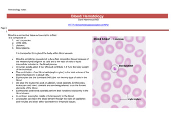
Hematology notesBlood/ HematologyDanil age 1Blood is a connective tissue whose matrix is fluid.It is composed of:1. red corpuscles,2. white cells,3. platelets,4. blood plasma.It is transported throughout the body within blood vessels. Blood is sometimes considered to be a fluid connective tissue because ofthe mesenchymal origin of its cells and a low ratio of cells to liquidintercellular substance, the blood plasma.In human adults about 5 liter of blood contribute 7-8 % to the body weightof the individual.The contribution of red blood cells (erythrocytes) to the total volume of theblood (haematocrit) is about 43%.Erythrocytes are the dominant (99%) but not the only type of cells in theblood.We also find leukocytes and, in addition, blood platelets. Erythrocytes,leukocytes and blood platelets are also being referred to as the formedelements of the blood.Erythrocytes and blood platelets perform their functions exclusively in theblood stream.In contrast, leukocytes reside only temporarily in the blood.Leukocytes can leave the blood stream through the walls of capillariesand venules and enter either connective or lymphoid tissues.
Hematology notesPage 2
Hematology notesPage 3Blood facts Approximately 8% of an adult's body weight is made up of blood.Females have around 4-5 litres, while males have around 5-6 litres. This difference is mainly due to the differences in body size between men and women.Its mean temperature is 38 degrees Celcius.It has a pH of 7.35-7.45, making it slightly basic (less than 7 is considered acidic).Whole blood is about 4.5-5.5 times as viscous as water, indicating that it is more resistant to flow than water.This viscosity is vital to the function of blood because if blood flows too easily or with too much resistance, it can strain the heart and lead to severe cardiovascularproblems.Blood in the arteries is a brighter red than blood in the veins because of the higher levels of oxygen found in the arteries.An artificial substitute for human blood has not been found.There are three major categories of plasma proteins, and each individual type of proteins has its own specific properties and functions in addition to their overall collective role:1.2.3.Albumins, which are the smallest and most abundant plasma proteins. Reductions in plasma albumin content can result in a loss of fluid from the blood and a gain of fluidin the interstitial space (space within the tissue), which may occur in nutritional, liver and kidney disease. Albumin also helps many substances dissolve in the plasma bybinding to them, hence playing an important role in plasma transport of substances such as drugs, hormones and fatty acids.Globulins, which can be subdivided into three classes from smallest to largest in molecular weight into alpha, beta and gamma globulins. The globulins include highdensity lipoproteins (HDL), an alpha-1 globulin, and low density lipoproteins (LDL), a beta-1 globulin. HDL functions in lipid transport carrying fats to cells for use in energymetabolism, membrane reconstruction and hormone function. HDLs also appear to prevent cholesterol from invading and settling in the walls of arteries. LDL carriescholesterol and fats to tissues for use in manufacturing steroid hormones and building cell membranes, but it also favours the deposition of cholesterol in arterial walls andthus appears to play a role in disease of the blood vessels and heart. HDL and LDL therefore play important parts in the regulation of cholesterol and hence have a largeimpact on cardiovascular disease.Fibrinogen, which is a soluble precursor of a sticky protein called fibrin, which forms the framework of blood clot. Fibrin plays a key role in coagulation of blood, which isdiscussed later in this article under Platelets.
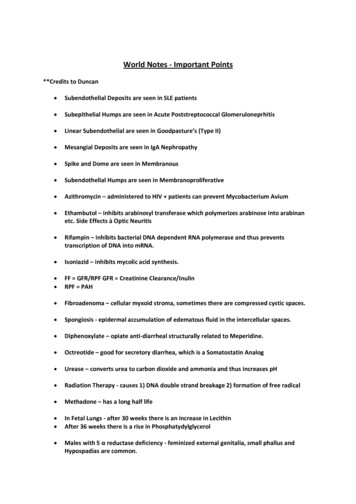
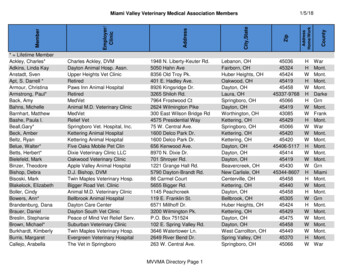
Hematology notesAmino acids These are formed from the break down of tissue proteins or from the digestion of digested proteins.Nitrogenous waste Being toxic end products of the break down of substances in the body, these are usually cleared from the bloodstream and are excreted by the kidneys at a ratethat balances their production.Page Nutrients Those absorbed by the digestive tract are transported in the blood plasma. These include glucose, amino acids, fats, cholesterol, phospholipids, vitamins and minerals.4Gases Some oxygen and carbon dioxide are transported by plasma. Plasma also contains a substantial amount of dissolved nitrogen.Electrolytes The most abundant of these are sodium ions, which account for more of the blood's osmolarity than any other solute.Hemopoiesis During fetal development, the formation of blood cells (hemopoiesis ) commences in wall of the yolk sac.After the second month of fetal development, the liver, and, slightly later, the spleen, become the dominant sites of hemopoiesis .From the 6th month, and dominating from the 7th month onwards, the formation of blood cells occurs in bone marrow, which is the major site offormation blood cells in normal adult humans.Yellow bone marrow, which harbours mainly adipocytes, dominates in the hollow of the diaphysis of adult long bones.Hemopoiesis occurs in red bone marrow, which is typically found between the trabeculae of spongy bone in the epiphysis of adult long bones.Both age and demands on hemopoiesis may effect the relative amounts of red and yellow bone marrow.Hemopoietic cells surround the vascular sinusoids and are supported by reticular connective tissue.In addition to the endothelial cells of the sinusoids and the reticulocytes of the connective tissue, macrophages are frequent in red bone marrow.ErythropoiesisErythropoiesis, the process of makingerythrocytes, begins with the formation ofproerythroblasts from hemopoietic stem cells.Over three to five days, several stages ofdevelopment follow as ribosomes proliferate andhemoglobin is synthesized.Finally, the nucleus is ejected, producing thedepression in the center of the cell.Young erythrocytes, called reticulocytes, stillcontaining some ribosomes and endoplasmicreticulum, pass into the bloodstream anddevelop into mature erythrocytes after anotherLeukopoiesisLeukopoiesis, the process of making leukocytes,is stimulated by various colony-stimulating factors(CSFs), hormones produced by mature whiteblood cells.The development of each kind of white blood cellbegins with the division of themopoietic stemcells into one of the following “blast” cells. Myeoblasts divide to form eosinophilic,neutrophilic, or basophilic myelocytes,which lead to the development of thethree kinds of granulocytes.Monoblasts lead to the development ofmonocytes.ThrombopoiesisThrombopoiesis, the process of makingplatelets, begins with the formation ofmegakaryoblasts from hemopoietic stem cells.The megakaryoblasts divide without cytokinesisto become megakaryocytes, huge cells with alarge, multilobed nucleus.The megakaryocytes then fragment intosegments as the plasma membrane infolds intothe cytoplasm.
Hematology notesone or two days. Lymphoblasts lead to the development oflymphocytes.ErythroproteinErythroprotein (EPO), a hormone producedmostly by the kidneys, stimulates bone marrowto produce erythrocytes.Page 5When inadequate amounts of oxygen aredelivered to body cells, a condition calledhypoxia, the kidneys increase EPO secretion,which, in turn, stimulates an increase inerythrocyte production.The average production rate of erythrocytes inhealthy individuals is two million cells persecond. Normal production requires adequateamounts of iron and vitamin B12 and folic acid.Hemopoietic CellsThe basis of hemopoiesis is a small population of self-replicating stem cells, which ultimately can generate all types of blood cells. Their progeny may develop intoeither lymphocytic stem cells or pluripotent haemal stem cells (colony-forming unit - stem cell - CFU-S). The latter type gives rise to stem cells which can form themajor groups of blood cells other than lymphocytes. Depending on their progeny it is possible to differentiate burst-forming unit of the erythroid line (BFU-E),colony-forming unit - granulocytes and macrophages (CFU-G/M), andcolony-forming unit - megakaryocytes (CFU-Mk).
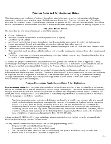
Hematology notesErythrocytes Page 6 The first identifiable stage of erythropoiesis is the proerythroblast - a large, slightly basophiliccell, which contains a large, lightly stained nulceus.Proerythroblasts proliferate to generate a sequence of cells which show a gradual decrease insize and condensation of their chromatin.They are named after changes in the staining characteristic of their cytoplasm (basophilicerythroblast, polychromatophilic and orthochromic normoblasts).The nucleus is finally extruded from the normoblast.The cell enters circulation as a reticulocyte, which still contains some organelles.Reticulocytes remain for a few days in either the bone marrow or the spleen to mature toerythrocytes.In some blood smears reticulocytes may be recognisable because of a very slight basophilicstaining - either homogeneous or in the form of a basophilic stippling.Granulocytes Myeloblast appear light-microscopically similar to proerythroblast.They proliferate to generate promyelocytes. Promyelocytes begin to accumulate nonspecific granules, but they are still able to divide.The maturation of their progeny, the myelocytes, is characterised by the accumulation of specific granules and changes in nuclear morphology.Metamyelocytes have a C-shaped nucleus.Blood Platelets (Thrombocytes) are, as mentioned above, fragments of the cytoplasm of megakaryocytes.Megakaryocytes are very large cells (up to 160 µm in diameter), which contain very large, highly lobulated, polyploid nuclei.Megakaryocytes are in turn the product of the differentiation of basophilic megakaryoblasts.Precursors of blood cells which are usually only found in the bone marrow can be found in peripheral blood in a variety of pathological conditions.If a Rh-negative mother has been immunised by erythrocytes of a Rh-positive foetus, a condition called Erythroblastosis fetalis may develop during subsequentpregnancies.It would show itself in the foetus or newborn by the presence of erythrocyte precursors in peripheral blood - although other, more severe symptoms should beobvious.Chronic myeloid leukemia is another condition - in this case showing itself by the presence of all types of granulocyte precursors in peripheral blood.
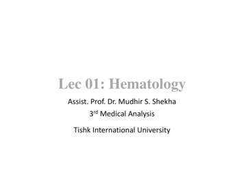
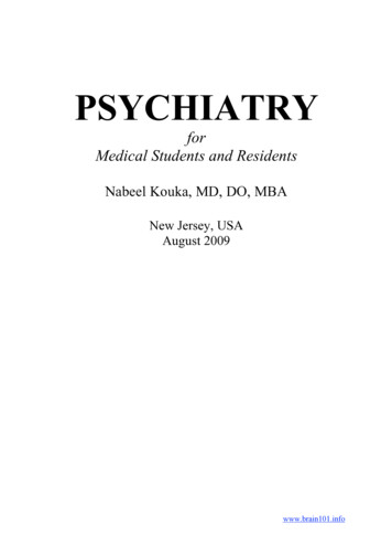
Hematology notes Page 7The nomenclature employed for haemopoietic cells (but not the number of stages recognized) is somewhat variable across texts.Note also that these cell types refer to stages of development along a morphologically more or less continuous spectrum.Most of the haemopoietic cells visible will be of the erythroblastic line. The only cell type of this line which is easy to distinguish in H&E stained sectionsare normoblasts.A very condensed nucleus is seen in late (orthochromic) normoblast. Granulocyte and erythrocyte precursors will mostly intermingle, but may bedistinguished by nuclear morphology and/or size. A bent nucleus is found in metamyelocytes - this shape is very pronounced in the last, immature form ofneutrophils, which are also called stab or band cells. If the cell(1) is large, with a distinct "clearing" in the otherwise pink cytoplasm and(2) has an ovoid or slightly indented nucleus, it is likely to be a myelocyte. Cells with large light nuclei and almost unstained cytoplasm are either reticulocytes ormacrophages. Macrophages are frequently associated with normoblasts, and together these cells form erythroblastic islands. The name for macrophages in theseislands, nurse cells, may tell you a bit about their function in addition to the scavenging of the expelled nuclei. If you still have some time and are desperate to get frustrated try to hunt up a nice basophilic erythroblast - a basophilic cell with homogeneously stainingnucleus that is somewhat smaller than the nuclei of granulocyte precursors.The formation of blood cells (cell types and acronyms are defined below)
Hematology notesAll the various types of blood cellsPage 8 are produced in the bone marrow (some 1011 of them each day in an adult human!).arise from a single type of cell called a hematopoietic stem cell — an "adult" multipotent stem cell.These stem cells are very rare (only about one in 10,000 bone marrow cells);are attached (probably by adherens junctions) to osteoblasts lining the inner surface of bone cavities;express a cell‐surface protein designated CD34;produce, by mitosis, two kinds of progeny:o more stem cells (A mouse that has had all its blood stem cells killed by a lethal dose of radiation can be saved by the injection of a single living stem cell!).o cells that begin to differentiate along the paths leading to the various kinds of blood cells.Which path is taken is regulated by the need for more of that type of blood cell which is, in turn, controlled by appropriate cytokines and/or hormones.For example, Interleukin-7 (IL-7) is the major cytokine in stimulating bone marrow stem cells to start down the "lymphoid" path leading to thevarious lymphocytes (mostly B cells and T cells).Some of the cytokines that drive the differentiation of the "myeloid" leukocytes are Erythropoietin (EPO), produced by the kidneys, enhances the production of red blood cells (RBCs).Thrombopoietin (TPO), assisted by Interleukin‐11 (IL‐11), stimulates the production of megakaryocytes. Their fragmentation produces platelets.Granulocyte‐macrophage colony‐stimulating factor (GM‐CSF), as its name suggests, sends cells down the path leading to both those cell types. In due course, one pathor the other is taken.o Under the influence of granulocyte colony‐stimulating factor (G‐CSF), they differentiate into neutrophils.o Further stimulated by interleukin‐5 (IL‐5) they develop into eosinophils.o Interleukin‐3 (IL‐3) participates in the differentiation of most of the white blood cells but pl
Hematology notes Page 3 Blood facts Approximately 8% of an adult's body weight is made up of blood. Females have around 4-5 litres, while males have around 5-6 litres. This difference is mainly due to the differences in body size between men and women. Its mean temperature is 38 degrees Celcius. It has a pH of 7.35-7.45, making it slightly basic (less than 7 is considered .