Transcription
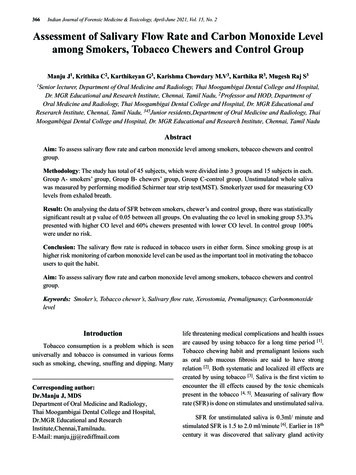
366Indian Journal of Forensic Medicine & Toxicology, April-June 2021, Vol. 15, No. 2Assessment of Salivary Flow Rate and Carbon Monoxide Levelamong Smokers, Tobacco Chewers and Control GroupManju J1, Krithika C2, Karthikeyan G3, Karishma Chowdary M.V3, Karthika R3, Mugesh Raj S31Senior lecturer, Department of Oral Medicine and Radiology, Thai Moogambigai Dental College and Hospital,Dr. MGR Educational and Research Institute, Chennai, Tamil Nadu, 2Professor and HOD, Department ofOral Medicine and Radiology, Thai Moogambigai Dental College and Hospital, Dr. MGR Educational andReserarch Institute, Chennai, Tamil Nadu, 345Junior residents,Department of Oral Medicine and Radiology, ThaiMoogambigai Dental College and Hospital, Dr. MGR Educational and Research Institute, Chennai, Tamil NaduAbstractAim: To assess salivary flow rate and carbon monoxide level among smokers, tobacco chewers and controlgroup.Methodology: The study has total of 45 subjects, which were divided into 3 groups and 15 subjects in each.Group A- smokers’ group, Group B- chewers’ group, Group C-control group. Unstimulated whole salivawas measured by performing modified Schirmer tear strip test(MST). Smokerlyzer used for measuring COlevels from exhaled breath.Result: On analysing the data of SFR between smokers, chewer’s and control group, there was statisticallysignificant result at p value of 0.05 between all groups. On evaluating the co level in smoking group 53.3%presented with higher CO level and 60% chewers presented with lower CO level. In control group 100%were under no risk.Conclusion: The salivary flow rate is reduced in tobacco users in either form. Since smoking group is athigher risk monitoring of carbon monoxide level can be used as the important tool in motivating the tobaccousers to quit the habit.Aim: To assess salivary flow rate and carbon monoxide level among smokers, tobacco chewers and controlgroup.Keywords: Smoker’s, Tobacco chewer’s, Salivary flow rate, Xerostomia, Premalignancy, CarbonmonoxidelevelIntroductionTobacco consumption is a problem which is seenuniversally and tobacco is consumed in various formssuch as smoking, chewing, snuffing and dipping. ManyCorresponding author:Dr.Manju J, MDSDepartment of Oral Medicine and Radiology,Thai Moogambigai Dental College and Hospital,Dr.MGR Educational and ResearchInstitute,Chennai,Tamilnadu.E-Mail: manju.jjj@rediffmail.comlife threatening medical complications and health issuesare caused by using tobacco for a long time period [1].Tobacco chewing habit and premalignant lesions suchas oral sub mucous fibrosis are said to have strongrelation [2]. Both systematic and localized ill effects arecreated by using tobacco [3]. Saliva is the first victim toencounter the ill effects caused by the toxic chemicalspresent in the tobacco [4, 5]. Measuring of salivary flowrate (SFR) is done on stimulates and unstimulated saliva.SFR for unstimulated saliva is 0.3ml/ minute andstimulated SFR is 1.5 to 2.0 ml/minute [6]. Earlier in 18thcentury it was discovered that salivary gland activity
Indian Journal of Forensic Medicine & Toxicology, April-June 2021, Vol. 15, No. 2is increased in smoking. But the observation was thatonly during the initial stages of smoking habit peoplehad increased salivary gland activity. [7]. Long termuse of tobacco causes alteration in the taste receptorsfunction leading to decrease in salivary reflux. Maincontent of tobacco is nicotine which acts on cholinergicreceptors of brain causing decrease in salivary secretion.[8]. This decrease in salivary flow leads to disorders suchas dysgeusia, pain and burning mouth, dental cariesand other oral infectious diseases. [9]. During smokingcarbon monoxide (CO) enters the blood circulationfrom cigarette smoke and forms carboxyhaemoglobin(COHb).Elimination of CO occurs primarily throughrespiration and a strong correlation exists betweenCObreath and COHb[10, 11, 12] enabling it to be used as aimportant tool for assessing smoking status.This study is conducted to assess salivary flow ratein different groups with many parameters along withassessment of carbon monoxide level in smokers andtobacco chewers.Materials and MethodologyThe study participants are the patients reported todepartment of oral medicine and radiology of a privatedental college in Chennai. The subjects were explainedabout the purpose and procedure of this study and consentwas obtained. The demographic details with history ofsmoking and chewing habit were also collected fromthe subjects. Ethical clearance was obtained from theinstitutional ethical committee.Inclusion criteria include subjects of age above 18years and who has a habit of smoking, chewing for atleast 6 months and the subjects who do not have anysystemic disease.367Exclusion criteria include subjects below 18 yearsof age, subjects with systemic diseases, salivary glanddisease, the patients under radiotherapy and the patientswho had quit their habit.The study has total of 45 subjects, which weredivided into 3 groups (A, B, and C) and 15 subjects ineach.Group A- subjects who have smoking habit only(smokers group),Group B- subject who have chewing habit only(chewers group)Group C- subject who are healthy and withoutsmoking or chewing habit (control group).Assessment of Salivary Flow Rate.Unstimulated whole saliva was measured byperforming modified Schirmer tear strip test(MST).Before performing the test, the participants were askedto sit upright in dental chair and relax. The subject werethen asked to swallow the salivary secretion in the mouthand told not to swallow during the test. Also the subjectwas asked to elevate the tongue during the procedure andwere retracted gently to avoid inappropriate wetting oftest strips. With the help of the cotton plier the test stripis held vertically and the rounded end place in the floorof the mouth either side of lingual frenum. Accordingto the length of the wetting, the reading were noted at5 minutes. Inference: reading 1 to 5mm/ 5minutes wastaken as severe, 6 to 10mm/ 5minutes as moderate, andabove 10mm to 24mm/ 5minutes as mild, 25mm to30mm/5mm as normal.Assessment of Xerostomia: This was done using aquestionnaire which is modified from Fox et al. (1987),and Pai et al. (2001), questionnaire [13, 14]. [Table.1]TABLE- 1 Modified questionnaire of assessment of xerostomia.1.Do you feel your mouth dry?2.Do you sip liquids to aid in swallowing dry food?3.Do you feel thirsty very frequently?4.Do you have difficulties swallowing any food?5.Does your mouth feels dry throughout the day?6.Do you chew gums/hard candies/minutest daily to relieve oral drynessMild xerostomiaModerate xerostomiaSevere xerostomia
368Indian Journal of Forensic Medicine & Toxicology, April-June 2021, Vol. 15, No. 2Based on the severity of the symptoms, subjectswere classified as mild, moderate and severe xerostomia.Assessment of Co Level:CO levels were monitored by breath carbonmonoxide monitor which is product of BEDFONTscientific Ltd. Approved by ISO13485 medicaldevices quality management. Smokerlyzer used formeasuring CO levels from exhaled breath accordingto manufacturer direction. It was an immediate, noninvasive and well established used to clarify fromsmokers to non-smokers. The smokerlyzer were placedin the patients mouth. The patient is asked to exhaleinto the smokerlyzer for 15seconds without pausing,inhalation and in a uniform place. Two readings wererecorded in 10 minutes time interval. The highest ofthese 2 CO levels and carboxyhaemoglobin percentageobtained were recorded. Inference 0-1(reading:0106ppm) no risk,1.1-3(reading:07-10ppm) lower risk, 3(reading:11-30ppm) high risk.Statistical AnalysisData was analysed using the statistical packagefor social service (spss) software, using student t-test,chi-square test ANOVA. These were applied to assessbetween the group differences. P-value of less than 0.05was considered as statistically significant. Significancelevel of 0.05 and confidence level of 95% wasconsidered.ResultsThe study included forty five subjects. On analysingthe given data among smoker group the mean age was37.8 16.88 (mean SD) having predominant malepopulation (100%). In chewers group the mean age was37.8 16.88 (mean SD) with 87% of male and 13% offemale population. The mean age of control group was36.13 11.69 (mean SD) with 53.3% of males and46.6% of the female population. In age distribution theP-value is 0.4784 which is not statistically significant atp 0.05 but in gender distribution p-value is 0.13124which is significant at p 0.05.[Table.2]Table.2. Gender Distribution.GENDERsmokingChewingcontrolRow [0.08]8 (12.00) [1.33]36FEMALE0 (0.00) [0.00]2 45 (Grand Total)The chi-square statistic is 8.6667. The p-value is .013124.The result is significant at p .05.On analysing various parameters like frequency, duration and quantity of tobacco consumption between thesmokers and chewers group, there was no significance in frequency and duration but a significance at p 0.05 in thequantity of tobacco consumption (1 - 5 packets/day) in smokers as 46.6% , and in chewers 87% was obtained.[Fig.1]Figure.1- QUANTITY CONSUMED
Indian Journal of Forensic Medicine & Toxicology, April-June 2021, Vol. 15, No. 2369On evaluating the oral mucosal lesions both the smokers and chewers group presented with 86.6% where as incontrol only 7%. The p value is 0.00001. Indicating significance at p 0.05 for oral lesions among smokers, chewersand control.[Fig.2.]Figure.2-MUCOSAL LESIONSBoth smokers and chewer’s presented with 20%of premalignant lesions where as none was in controlgroup. The p value is 0.04908 which is significant atp 0.05.On evaluating periodontal lesions among the 3groups: 26.66% of smokers and 40% of chewers and33.3% in control presented with the lesion with a P valueof 0.7408, there was no significance at p 0.05.Likewise on evaluating the dental caries therewas no significance, 53.3% in smokers group, 80% inchewers group and 86.6% in control group presentedwith dental caries with a p value of (0.7408).On analysing the data of SFR between smokers andcontrol group using student t test the mean value was-3.600 with the p value of 0.003 [Table.3]. Likewise themean value was -2.533 between chewers and controlwith p value of 0.012 [Table.4.]. There was statisticallysignificant result at p value of 0.05 between smokers andcontrol group, and chewers and control group.Table.3. SFR between smokers and control groupMean-3.600SD3.961Std. ErrorMean1.02395%ConfidenCeInterval of the 003
370Indian Journal of Forensic Medicine & Toxicology, April-June 2021, Vol. 15, No. 2Table.4. SFR between chewers and control groupMean-2.533SDStd. ErrorMean3.420.88395%ConfidenCeInterval of the 012Xerostomia:On analysing the data for xerostomia between smokers and control the mean value was 0.267 with the p valueof 0.484. Likewise the mean value between chewers group and control is 0.133 with the p value of 0.653. There wasa statistically significant difference between smokers and control group. But no significance between chewers andcontrol group.CO Level:On evaluating the co level the smoking group (53.3%)presented with higher CO level (high risk) In chewers(60%) presented with lower CO level(lower risk). The control group (100%) were at no risk. [fig.3.]Figure.3-CO LEVELDiscussionIn maintaining the oral environment saliva hasa important role [15]. Taking this into account thealterations in salivary flow rate either qualitatively orquantitatively may lead to local adverse effects such asdental caries, oral mucositis, oral infections, chewingdisorders, or extra oral adverse effects like dysphagia,halitosis, weight loss [16,17,18]. Measurement of salivarysecretion can be done through various methods suchas unstimulated whole saliva and stimulated saliva.Unstimulated whole saliva reflects basal salivary flowrate, it is present in our mouth for 14 hours and providesprotection to the oral tissues while stimulated saliva ispresent during food intake and for 2 hours [19]. In ourstudy, we have chosen to measure unstimulated saliva,as it is easy, non-invasive and comfortable procedure forthe patients.Tobacco both in smoked and smokeless formcontains many toxic compositions which causes changesfunctionally and structurally in saliva [20].In the present study SFR was reduced in smokersand chewrer’s group when compared with the control.This finding is in accordance with the study conductedby Rooban et al[21](2006) and Alpana Kanwar et al[22](2013) on long term effects of tobacco on resting whole
Indian Journal of Forensic Medicine & Toxicology, April-June 2021, Vol. 15, No. 2mouth salivary flow where there was a significantdifference between two groups of smoked and smokelesstobacco. This might be due to effects of nicotine REF8Other factors that influence salivary flow rate are genderand smoking status, women show lower salivary flowrate[23, 24, 25]. The relation between salivary secretionand smoking is controversial, as normal secretion andpresence of hyposalivation have both been reported [26,27, 28, 29, 30].In the present study xerostomia was observed insmokers but not in chewers and control group. This isin accordance with the study conducted by Maryam Rodet al [5] in which smokers had reduced salivary flow rateand xerostomia.Oral mucosal lesions were seen in both smoker’sand chewer’s at 86.6% where as only 7% in control.This shows that the habit of using tobacco has a higherchance of oral mucosal lesions. According to the studyconducted by Sujatha et al(2012)[31] detectable mucosalchanges were seen in 38.2% of patients who usedsmoked form of tobacco when compared with smokelesstobacco users no changes were seen in 43.3%. There wasa significant correlation of occurrence of lesions withthe duration of habit. In contrast in the present studyfrequency and duration did not have any significanceand only quantity of tobacco consumed add a significantvalue for 5 packets per day in both smokers and chewersgroup. Dose response relationship is important becauseit gives a evidence for educating tobacco users about theill effects of such habits and to reduce the quantity orcompletely stop such habits [31].In a study conducted by C H-Lee(2003) et al [32] riskof premalignant lesions was seen in non-smokers whohad the habit of chewing quid without tobacco whencompared with those who used tobacco. Likewise inour study both the smokers and chewers presented withpremalignant lesions and none was presented in controlgroup.This shows that tobacco is the important riskfactor in causing premalignant lesions.In our study there was no significance for boththe periodontal lesions and dental caries among the3 groups but comparatively chewer’s group had aslight predominance for both the conditions. This is inaccordance with the study conducted by Offenbatcheret al (1985) [33] where using smokeless tobacco was a371risk factor for the prevalence of gingival pathology anddental caries. In a study conducted by Sandberg et al(2011) [34] on assessing carbon monoxide level smokersgroup were at the higher risk group than non-smokers.Likewise in the present study smokers were at a highrisk and this shows that breath carbon monoxide levelassessing tool can be used to motivate the smoker’s andhelp to quit the habit.ConclusionFrom the present study we can conclude that thesalivary flow rate is reduced in tobacco users in eitherforms smoke and smokeless along with smoker’s beingat a higher risk for carbon monoxide level .It is alsofound that using tobacco makes oral mucosa vulnerableto many oral mucosal and dental diseases. Breath carbonmonoxide level monitor can be used as an important toolin motivating the tobacco users to quit the habit as it iseasy and non invasive.Since this is a preliminary studyin which the sample size is small with multiple factors infuture it can be carried on a larger sample.Conflict of Interest: Nil.Source of Funding: NilEthical Clearance: Obtained.References1.Jaffe JH. Drug Addiction and Drug Abuse, In:Gilman AG, Goodman LS, Ral, TW, Murad F(Eds). The Pharmacological Basis of Therapeutics.New York MacMillan Publishing Co, 1985. 532-81.2.Sarode, S. C., Sarode, G. S., &Tupkari, J. V.Oral potentially malignant disorders: A proposalfor terminology and definition with review ofliterature. Journal of oral and maxillofacialpathology : JOMFP, 2014. 18(Suppl 1), S77–S80.3.Jethwa, A. R., &Khariwala, S. S. Tobacco-relatedcarcinogenesis in head and neck cancer. Cancermetastasis reviews, 2017. 36(3), 411–423.4.Gupta, B., & Johnson, N. W. Systematic review andmeta-analysis of association of smokeless tobaccoand of betel quid without tobacco with incidenceof oral cancer in South Asia and the Pacific. PloSone, 2014. 9(11), e113385.5.Rad, M., Kakoie, S., NiliyeBrojeni, F.,&Pourdamghan, N. Effect of Long-term Smoking
372Indian Journal of Forensic Medicine & Toxicology, April-June 2021, Vol. 15, No. 2on Whole-mouth Salivary Flow Rate and OralHealth. Journal of dental research, dental clinics,dental prospects, 2010. 4(4), 110–114.6.Standring S, Ellis H, Healy JC, Johnson D, WilliamsA. Gray’s Anatomy, The Anatomical Basis ofClinical Practice. 39thedn. Elsevier ChurchillLivingstone, 2005; p. 581-608.7.Larson PS, Haag HB, Silvette H. Tobacco,Experimental and Clinical Studies. Baltimore, TheWilliams and Wilkins Company. 1961.8.Bouquot DJ, Schroeder K. Oral effect of tobaccoabuse. J Am Dent Inst ContEduc, 1992. 43:3-17.9.Wu Ava J, Ship JA, Bethesda L, Arbor A, Mich.A characterization of major salivary gland flowrates in the presence of medications and systemicdiseases. Oral Surgery, Oral Medicine, OralPathology, Oral Radiology, and Endodontology,1993. 76:301‑6.10. Wald NJ, Idle M, Boreham J, Bailey A. Carbonmonoxide in breath in relation to smoking andcarboxyhaemoglobin levels. Thorax,1981. 36:366–369.11. Jarvis MJ, Russell MA, Saloojee Y. Expired aircarbon monoxide: a simple breath test of tobaccosmoke intake. Br Med J 1980. 281: 484–485.12. Andersson MF, Moller AM. Assessment of carbonmonoxide values in smokers: a comparison of carbonmonoxide in expired air and carboxyhaemoglobinin arterial blood. Eur J Anaesthesiol 2010. 27: 812–818.13. Fox, et al. Subjective reports of Xerosternia andobjective measure of salivary gland performance. JDent Association, 1987. 115:581–84.14. Pai S, Ghezz EM, Ship JA. Development of avisual analogue scale questionnaire for subjectioveassessment of saliverydysfuncton.Oral Surgery,Oral Medicine, Oral Pathology, Oral Radiology,and Endodontology, 2001. 3:311–16.15. Atkinson JC, Baum BJ. Salivary enhancement:current status and future therapies. Journal ofdental education; 2001. 105: 1096-101.16. Atkinson JC, Wu A. Salivary gland dysfunction:causes, symptoms, treatment. J American DentalAssociation, 1994. 125: 409-16.17. Ship JA, Pillemer SR, Baum BJ. Xerostomia andthe geriatric patient. Journal of American GeriatricSociety, 2002. 50: 535-43.18. Valdez, I. H., & Fox, P. C. Interactions of thesalivary and gastrointestinal systems. II. Effects ofsalivary gland dysfunction on the gastrointestinaltract. Digestive diseases (Basel, Switzerland), 1991.9(4), 210–218.19. Sreebny, L.M. Saliva in health and disease: anappraisal and update. International Dental Journal,2000. 50: 140-161.20. Sreebny LM, Valdini A. Xerostomia. A neglectedsymptom. Archives of internal medicine, 1987.147: 1333-7.21. Rooban T, Mishra G, Elizabeth J, Ranganathan K,Saraswthi TR. Effect of habitual arecanut chewingon resting whole mouth salivary flow rate and PH.Indian Journal of Medical Sciences, 2006. 60:95105.22. Kanwar A, Sah K, Grover N, Chandra S, SinghRR. Long-term effect of tobacco on resting wholemouth salivary flow rate and pH: An institutionalbased comparative study. European Journal ofGeneral Dentistry, 2013. 2:296-9.23. Percival, R. S., Challacombe, S., & Marsh, P. D.Flow Rates of Resting Whole and Stimulated ParotidSaliva in Relation to Age and Gender. Journal ofDental Research, 1994. 73(8), 1416–1420.24. Mazengo MC, Söderling, Alakuijala P, TieksoJ, Tenovuo J, Simell O, et al. Flow rate andcomposition of whole saliva in rural and urbanTanzania with special reference to diet, age, andgender. Caries Research, 1994. 28: 468-76.25. Ikebe K, Sajima H, Kobayashi S, Hata K, MoriiK, Nokubi T, et al, Association of salivary flowrate with oral function in a sample of communitydwelling older adults in Japan.Oral Surgery, OralMedicine, Oral Pathology, Oral Radiology, andEndodontology, 2002. 94: 184-90.26. Scott J, Woods K, Baxter P. Salivary flow rate,protein and electrolyte concentrations in chronicalcoholic patients. Journal deBiologieBuccale,1988. 16: 215-8.27. Dutta SK, Dukehart M, Narang A, Latham PS.Functional and structural changes in parotid glandsof alcoholic cirrhotic patients. Gastroenterology,1989. 96: 510-8.28. Bagán JV, Alapont L, Sanz C, et al. Dental andsalivary alterations in patients with liver cirrhosis:a study of 100 cases, MedicinaClinica, 1998.111(4):125-128.
Indian Journal of Forensic Medicine & Toxicology, April-June 2021, Vol. 15, No. 229. Jhonson NW, Bain CA, and EU-Working Groupon Tobacco and Oral Health. Tobacco and oraldisease. British Dental Journal, 2000. 189: 200-6.30. Enberg N, Alho H, Loimaranta V, LenanderLumikari M. Saliva flow rate, amylase activity,and protein and electrolyte concentrations in salivaafter acute alcohol consumption.Oral Surgery,Oral Medicine, Oral Pathology, Oral Radiology,and Endodontology, 2001. 92: 292-8.31. Sujatha, D., Hebbar, P. B., & Pai, A. Prevalence andcorrelation of oral lesions among tobacco smokers,tobacco chewers, areca nut and alcohol users. AsianPacific journal of cancer prevention : APJCP,2012. 13(4), 1633–1637.37332. Lee, C., Ko, Y., Huang, H. et al., The precancerrisk of betel quid chewing, tobacco use andalcohol consumption in oral leukoplakia and oralsubmucous fibrosis in southern Taiwan. BritishJournal of Cancer, 2003. 88, 366–372.33. Offenbacher, S., & Weathers, D. R., Effects ofsmokeless tobacco on the periodontal, mucosal andcaries status of adolescent males. Journal of oralpathology, 1985. 14(2), 169–181.34. Sandberg A, Sko ld CM, Grunewald J, Eklund A,Wheelock A M Assessing Recent Smoking Statusby Measuring Exhaled Carbon Monoxide Levels.PLoS ONE, 2011. 6(12): e28864.
366 Indian Journal of Forensic Medicine & Toxicology, April-June 2021, Vol. 15, No. 2 Assessment of Salivary Flow Rate and Carbon Monoxide Level among Smokers, Tobacco Chewers and Control Group Manju J1, Krithika C2, Karthikeyan G 3, Karishma Chowdary M.V , Karthika R 3, Mugesh Raj S 1Senior lecturer, Department of Oral Medicine and Radiology, Thai Moogambigai Dental College and Hospital,