Transcription
Oh and Yi BMC Bioinformatics 2016, 17(Suppl 6):220DOI 10.1186/s12859-016-1079-5RESEARCHOpen AccessPrediction of scaffold proteins based onprotein interaction and domainarchitecturesKimin Oh and Gwan-Su Yi*From The ACM Ninth International Workshop on Data and Text Mining in Biomedical InformaticsMelbourne, Australia. 23 October 2015AbstractBackground: Scaffold proteins are known for being crucial regulators of various cellular functions by assemblingmultiple proteins involved in signaling and metabolic pathways. Identification of scaffold proteins and the study oftheir molecular mechanisms can open a new aspect of cellular systemic regulation and the results can be appliedin the field of medicine and engineering. Despite being highlighted as the regulatory roles of dozens of scaffoldproteins, there was only one known computational approach carried out so far to find scaffold proteins frominteractomes. However, there were limitations in finding diverse types of scaffold proteins because their criteriawere restricted to the classical scaffold proteins. In this paper, we will suggest a systematic approach to predictmassive scaffold proteins from interactomes and to characterize the roles of scaffold proteins comprehensively.Results: From a total of 10,419 basic scaffold protein candidates in protein interactomes, we classified them intothree classes according to the structural evidences for scaffolding, such as domain architectures, domaininteractions and protein complexes. Finally, we could define 2716 highly reliable scaffold protein candidates andtheir characterized functional features. To assess the accuracy of our prediction, the gold standard positive andnegative data sets were constructed. We prepared 158 gold standard positive data and 844 gold standard negativedata based on the functional information from Gene Ontology consortium. The precision, sensitivity and specificityof our testing was 80.3, 51.0, and 98.5 % respectively. Through the function enrichment analysis of highly reliablescaffold proteins, we could confirm the significantly enriched functions that are related to scaffold protein binding.We also identified functional association between scaffold proteins and their recruited proteins. Furthermore, wechecked that the disease association of scaffold proteins is higher than kinases.Conclusions: In conclusion, we could predict larger volume of scaffold proteins and analyzed their functionalcharacteristics. Deeper understandings about the roles of scaffold proteins from this study will provide a higheropportunity to find therapeutic or engineering applications of scaffold proteins using their functional characteristics.* Correspondence: gsyi@kaist.ac.krDepartment of Bio and Brain Engineering, KAIST, Daejeon, Korea 2016 Oh and Yi. Open Access This article is distributed under the terms of the Creative Commons Attribution 4.0International License (http://creativecommons.org/licenses/by/4.0/), which permits unrestricted use, distribution, andreproduction in any medium, provided you give appropriate credit to the original author(s) and the source, provide a link tothe Creative Commons license, and indicate if changes were made. The Creative Commons Public Domain Dedication o/1.0/) applies to the data made available in this article, unless otherwise stated.
Oh and Yi BMC Bioinformatics 2016, 17(Suppl 6):220BackgroundCells regulate and integrate various functional modulesto monitor external and internal states, and to executethe appropriate physiological responses. Generally, cellscan monitor environmental stimuli using sensors likereceptors. This information is then processed by intracellular signaling networks to control various cellularoutputs. Scaffold proteins are known as an importantcontroller in this process [1]. Scaffold proteins are signaling organizers which can modulate signaling specificity,integration, crosstalk, feedback, and multiplicity by actingas a physical platform to assemble signaling components[2, 3]. Through these regulations, scaffold protein can leadto dynamic signaling outputs [4]. Scaffold proteins areinvolved not only signaling processes but also in theassembly-line processes and cell-cell communications [1].Scaffold proteins also control enzymatic activities by conformational fine-tuning. Scaffold proteins can engage theirinteracting partners and transport them into specific cellular compartments [5]. To sum up, scaffold proteins basically need to assemble multiple proteins by protein-proteininteraction using interacting domain to enforce proximity.Mainly, scaffold proteins regulate spatial organization ofreactions and control dynamics by recruiting modifiers oracting as catalysts. They also act as a signaling/metabolismorganizer. Through these functionalities of scaffold proteins, it is possible to combine the use of these elements,protect activated signaling molecules from inactivation,and control dynamic signaling output.As mentioned, the characteristics of scaffold proteinscould be applied as therapeutic targets to treat humandiseases and industrial applications to synthesize desiredchemical products by engineering. There has been encouraging example of scaffold proteins as therapeuticapplications. Some studies have suggested IQGAP1 proteins are highly expressed in cancer cell lines [6] andplays a role for scaffold protein IQGAP1 in enhancingtumorigenesis, but IQGAP1 knockout mice are viableand fertile, do not show any defects in normal epitheliumand heal wounds normally [7]. Thus, IQGAP1 is a potential tumor-required scaffold protein that is dispensable forhomeostasis. So, they made scaffold-kinase interactionblockade (SKIB). SKIB acts using a mechanism distinctfrom direct kinase inhibition and may be a strategy totarget overactive oncogenic kinase cascades in cancer [8].Like this example, aberrant regulation of these variouscellular functions can lead to the development of manytypes of diseases, because scaffold proteins act as systemicregulators in cellular network.In spite of the importance of scaffold protein, only afew have been discovered on an individual basis andtheir regulatory roles are largely unknown. Zeke et al.provide a definition for classical scaffold proteins. Classical scaffold protein can be defined as proteins that: (i)Page 436 of 442lack intrinsic catalytic activity relevant for signaling; (ii)have at least two binding partners with catalytic activityrelevant for signaling; and (iii) have binding partners thatinteract with each other in a direct or indirect way [4].Fidel and Mario firstly predicted potential scaffold proteins from interactomes according to the criteria byZeke et al. [9]. However, there was a limitation to finddiverse scaffold proteins because their criteria were restricted to the classical scaffold proteins. In this study,we searched known scaffold proteins from articles anddatabase and used that knowledge to give reliability toscaffold proteins predicted from interactomes.We newly defined criteria for finding scaffold proteinsfocused on structural features to act as scaffold proteins.We extracted 10,127 proteins which have multiple interacting partners from protein interactomes and defined2716 reliable scaffold proteins according to our novelcriteria. We carried out the functional association between scaffold proteins and their recruited proteins andthe disease association were tested. Through functionalenrichment analysis, we could identify the informationof their known function and additional novel implications.As a result, our discovery can help further investigation tostudy or utilize scaffold proteins for engineering andtherapeutics.MethodsData collectionCollection of interactome dataTo predict scaffold proteins from interactomes by usingstructural features, we collected protein-protein interaction (PPI), domain-domain interaction (DDI), andprotein-domain, and protein complex data. The proteindomain information were taken from the Pfam database[10]. PPI and DDI data were collected from integratedPPI database [11] and integrated DDI database (IDDI)[12] respectively. Moreover, we downloaded the proteincomplex datasets from COFECO [13].Collection of functional categoriesFrom the UniProtKB, we first obtained totalhuman proteins in SwissProt [14]. Disease-associated genes werecollected from three databases: OMIM, PharmGKB [15],KEGG DISEASE [16]. Because the naming of diseasestatus vary among the source databases, we standardizedthe disease names by extracting the Unified MedicalLanguage System (UMLS) [17] IDs using MetaMap. TheUMLS IDs were converted to ICD-10-CM (InternationalClassification of Diseases, 10th Revision, Clinical Modification) once more, using the mapped information thatwere provided in the UMLS. Drugs and their targetsdata were collected from DrugBank [18]. To comparethe functional associations between scaffolds and theirpartner proteins, we prepared data of localization and
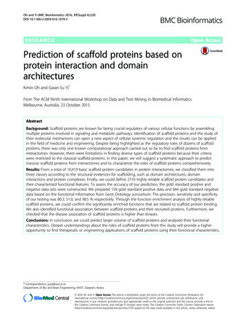
Oh and Yi BMC Bioinformatics 2016, 17(Suppl 6):220pathway. Localization data is collected from Gene Ontology [19] Cellular Compartment. Each identifier of theGene Ontology Cellular Compartment is re-organizedinto 17 cellular compartments according to the hierarchical structure (cell surface, chromosome, cytoplasm, cytoplasmic membrane-bounded vesicle, cytoskeleton, cytosol,endoplasmic reticulum, endosome, ER-Golgi intermediatecompartment, extracellular region, Golgi apparatus, mitochondrion, nucleus, plasma membrane, ribosome, sarcoplasmic reticulum, vacuole). Pathway data is collectedfrom four different pathway databases (KEGG [20], PID[21], Reactome [22], and WikiPathway [23]) and definedpathway names and their components were extracted.Page 437 of 442multiple competitive interacting partners using samedomain regions.Characterization of scaffold protein candidatesGene annotation enrichment analysisWe used the DAVID [24] tools to analyze functionalcharacteristics of collected scaffold protein candidates.The functional meaning of the scaffold protein candidates was interpreted using function enrichment analysistool in DAVID. We analyzed functional implications inGO molecular function, GO cellular compartment, GObiological process and Pfam families. The p-values wereadjusted by multiple testing corrections using Benjaminiand Hochberg’s method [25].Collection of known scaffold proteinsTo test reliability of our prediction, we defined the goldstandard positive and the negative set using basic textmining and functional term filtering. For the gold standard positive set, we collected scaffold protein candidatesfrom multiple sources. First, we manually gathered scaffold proteins from review articles. Second, we foundcandidates using query search from functional descriptions of UniProt database and title/abstract of PubMed.From those candidates, we filtered out candidates whichalready have their known molecular functions as scaffoldactivities and complex assemblies. For the gold standardnegative set, we excluded proteins which have molecularfunctions and biological functions related to known scaffold proteins.Criteria for predicting novel scaffold proteinsWe proposed criteria for finding scaffold protein candidates: (i) direct interaction with at least two proteins, (ii)domain-domain interaction between scaffold and twopartner proteins using different domain regions, and (iii)scaffold and two partner proteins should be componentsin the same protein complex (Fig. 1). This criteria is different from Zeke’s definition of classical scaffold protein[4]. Our criteria can filter out hub proteins which haveFunctional association analysisWe tested the hypothesis of having no associationbetween scaffold proteins and disease related genes (disease genes or drug targets). To use chi-square statistics,we made contingency table. Observed frequency is compared to expected frequency. If there was no associationbetween scaffold proteins and disease related genes, thenthe expected frequency should be almost equal to theobserved frequency, and the value of the chi-square statistic would be small and the probability (p-value) wouldbe large.ResultsStatisticsData statisticsWe collected various kinds of resources and constructeddatabase using Oracle 10 g. All proteins were filtered inHomo sapiens and Swiss-Prot which are manually annotated and reviewed. Protein-protein interaction data wasfiltered by experimental detection methods. Domaindomain interactions were selected which have 3D structural evidences (Table 1-a). We predicted scaffold proteincandidates and classified into three types, according to theeligibility criteria. Actually our novel criteria means type Icase, however we allowed to classify into type II and typeIII, because our resources of domain, DDI, and proteincomplexes were not completely detected (Table 1-b). Bothcriteria 2 and 3 make scaffold proteins possible to be existwith their partner proteins together simultaneously.Performance testFig. 1 Structural criteria for predicting scaffold protein candidatesTo evaluate the ability of the prediction performance, weused a statistical measurement. We defined the goldstandard positive and negative scaffold protein set andcalculated the number of true positive, false positive,true negative and false negative. Using these four outcomes, we made 2 2 contingency table and we obtainedprecision, sensitivity and specificity of our tests (Table 2).
Oh and Yi BMC Bioinformatics 2016, 17(Suppl 6):220Page 438 of 442Table 1 Statisticsa) Statistics of collected dataTypeSourceStatisticsProteinUniProtProtein: 20233Articles: 269469Protein-proteininteractionComBiCom (BIND, BioGRID,DIP, HPRD, IntAct, MIPS)Proteins: 82894PPIs: 73743DomainPfamDomains: 4895Proteins: 17316Domain-domaininteractioniDDI (3DID, iPfam)Domains: 3214DDIs: 17770Protein complexCOFECOProtein complexes:3317Proteins: 4597PathwayKEGG, NCI-PID, Reactome,WikiPathwayPathways: 2620Proteins: 8413Cellular locationGene OntologyCellular CompartmentGO terms: 635Proteins: 9820Disease geneOMIM, PharmGKB, KEGGDiseaseDisease: 502Genes: 4950Drug targetDrugBankDrugs: 1574Proteins: 1077Gold standard setUniProt, PubMed,Gene OntologyPositive: 104Negative: 844KinasePhosphoELM, PhosphositeKinase: 468b) Statistics of scaffold protein candidatesClassCriteria 1Criteria 2Criteria 3# of scaffoldproteinsType IOOO616Type IIOOX1792Type IIIOXO308The precision, sensitivity and specificity of our tests were80.3, 51.0, and 98.5 %, respectively.showed that 87 GOCC, 284 GOBP, 85 GOMF, and 41Pfam terms are significantly enriched. We could findsignificant functional implications in the scaffold proteinslike ‘metabolic process’, ‘phosphorylation’, ‘cell death’, ‘cellproliferation’, ‘apoptosis’, ‘signaling pathway’, ‘complex assembly’. According to the cellular component result, scaffold proteins had significant enrichments on the allcellular compartments. As we expected, binding that arerelated to the various molecular functions were significantly enriched. Interestingly, some molecular functions(‘transcription regulator activity’, ‘nucleotide binding’,‘uniquitin protein ligase binding’) could show that scaffoldproteins might have special cellular functions such asassembling transcription factor complex or ubiquitin ligase complex. Furthermore, ‘kinase activity’ shows that thescaffold proteins canalso have catalytic activities and thisis distinguished from characteristics of classical scaffoldproteins. Well-known modular PPI domains are enrichedfrom Pfam family and it supports binding functions ofscaffold proteins (Fig. 2).Functional similarity between scaffold and partner proteinsWe compared functional information of scaffold proteins with their partner proteins. In case of Type I,93.0 % of the total scaffold proteins in Type I have cellular localization information and 99.3 % are matched withpartner’s information (Table 3-a). In the same way,86.2 % of total scaffold proteins in Type I belong topathways and 96.1 % have partner proteins which havesame pathway information (Table 3-b). This result showsthe possibilities to predict novel cellular functions ofscaffold proteins and partner proteins from their knowninformation.Disease associationFunctional characteristics of predicted scaffold proteinsEnriched functionsWe carried out a function enrichment analysis for thecandidates of scaffold proteins using the GO cellular component (GOCC), GO biological process (GOBP), GO molecular function (GOMF) and Pfam family at Bonferronicorrected p-value of 0.001. Functional enrichment resultSome studies have suggested the scaffold proteinIQGAP1 as a therapeutic target for inhibiting tumorigenesis. Like this example, scaffold proteins could bedisease markers or drug targets because of their important role as a systemic regulator. Hence we tested associations between scaffold proteins and disease relatedgenes. Additionally, we tested associations between setTable 2 2 2 contingency table for evaluating the performance of predictionTrue conditionPredicted condition(2716)Total populationCondition positive (158)Condition negative (844)Prevalence 17.1 %Predicted conditionpositive6713Precision (Positive predictive value)83.8 %Predicted conditionnegative91831False omission rate 9.8 %Accuracy (89.6 %)Sensitivity (True positive rate) Fall-out (False positive rate)42.4 %1.5 %Miss rate (False negative rate) Specificity (True negative rate)57.6 %98.5 %
Oh and Yi BMC Bioinformatics 2016, 17(Suppl 6):220Page 439 of 442Fig. 2 Enrichment of Gene Ontology annotations and Pfam families. The four histograms show significantly enriched Gene Ontology annotationsand Pfam domain families for the 2716 scaffold protein candidates. The x-axis represents the number of scaffold protein candidates belonging tothe respective categoryTable 3 Similarity between scaffold protein candidates andpartner proteinsa) Similarity of cellular localizationType# of scaffold proteinsTotalKnownMatched with partner’s informationType I616573 (93.0)569 (99.3)Type II17981372 (76.3)1285 (93.7)Type III308264 (85.7)251 (95.1)of kinases and disease related genes for comparison.Among 616 scaffold proteins in Type I, 188 scaffoldproteins are known as disease genes and 61 scaffoldproteins are drug targets. In kinase case, 136 kinases areknown as disease genes and 92 kinases are drug targetsamong total 468 kinases. We made contingency tablesabout observed and expected frequency. From thesecontingency tables, we could calculate chi-square values.Table 4 shows that the disease association of scaffoldproteins is higher than kinases. Conversely, drug targetassociation of scaffold proteins is lower than kinases,but this result is obvious because kinases have beenTable 4 Disease and drug target association of scaffold proteincandidates and kinasesb) Similarity of related pathwayDisease associationDrug target associationTypeScaffoldKinaseScaffoldKinaseType I# of scaffold proteinsTotalKnownMatched with partner’s informationRisk ratio1.261.191.913.94616531 (86.2)497 (93.6)Odd ratio1.371.272.014.66Type II17981178 (65.5)856 (72.7)Chi-square value12.65.4789.12195.35Type III308212 (68.8)142 (67.0)p-value3.85E-041.93E-022.72E-072.16E-44
Oh and Yi BMC Bioinformatics 2016, 17(Suppl 6):220researched as drug target candidates until now. Ourresult shows that scaffold proteins have associationwith diseases and drug targets, so it gives us the reason tostudy scaffold proteins as therapeutic targets.Case studyAs mentioned, predicted scaffold proteins show highassociation with disease gene and drug targets. Throughthe additional analysis, we selected two cases that are related to disease condition from scaffold protein candidates(Fig. 3). AXIN1 is already known as a scaffold protein [26]and in our prediction, it interacts with GSK3B andCTNNB1. We analyzed a microarray dataset in case–control designed from the NCBI Gene Expression Omnibusfor type 2 diabetes (GSE29231) [27]. The statistical analysis of gene differential expression was computed andthen the p-values of each gene were obtained using theBenjamini & Hochberg method. AXIN1 is down regulatedin diabetes condition and CTNNB1 activation is associated with an increment in glucose uptake [28]. From theseevidences, we could make hypothesis that type 2 diabetesis caused by a decrement of glucose import because activation of CTNNB1 is inhibited by lower expression ofAXIN1.Our prediction identified PIK3R1 as a scaffold proteincandidate by recruiting GAB1 and PIK3CA. We couldfind protein expression level of PIK3R1 in both thePage 440 of 442normal cell and the cancer cell using Human ProteinAtlas [29]. Protein expression of PIK3R1 was not detected in normal breast cell, however it was highlyexpressed in breast cancer cell. PIK3CA is known as agene related malignant neoplasm of breast [30] and inhibits apoptosis function. From these evidences, wecould make hypothesis that cancer-specific high expression of PIK3R1 increases activation of PIK3CA and as aresult, negatively regulated apoptotic function cause cancer in breast.DiscussionUsing massive data from high-throughput screening, wecould predict plenty of candidate proteins which may actas scaffolds. Many of them are not known as scaffoldproteins but they have possibilities to recruitpartner proteins and regulate their functions. Although our textmining methods can be improved, known scaffold proteins extracted from articles and database might be quitehelpful to corroborate the reliability of scaffold proteinsthat are predicted from interactomes. In this study, weused highly reliable data of protein-protein interactionand domain-domain interaction. Because there are manypredicted information of protein domain, proteinprotein interaction and domaindomain interaction, thereis a chance to expand predicted scaffold proteins withscores of reliabilities. If we could utilize functionalFig. 3 Models of AXIN1 scaffold protein and PIK3R1 scaffold protein candidate. a AXIN1 is a known scaffold protein and AXIN1 interacts withGSK3B and CTNNB1 using RGS and Axin b-cat bind domain respectively. CTNNB1 is related activation of glucose import. Through gene expressionanalysis, AXIN1 is down regulated in type 2 diabetes. b PIK3R1 is predicted as a scaffold protein. RIK3R1 can recruit GAB1 and PIK3CA using SH2domains. PIK3CA is known as a gene related to malignant neoplasm of blast and inhibits apoptotic function. Protein expression of PIK3R1 is notdetected in normal breast cell, however it is highly expressed in breast cancer cell
Oh and Yi BMC Bioinformatics 2016, 17(Suppl 6):220information or condition specific data, predicted scaffoldproteins might be classified into various types by theirfunctional characteristics, such as localization, pathwayregulation or crosstalk. These functional characteristicsalso can be used as a measurements of the reliabilityscores. Some of known scaffold proteins recruit morethan two proteins, but we restricted scaffold protein withtwo partner proteins, because there are so many possiblecombinations of partner proteins sets. We can filter andfind scaffold proteins which can recruit more than twoproteins from our predictions.ConclusionScaffold proteins can precisely control the specificityand dynamics of information transfer. Furthermore,scaffold proteins have versatility due to their modularity, which allows recombination of protein domains tobuild new signaling pathways. In the past, scaffold proteins were discovered only by chance via experimentsaimed at studying the function of signaling enzymes orreceptors. We carried out extraction of scaffold proteins from articles and database and prediction frominteractomes according to the new criteria we proposed. Through functional enrichment analysis, weidentified not only the known functional implicationsof scaffold proteins but novel enriched terms. Usingfunctional characteristics of partner proteins, we alsopredicted new function of scaffold proteins. Finally, wefound that scaffold proteins were highly associated withdiseases and drug targets like kinases. Through futurestudies, more can be understood about the role of scaffold proteins, and scaffolds can be used to generatenew and predictable pathway to program useful cellularbehaviors. In this respect, this study can support further researches for discovering the target of molecularengineering and therapy.Ethics approval and consent to participateNot applicable.Consent for publicationNot applicable.Availability of data and materialsThe datasets supporting the conclusions of this articleare included within the article and its additional files(Additional files 1 and 2).Additional filesAdditional file 1: It contains gold standard positive set, gold negativeset and predicted scaffold proteins with type. (XLSX 106 kb)Additional file 2: It contains results of functional enrichment usingDAVID. (P-value 0.001). (XLSX 209 kb)Page 441 of 442Competing interestsThe authors declare that they have no competing interests.Authors’ contributionsKO designed and conducted the experiments and wrote the manuscript.GSY designed and supervised the experiments and wrote the manuscript.KO and GSY discussed the results, implications and commented on themanuscript at all stages. All authors read and approved the final manuscript.AcknowledgmentsThis work was supported by the Bio-Synergy Research Project (NRF2012M3A9C4048759), the Converging Research Center Program (ProjectNo.2015054201), and the KAIST Future Systems Healthcare Project funded bythe Ministry of Science, ICT and Future Planning.DeclarationsPublication charges for this article have been funded by the Bio-Synergy ResearchProject (NRF-2012M3A9C4048759) of the Ministry of Science, ICT and FuturePlanning through the National Research Foundation.This article has been published as part of BMC Bioinformatics Volume 17Supplement 6, 2016: Proceedings of the ACM Ninth International Workshopon Data and Text Mining in Biomedical Informatics. The full contents of thesupplement are available online at http://dl.acm.org/citation.cfm?id 2811186.Published: 28 July 2016References1. Good MC, Zalatan JG, Lim WA. Scaffold proteins: hubs for controlling theflow of cellular information. Science. 2011;332(6030):680–6.2. Pan CQ, Sudol M, Sheetz M, Low BC. Modularity and functional plasticityof scaffold proteins as p(l)acemakers in cell signaling. Cell Signal.2012;24(11):2143–65.3. Palfy M, Remenyi A, Korcsmaros T. Endosomal crosstalk: meeting pointsfor signaling pathways. Trends Cell Biol. 2012;22(9):447–56.4. Zeke A, Lukacs M, Lim WA, Remenyi A. Scaffolds: interaction platformsfor cellular signalling circuits. Trends Cell Biol. 2009;19(8):364–74.5. Shaw AS, Filbert EL. Scaffold proteins and immune-cell signalling.Nat Rev Immunol. 2009;9(1):47–56.6. White CD, Brown MD, Sacks DB. IQGAPs in cancer: a family of scaffoldproteins underlying tumorigenesis. FEBS Lett. 2009;583(12):1817–24.7. Jameson KL, Mazur PK, Zehnder AM, Zhang J, Zarnegar B, Sage J, Khavari PA.IQGAP1 scaffold-kinase interaction blockade selectively targets RAS-MAPkinase-driven tumors. Nat Med. 2013;19(5):626–30.8. Stuart DD, Sellers WR. Targeting RAF-MEK-ERK kinase-scaffold interactions incancer. Nat Med. 2013;19(5):538–40.9. Ramirez F, Albrecht M. Finding scaffold proteins in interactomes. Trends CellBiol. 2010;20(1):2–4.10. Finn RD, Bateman A, Clements J, Coggill P, Eberhardt RY, Eddy SR, Heger A,Hetherington K, Holm L, Mistry J, et al. Pfam: the protein families database.Nucleic Acids Res. 2014;42(Database issue):D222–30.11. Youngwoong H, Choong-Hyun S, Min-Sung K, Gwan-Su Y. "Combineddatabase system for binary protein interaction and co-complex association."In: IEEE 10.1109/IACSIT-SC.2009.42, 538-54212. Kim Y, Min B, Yi GS. IDDI: integrated domain-domain interaction and proteininteraction analysis system. Proteome Sci. 2012;10 Suppl 1:S9.13. Sun CH, Kim MS, Han Y, Yi GS. COFECO: composite function annotationenriched by protein complex data. Nucleic Acids Res. 2009;37(Web Server issue):W350–5.14. UniProt C. UniProt: a hub for protein information. Nucleic Acids Res.2015;43(Database issue):D204–12.15. Whirl-Carrillo M, McDonagh EM, Hebert JM, Gong L, Sangkuhl K, Thorn CF,Altman RB, Klein TE. Pharmacogenomics knowledge for personalizedmedicine. Clin Pharmacol Ther. 2012;92(4):414–7.16. Kanehisa M, Goto S, Furumichi M, Tanabe M, Hirakawa M. KEGG forrepresentation and analysis of molecular networks involving diseases anddrugs. Nucleic Acids Res. 2010;38(Database issue):D355–60.17. Bodenreider O. The Unified Medical Language System (UMLS):integrating biomedical terminology. Nucleic Acids Res.2004;32(Database issue):D267–70.
Oh and Yi BMC Bioinformatics 2016, 17(Suppl 6):220Page 442 of 44218. Law V, Knox C, Djoumbou Y, Jewison T, Guo AC, Liu Y, Maciejewski A,Arndt D, Wilson M, Neveu V, et al. DrugBank 4.0: shedding new light ondrug metabolism. Nucleic Acids Res. 2014;42(Database issue):D1091–7.19. Gene Ontology C. Gene Ontology Consortium: going forward. Nucleic AcidsRes. 2015;43(Database issue):D1049–56.20. Kanehisa M, Goto S. KEGG: kyoto encyclopedia of genes and genomes.Nucleic Acids Res. 2000;28(1):27–30.21. Schaefer CF, Anthony K, Krupa S, Buchoff J, Day M, Hannay T,Buetow KH. PID: the Pathway Interaction Database. Nucleic Acids Res. 2009;37(Database issue):D674–9.22. Croft D, Mundo AF, Haw R, Milacic M, Weiser J, Wu G, Caudy M, Garapati P,Gillespie M, Kamdar MR, et al. The Reactome pathway knowledgebase.Nucleic Acids Res. 2014;42(Database issue):D472–7.23. Kelder T, van Iersel MP, Hanspers K, Kutmon M, Conklin BR, Evelo CT,Pico AR. WikiPathways: building research communities on biologicalpathways. Nucleic Acids Res. 2012;40(Database issue):D1301–7.24. Dennis Jr G, Sherman BT, Hosack DA, Yang J, Gao W, Lane HC, Lempicki RA.DAVID: Database for Annotation, Visualization, and Integrated Discovery.Genome Biol. 2003;4(5):3.25. Benjamini Y, Hochberg Y. Controlling the False Discovery Rate - a Practicaland Powerful Approach to Mult
as a physical platform to assemble signaling components [2, 3]. Through these regulations, scaffold protein can lead to dynamic signaling outputs [4]. Scaffold proteins are involved not only signaling processes but also in the assembly-line processes and cell-cell communications [1]. Scaffold proteins also control enzymatic activities by con-