Transcription
REGULAR ARTICLEEvidence of thrombotic microangiopathy in children with SARS-CoV-2across the spectrum of clinical presentations1Immune Dysregulation Frontier Program, Department of Pediatrics, 2Division of Oncology, Department of Pediatrics, and 3Division of Hematology, Children’s Hospital ofPhiladelphia, University of Pennsylvania Perelman School of Medicine, Philadelphia, PA; 4Department of Pathology and Laboratory Medicine and 5Department of Microbiology,University of Pennsylvania Perelman School of Medicine, Philadelphia, PA; and 6Division of Allergy and Immunology, Department of Pediatrics, 7Division of Rheumatology,Department of Pediatrics, 8Division of Emergency Medicine, Department of Pediatrics, 9Division of Infectious Diseases, Department of Pediatrics, 10Division of Critical CareMedicine, Department of Anesthesiology and Critical Care Medicine, 11Division of Cardiology, Department of Pediatrics, and 12Division of Nephrology, Department of Pediatrics,Children’s Hospital of Philadelphia, University of Pennsylvania Perelman School of Medicine, Philadelphia, PAKey Points sC5b9 plasma levelsare elevated in childrenwith SARS-CoV-2infection, even if theyhave minimal symptomsof COVID-19. A high proportion ofchildren with SARSCoV-2 infection metclinical criteria for TMA.Most children with severe acute respiratory syndrome coronavirus 2 (SARS-CoV-2) infectionhave mild or minimal disease, with a small proportion developing severe disease ormultisystem inflammatory syndrome in children (MIS-C). Complement-mediated thromboticmicroangiopathy (TMA) has been associated with SARS-CoV-2 infection in adults but has notbeen studied in the pediatric population. We hypothesized that complement activation playsan important role in SARS-CoV-2 infection in children and sought to understand if TMA waspresent in these patients. We enrolled 50 hospitalized pediatric patients with acute SARS-CoV-2infection (n 5 21, minimal coronavirus disease 2019 [COVID-19]; n 5 11, severe COVID-19) orMIS-C (n 5 18). As a biomarker of complement activation and TMA, soluble C5b9 (sC5b9,normal 247 ng/mL) was measured in plasma, and elevations were found in patients withminimal disease (median, 392 ng/mL; interquartile range [IQR], 244-622 ng/mL), severe disease(median, 646 ng/mL; IQR, 203-728 ng/mL), and MIS-C (median, 630 ng/mL; IQR, 359-932 ng/mL)compared with 26 healthy control subjects (median, 57 ng/mL; IQR, 9-163 ng/mL; P , .001).Higher sC5b9 levels were associated with higher serum creatinine (P 5 .01) but not age. Of the19 patients for whom complete clinical criteria were available, 17 (89%) met criteria for TMA.A high proportion of tested children with SARS-CoV-2 infection had evidence of complementactivation and met clinical and diagnostic criteria for TMA. Future studies are needed todetermine if hospitalized children with SARS-CoV-2 should be screened for TMA, if TMA-directedmanagement is helpful, and if there are any short- or long-term clinical consequences ofcomplement activation and endothelial damage in children with COVID-19 or MIS-C.IntroductionDuring the coronavirus disease 2019 (COVID-19) pandemic caused by severe acute respiratorysyndrome coronavirus 2 (SARS-CoV-2), three distinct clinical phenotypes have emerged in children.Submitted 22 September 2020; accepted 29 October 2020; published online 8December 2020. DOI 10.1182/bloodadvances.2020003471.*C.D. and K.O.M. contributed equally to this work.†H.B., E.M.B., and D.T.T. contributed equally to this work.Presented in abstract form at the 62nd annual meeting of the American Society ofHematology, 5-8 December 2020.8 DECEMBER 2020 x VOLUME 4, NUMBER 23Requests for data sharing may be submitted to the corresponding author (HamidBassiri; e-mail: bassiri@email.chop.edu).The full-text version of this article contains a data supplement. 2020 by The American Society of Hematology6051Downloaded from df/4/23/6051/1794129/advancesadv2020003471.pdf by guest on 17 December 2020Caroline Diorio,1,2,* Kevin O. McNerney,1,2,* Michele Lambert,1,3 Michele Paessler,1,4 Elizabeth M. Anderson,5 Sarah E. Henrickson,1,6Julie Chase,1,7 Emily J. Liebling,7 Chakkapong Burudpakdee,1 Jessica H. Lee,1 Frances B. Balamuth,8 Allison M. Blatz,9 Kathleen Chiotos,9,10Julie C. Fitzgerald,1,10 Therese M. Giglia,11 Kandace Gollomp,1,3 Audrey R. Odom John,9 Cristina Jasen,6 Tomas Leng,1,2 Whitney Petrosa,1Laura A. Vella,9 Char Witmer,3 Kathleen E. Sullivan,1,6 Benjamin L. Laskin,12 Scott E. Hensley,5 Hamid Bassiri,1,9,† Edward M. Behrens,1,7,†and David T. Teachey1,2,†
High rates of thrombosis and thrombotic-related complicationshave been reported in adult patients with severe COVID-19.12,13These complications have been reported in healthy young people,without prior comorbidities, raising the concern that thromboticcomplications could be directly caused or exacerbated by SARSCoV-2 infection.12 Studies in adults have invoked thromboticmicroangiopathy (TMA) as a potential cause for severe manifestations of COVID-19.14-16 TMA results from endothelial cell damageto small blood vessels, leading to hemolytic anemia, thrombocytopenia, and, in some cases, organ damage.17-21 TMA has beenreported in postmortem studies of adult patients with COVID-19.22One proposed mechanism for SARS-CoV-2–mediated TMA is viacomplement activation.15-18 Complement dysregulation results inunregulated formation of the C5b9 membrane attack complex,leading to the clinical manifestations of TMA. Soluble C5b9(sC5b9) is a clinically available biomarker and has been implicatedas an indicator of severity in hematopoietic stem cell transplant–associated TMA (HSCT-TMA), as patients with markedlyelevated sC5b9 have increased mortality.23 In mouse models of therelated coronaviruses (SARS-CoV and Middle East respiratorysyndrome coronavirus), knockout or blockade of components of thealternative complement pathway led to amelioration of severerespiratory syndromes and a decrease in cytokine production.24,25In an initial small cohort of children, we previously showed thatsC5b9 levels may be elevated across the spectrum of manifestations of SARS-CoV-2 infections.11 We did not, however, evaluatewhether these children met the clinical criteria for diagnosis of TMAnor did we compare these values vs those of healthy controlsubjects. We hypothesized that sC5b9 was a marker of TMA inpatients with evidence of SARS-CoV-2 infection. Here, we presentserum levels of sC5b9 in healthy control subjects and in childrenwith acute SARS-CoV-2 or MIS-C, and evaluate these cohorts forthe clinical and laboratory findings of TMA.MethodsStudy design and populationWe prospectively enrolled patients admitted to the Children’sHospital of Philadelphia (CHOP) during the COVID-19 pandemicwho had a positive SARS-CoV-2 reverse transcriptase polymerasechain reaction (RT-PCR) from upper respiratory tract mucosa, orwho met clinical criteria for MIS-C. The enrollment criteria havebeen previously described.11 Patients were classified into 3 groups:minimal COVID-19, severe COVID-19, or MIS-C. Minimal COVID-196052DIORIO et alwas defined as patients with an incidental finding of COVID-19during routine testing before admission or a procedure, or thosewith mild COVID-19 symptoms that did not require noninvasivemechanical ventilation (including high-flow nasal cannula, continuous positive airway pressure, or bilevel positive airway pressure).Severe COVID-19 was defined as patients requiring new noninvasive or invasive mechanical ventilation, or escalation above theirbaseline invasive or noninvasive mechanical ventilation. MIS-C wasdefined per the Centers for Disease Control and Prevention criteriaas patients who had fever, laboratory evidence of inflammation,illness with at least 2 organ systems involved (cardiac, renal,respiratory, hematologic, gastrointestinal, dermatologic, or neurologic), no alternative plausible diagnosis and positive SARS-CoV-2infection by RT-PCR, serology (performed in a Clinical LaboratoryImprovement Amendments/College of American Pathologists[CLIA/CAP] laboratory), or proven COVID-19 exposure to a closecontact within 4 weeks before onset of symptoms.26 Patients wereprospectively classified into one of these groups by physicians withexpertise in pediatric hematology/oncology (C.D. and D.T.T.),pediatric infectious diseases (H.B.), and pediatric rheumatology(E.M.B.). Adjudicators were blinded to sC5b9 levels and TMAclinical criteria at the time of classification into these groups. Allpatients who consented were enrolled in a consecutive manner. Ifpatients were subsequently found to not meet criteria (ie, falsepositive SARS-CoV-2 infection or different infectious etiologydetermined as cause of MIS-C symptoms), they remained enrolledbut were excluded from analyses.Subject samples were compared with those of normal controlsamples obtained from the coagulation laboratory at CHOP.Discarded plasma collected from otherwise healthy children whohad been evaluated for symptoms of a bleeding disorder (eg,epistaxis, menorrhagia) was used for the control groups. A limitedchart review was performed to confirm that these patients had nocomorbid medical illness or medical problems. Children who werefound to have comorbid medical issues, other than underlyingbleeding disorders, were excluded.Patients were categorized as having TMA based on the following 7criteria, adapted from Gloude et al27: elevated lactate dehydrogenase (LDH) levels greater than the upper limit of normal for age,schistocytes on blood smear, new thrombocytopenia below thenormal range for age, new anemia below the normal range for age,evidence of proteinuria ( 11 proteinuria [30 mg/dL] on randomurinalysis or random urine protein/creatinine ratio 2 mg/mg),hypertension (blood pressure .99th percentile for age, sex, andheight if aged ,18 years, 140 mm Hg systolic or 90 mm Hgdiastolic if aged 18 years, at least twice during the admission, or ifreceiving antihypertensive therapy for new hypertension), andelevated sC5b9. To meet criteria for TMA, patients had to meet atleast 5 of the 7 criteria during their hospital admission for COVID-19or MIS-C. Hematoxylin and eosin–stained peripheral blood smearswere independently examined for schistocytes by a hematologistand hematopathologist, who were blinded to the categorization andclinical histories of patients, as well as to the other examiners’findings. In a subsequent analysis, a simple set of clinical criteria wasproposed to define TMA in settings in which sC5b9 and otherdiagnostics may be unavailable. These criteria included the presenceof thrombocytopenia, microangiopathic hemolytic anemia (anemia forage and schistocytes on a peripheral blood smear), and organdysfunction. Organ dysfunction was defined as renal dysfunction,8 DECEMBER 2020 x VOLUME 4, NUMBER 23Downloaded from df/4/23/6051/1794129/advancesadv2020003471.pdf by guest on 17 December 2020Most children with acute SARS-CoV-2 infection are asymptomaticor develop mild symptoms.1-3 A small proportion of acutely infectedchildren, typically adolescents with significant comorbidities, develop severe respiratory symptoms requiring hospitalization oradmission to the pediatric intensive care unit.1,2,4 In addition, somechildren develop multisystem inflammatory syndrome in children(MIS-C), a hyperinflammatory syndrome characterized by fever,inflammation, and organ dysfunction in the setting of recent SARSCoV-2 infection. With few exceptions, MIS-C seems to uniquelyaffect children.5-8 Severe cases of MIS-C present with shock andcardiovascular collapse, with 60% to 80% of children requiringpediatric intensive care unit care in recent large studies.5,6,9 MIS-Cseems to be a postinfectious immune-mediated phenomenon distinctfrom other manifestations of SARS-CoV-2.10,11 The pathophysiologyof these presentations is currently unknown.
Two different sensitivity analyses with highly stringent criteria wereperformed. First, we evaluated all patients included in the clinicalcohort for TMA based on the aforementioned criteria. In thisanalysis, we imputed all missing data as negative. In the secondsensitivity analysis, sC5b9 was excluded in the criteria for TMA. Weused the remaining 6 clinical criteria to classify all patients as havingclinical TMA (met 5 or 6 criteria), being indeterminate (4 criteria), ornot meeting criteria for TMA (#3 criteria). In patients who weredeemed indeterminate, we looked at whether sC5b9 elevationwould lead them to meet criteria for TMA.Data collectionData were abstracted from the patient chart on to a standardizedcase report form using a Research Electronic Data Capturedatabase (Vanderbilt University, Nashville, TN) hosted at CHOP.30Data were abstracted by a physician or a research assistant. All dataelements extracted were validated by a physician. Data collectedincluded demographic information, comorbid conditions, and mostextreme laboratory values for laboratory studies of interest. Clinicalcriteria for TMA were abstracted as outlined earlier.Blood collection and assaysBlood draws were obtained in conjunction with the first clinicalblood draw after consent was obtained, within the first 2 weeks ofthe positive SARS-CoV-2 test or admission for MIS-C. For mostpatients, blood samples were obtained within 48 hours of admission.Blood was collected in a lithium heparin tube and processed intoplasma and cell pellets. Separated components were frozen forbatched analysis.Proinflammatory cytokine profilingTen proinflammatory cytokines were measured (interferon-g, interleukin IL-1b [IL-1b], IL-2, IL-4, IL-6, IL-8, IL-10, IL-12p70, IL-13,and tumor necrosis factor-a) using V-Plex Pro-inflammatory Panel 1Human Kits (catalog #K15049D; Meso Scale Diagnostics, Rockville, MD). All assays were performed in duplicate, according to themanufacturer’s protocol. Assays were read and analyzed ona QuickPlex SQ120 (Meso Scale Diagnostics). Of particularinterest to TMA is the neutrophil chemotactic factor IL-8, due toits role as a marker of endothelial damage.31The upper limit of normal for the sC5b9 assay was set as 247 ng/mL,based on the upper limit of normal defined by two CLIA/CAP clinicalassays (250 ng/mL, Machaon Diagnostics, Oakland, CA; 244 ng/mL,Cincinnati Children’s Hospital, Cincinnati, OH). We validated thisupper limit of normal on our research-based ELISA by runningsamples from 26 healthy control subjects; the mean value from thesecontrols was 84.5 ng/mL, and two standard deviations above thismean was 247 ng/mL.SARS-CoV-2–specific antibodiesComplement is believed to be activated by coronaviruses inlarge part via the alternative pathway.24,25 Furthermore, in thecomplement-mediated TMA syndromes, complement is activatedby the loss of negative regulation of the alternative pathway.30 Wepreviously showed that most patients, and specifically patients withMIS-C, had elevations in anti–SARS-CoV-2 antibody titers.31 Weconsidered whether sC5b9 elevations may be related to classicalpathway activation from antiviral antibody–antigen complexes ratherthan the alternative complement activation of TMA. SARS-CoV2–specific antibodies were measured by using ELISA as previouslydescribed.32 Serum IgG, IgA, and IgM antibodies against the receptorbinding domain (RBD) of the spike protein were measured.32,33Optical densities at the 450 nm wavelength were obtained ona SpectraMax 190 Microplate Reader (Molecular Devices, SanJose, CA). Serum antibody titers are expressed as the reciprocalserum dilution at a set optical density based on a standard from themonoclonal antibody CR3022 starting at 0.5 mg/mL.Statistical analysesAnalysis and visualizations were performed by using Prism 8 (GraphPad, San Diego, CA) and Stata/IC (StataCorp, College Station, TX).Descriptive statistics were used to summarize characteristics in eachgroup. Pairwise comparisons were performed by using Kruskal-Wallistesting. The Mann-Whitney U test was used to compute P valuesbetween 2 groups, and post hoc Dunn’s test for multiple comparisonswas used to compute P values. Pearson’s r was used to calculatecorrelations between sC5b9 and clinical variables and to examine therelationship between sC5b9 and log-transformed viral titers. The x2test was used to compare count data.Study approvalThis study was conducted according to the guidelines of theDeclaration of Helsinki. Due to the COVID-19 pandemic, verbalconsent was obtained from a legally authorized representative, withconsent forms signed by the consenting physician or researchassistant and a physical copy provided to the consenting party.Assent was obtained from children aged .7 years, if appropriate.The study protocol was approved by the Institutional Review Board(IRB) at CHOP. For healthy control subjects, protected healthinformation was not recorded. The CHOP IRB determined that thelimited chart review of this control cohort met the IRB exemptioncriteria per 45 CFR 46.104(d) 4(ii) and waiver of Health InsurancePortability and Accountability Act of 1996 authorization.sC5b9 assayPlasma samples were assayed for sC5b9 at 2 dilutions by usinga human C5b9 enzyme-linked immunosorbent assay (ELISA) set(#558315; BD Biosciences, San Jose, CA) according to themanufacturer’s protocols. All samples were assayed in triplicate.Standard curves were used to derive sC5b9 levels from best fit curves.8 DECEMBER 2020 x VOLUME 4, NUMBER 23ResultsBetween 3 April 2020, and 7 July 2020, a total of 112 patientswere screened and 58 were enrolled. Fifty patients were included inthis analysis for whom complete sC5b9 data were available(supplemental Figure 1). All included patients had either a positiveTHROMBOTIC MICROANGIOPATHY IN PEDIATRIC SARS-CoV-26053Downloaded from df/4/23/6051/1794129/advancesadv2020003471.pdf by guest on 17 December 2020liver dysfunction (bilirubin levels .2 times the upper limit of normal forage, alanine aminotransferase or aspartate aminotransferase levels.3 times the upper limit of normal), or cardiac dysfunction (troponinlevels greater than the upper limit of normal or requirement forinotropic support). The glomerular filtration rate (GFR) was calculatedby using the original Schwartz formula.28 Patients were classified ashaving renal dysfunction according to the Kidney Disease: Improving Global Outcomes (KDIGO) criteria.29 Baseline creatinine wasdefined as the lowest creatinine level in the 3 months beforeadmission or, if unavailable, was calculated per KDIGO definitions.Acute kidney injury (AKI) was defined as KDIGO stage 2 or higher (atleast a twofold increase from baseline).
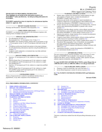
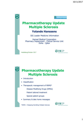
Laboratory data are presented in Table 2. If ,50% of the patients inthe group had data available for a given laboratory value, these datawere excluded from the table. Median D-dimer values tended to behigher in patients with MIS-C than in those with severe COVID-19.Patients with severe COVID-19 and MIS-C had highly elevatedinflammatory markers, including ferritin and C-reactive protein.Patients with MIS-C also had evidence of acute cardiac injury withelevated troponin and B-type natriuretic peptide protein levels.The median sC5b9 level in the healthy control subjects (57 ng/mL;interquartile range [IQR], 9-163 ng/mL) differed significantly fromthat in patients with minimal disease (392 ng/mL; IQR, 244-622 ng/mL),severe disease (646 ng/mL; IQR, 203-728 ng/mL), and MIS-C(630 ng/mL; IQR, 359-932 ng/mL) (P , .001 in each case)(Figure 1A). There were no statistically significant differencesbetween patients with minimal disease, severe disease, or MIS-C.One patient with minimal symptoms of COVID-19 but a very highsC5b9 (1568 ng/mL) was concomitantly diagnosed with systemiclupus erythematosus. To ensure that comorbidities such as lupusthat can be associated with complement dysfunction were notconfounding this relationship, we repeated this analysis butexcluded all patients with a diagnosis of lupus, cancer, sickle celldisease, renal disease, or inflammatory disease. Levels of sC5b9 inpatients with minimal COVID-19, severe COVID-19, and MIS-Cremained elevated compared with levels from healthy controlsubjects (P , .0001) (supplemental Figure 2).Clinical TMA and sC5b9 elevations are closely associated withrenal damage.17 We compared plasma sC5b9 levels betweenthose with AKI (defined as KIDGO stage 2 or above) and thosewithout AKI (KDIGO stage 1 or less), and found significantly highersC5b9 levels in those with AKI (717 ng/mL; IQR, 404-1232 ng/mL)than in those without AKI (433 ng/mL; IQR, 232-706 ng/mL) (P 5.0374) (Figure 1B). We next evaluated correlations betweensC5b9, creatinine, GFR, and blood urea nitrogen in the entirecombined cohort with SARS-CoV-2 or MIS-C. Because creatininelevels in children increase with age, the correlation between sC5b9and age was also examined. Elevations in sC5b9 correlated ina statistically significant manner with the maximum creatinine (r 50.36; P 5 .01), blood urea nitrogen (r 5 0.29; P 5 .04), and GFR(r 5 –0.36; P 5 .01) measured during hospitalization (Figure 1C-E)but not with age (P 5 .512). In our cohort, 10% of minimal COVID-19,36% of severe COVID-19, and 28% of MIS-C patients hadevidence of AKI (Table 1).In patients with severe COVID-19 and MIS-C, we examinedcorrelations between sC5b9 and markers of inflammation, hemolysis, and coagulopathy, including ferritin, C-reactive protein, LDH,prothrombin time, partial thromboplastin time, fibrinogen, D-dimer,6054DIORIO et alaspartate transaminase, hemoglobin, and platelets. Peak valueswere used for all except hemoglobin, in which the lowest value wasused, and for fibrinogen and platelets, in which both peak value andlowest value were calculated. This analysis was limited to those withsevere COVID-19 and MIS-C, due to a high proportion of missingdata in children with minimal disease. Heatmaps of Pearson rcorrelations and P values are shown in Figure 2. sC5b9 did notcorrelate significantly with any of these variables. Clinical variables,including markers of coagulopathy and cytopenias, clusteredtogether.Peripheral blood smears were available on 34 patients. Schistocytes were present in 5 (45%) of 11 patients with minimal COVID-19,7 (87%) of 8 patients with severe COVID-19, and 13 (87%) of 15patients with MIS-C (x2 5 6.59; P 5 .037) (supplemental Figure 3).We retrospectively evaluated if patients met expanded clinicalcriteria for TMA in the subgroup of patients who had a blood smear,complete blood count, and LDH level available (n 5 19). Of thosepatients, 17 (89%) met clinical criteria for TMA. sC5b9 levels wereelevated both in patients who did and did not meet criteria for TMA(supplemental Figure 4). Some patients in the severe COVID-19and minimal COVID-19 cohorts had comorbidities; these are listedin Table 3. Of note, all patients in the MIS-C group were previouslyhealthy or had minimal comorbidities. Of those who met criteria forAKI in Figure 1B, 7 of 9 patients had all criteria for TMA assessed,and all 7 met criteria for TMA.We found that 13 (38%) of 34 patients met simple clinical criteriafor TMA (microangiopathic hemolytic anemia, thrombocytopenia,and evidence of organ damage). In patients who met simpleclinical criteria for TMA, the median sC5b9 level was 420 ng/mL,compared with 634 ng/mL in patients who did not meet simplecriteria for TMA (P 5 .60). Urinalyses were available on 28 ofthese patients, and proteinuria was present in 9 of 13 patientswho met criteria for simple TMA and 10 of 15 patients who did not(x2 5 2.92; P 5 .09).Supplemental Table 1 presents similar data on patients in whomfull peripheral blood smears, LDH levels, and complete bloodcount were not available. We performed a sensitivity analysis andevaluated whether all patients, regardless of whether completecriteria were available, met criteria for TMA. For this analysis, sC5b9elevations were included in the clinical criteria for TMA. All missingdata were counted as negative. In total, 24 (48%) of 50 patients metcriteria for TMA, including 21% of patients with minimal disease,82% of patients with severe disease, and 61% of patients with MIS-C(supplemental Table 2). Summary data for patients according todisease category are presented in supplemental Tables 3-5.We considered that sC5b9 elevations could be caused by anunknown pathophysiological process in SARS-CoV-2 patientsother than TMA. We therefore performed a sensitivity analysis inwhich sC5b9 was not included in the criteria for TMA. To test themost stringent hypothesis, missing data were imputed to benegative. Fifteen (30%) of 50 patients still met criteria for TMA, 9patients were indeterminate, and 26 patients did not meet criteriafor TMA. Of the 9 indeterminate patients, all 9 had elevations insC5b9 that would have led them to meet criteria for TMA if sC5b9had been included.To probe if the complement activation of SARS-CoV-2 infectioncould be related to immune complex and classical activation, we8 DECEMBER 2020 x VOLUME 4, NUMBER 23Downloaded from df/4/23/6051/1794129/advancesadv2020003471.pdf by guest on 17 December 2020SARS-CoV-2 PCR or a positive SARS-CoV-2 antibody test.Patients were classified into the 3 categories described earlier:minimal COVID-19 (n 5 21), severe COVID-19 (n 5 11), or MIS-C(n 5 18). In addition, remnant plasma on healthy control subjectswas collected (n 5 26). Demographic characteristics of thepatients are presented in Table 1. Clinical characteristics wereconsistent with previously reported pediatric patients with MIS-Cand COVID-19.5,7 Twenty of these patients (n 5 5, minimal COVID-19;n 5 9, severe COVID-19; and n 5 6, MIS-C) were included ina previously published analysis, focused on comparing clinicalfeatures and cytokines between groups.11 Study identifiers havebeen included to allow for comparison across reports.
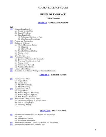
Table 1. Demographic data associated with patients included in the sampleVariableAge, median (IQR), yMinimal COVID-19 (n 5 21)Severe COVID-19 (n 5 11)MIS-C (n 5 18)13 (5-17)15 (14-17)9 (7-13)Sex, n (%)Female9 (43)6 (55)9 (50)12 (57)5 (45)9 (50)White8 (38)5 (45)8 (44)Black10 (48)3 (27)7 (39)Other3 (14)2 (18)2 (11)Declined or not reported01 (9)1 (6)4 (19)3 (27)3 (17)17 (81)7 (64)14 (78)MaleRace, n (%)HispanicNot HispanicDeclined065 (19.9-90.23); n 5 19BMI percentile, median (IQR)1 (9)1 (6)82 (65-95)93 (80-98)11 (100)13 (72)Pediatric intensive care unit admission, n (%)Yes2 (10)No19 (90)05 (28)Extracorporeal membrane oxygenation, n (%)Yes0No21 (100)2 (18)09 (82)18Inotropic support, n (%)Yes8 (73)12 (67)21 (100)03 (27)6 (33)Yes1 (5)*9 (82)4 (22)No20 (95)2 (18)14 (78)Yes5 (24)5 (45)3 (17)No16 (76)6 (55)15 (83)Yes2 (10)4 (36)5 (28)No16 (76)7 (64)13 (72)NoInvasive mechanical ventilation, n (%)Liver dysfunction,† n (%)Renal dysfunction,‡ n (%)Not available3 (14)Dialysis, n (%)YesNo01 (9)021 (100)10 (91)18 (100)Yes11 (52)9 (82)10 (56)No10 (48)2 (18)8 (44)Hypertension,§ n (%)Proteinuria, n (%)Yes7 (33)8 (72)12 (66)No7 (33)3 (27)4 (22)Not available7 (33)04 (22)*One patient with minimal COVID-19 symptoms was admitted during a trauma team activation and was intubated for airway protection.†Defined as bilirubin levels .2 times the upper limit of normal for age, alanine aminotransferase or aspartate aminotransferase levels .3 times the upper limit of normal.‡Defined according to KDIGO criteria.§Defined as blood pressure .99th percentile for age, height, and sex.8 DECEMBER 2020 x VOLUME 4, NUMBER 23THROMBOTIC MICROANGIOPATHY IN PEDIATRIC SARS-CoV-26055Downloaded from df/4/23/6051/1794129/advancesadv2020003471.pdf by guest on 17 December 2020Ethnicity, n (%)
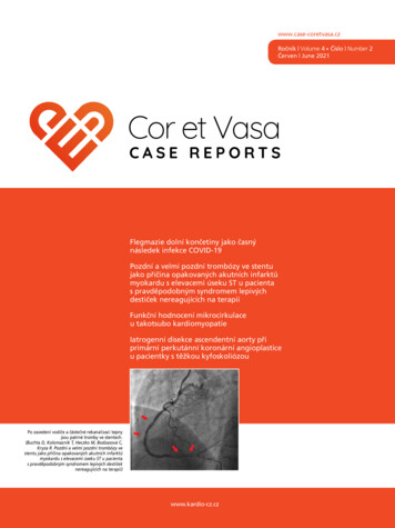
Table 1. (continued)Minimal COVID-19 (n 5 21)VariableSevere COVID-19 (n 5 11)MIS-C (n 5 18)Outcome, n (%)Discharged21 (100)7 (64)18 (100)Remains hospitalized02 (18)0Death02 (18)0*One patient with minimal COVID-19 symptoms was admitted during a trauma team activation and was intubated for airway protection.†Defined as bilirubin levels .2 times the upper limit of normal for age, alanine aminotransferase or aspartate aminotransferase levels .3 times the upper limit of normal.‡Defined according to KDIGO criteria.§Defined as blood pressure .99th percentile for age, height, and sex.Levels of IL-8, measured as a marker of endothelial dysfunction,differed significantly between patients with MIS-C (P 5 .0166) andpatients with severe COVID-19 (P 5 .0079), when compared withminimal COVID-19 patients, but not between patients with MIS-Cand severe disease (P 5 .99) (supplemental Figure 5). Levels of IL-8did not correlate with elevations in sC5b9 (P 5 .184). Levels ofinterferon-g differed significantly between patients with minimaldisease and MIS-C (P 5 .01) but not between those with MIS-Cand severe disease (P 5 .13) or between those with minimaldisease and severe disease (P . .99).Table 2. Median, IQR, and number of patients of the most extreme laboratory values during the admission for patients included in the sampleMinimal COVID-19Value (reference range)Median (IQR)Severe COVID-19nMedian (IQR)MIS-CnMedian (IQR)nCoagulationD-dimer, highest (0.27-0.60 mg/mL FEU)—2.53 (0.6-20.5)114.93 (3.42-5.75)17PT, highest (11.6-13.8 s)—16 (13.2-31)1115.4 (14.4-17.4)18PTT, highest (22-36 s)—52.4 (37.5-78.8)1131.8 (28.4-36.6)17Lowest—297 (239-332)10297 (237-370)17Highest—488 (305-890)10572 (484-721)1718Fibrinogen (172-471 mg/dL)ChemistryAST, highest (10-30 U/L)59 (40-72)17123 (54-627)1188 (63-104)Creatinine, highest (0.3-0.8 mg/dL)0.4 (0.3-0.8)190.7 (0.3-2.3)110.6 (0.5-1.3)18193 (108-221)18111 (40-243)11136 (65-182)181923 (13-34)1121.5 (16-39)18GFR, minimum (mL/min)BUN, highest (7-18 mg/dL)12 (7-23)HematologyPlatelets (150-400 K/mL)Lowest220 (127-295)21128 (78-165)11141 (121-194)18Highest414 (165-363)21311 (223-347)11414 (275-483)189.2 (7.4-11.9)218.9 (6.7-11.1)118.9 (7.9-10)18419 (164-2747)10806 (665-1162)1730.3 (7-34.9)1123.7 (19-34.6)1859 (43-82)17H
University of Pennsylvania Perelman School of Medicine, Philadelphia, PA; and 6Division of Allergy and Immunology, Department of Pediatrics, 7Division of Rheumatology, . (CHOP) during the COVID-19 pandemic who had a positive SARS-CoV-2 reverse transcriptase polymerase chain reaction (RT-PCR) from upper respiratory tract mucosa, or .