Transcription
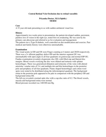
Central Retinal Vein Occlusion due to retinal vasculitisPriyanka Doctor, M.S.(Ophth.)MERSICaseA 53 year old male presenting to us with sudden unilateral visual lossHistoryApproximately two weeks prior to presentation, the patient developed sudden, persistent,painless loss of vision in the right eye, noted first on awakening. He was seen by hisprimary care physician and referred to us for evaluation and management.The patient was a Type II diabetic, well controlled on diet modification and exercise. Pastmedical and family history were otherwise unremarkable.ExaminationThe visual acuity in OD and OS were Finger counting at 4 meters and 20/20 respectively.There was a 2 afferent papillary defect OD and the anterior segment OU wasunremarkable with open angle in all four quadrants on gonioscopy and normal IOP OU.Fundus examination revealed a hyperemic disc OD, with filled cup and blurred discmargins. Blood vessels overlying the disc were dilated and tortuous with splinterhemorrhages. There was generalized dilatation and tortuosity of the veins with alteredarteriolar: venular ratio of 2:4, and multiple dot and blot hemorrhages in the posteriorpole and periphery of all four quadrants. Flame shaped hemorrhages and cotton woolspots were noted to be distributed along the inferotemporal quadrant. The backgroundretina in the posterior pole appeared to be pale in comparison with the periphery OD andbackground retina OS.The left eye revealed a normal optic disc with a cup-disc ratio of 0.2. The blood vessels,macula and background retina were normal.Blood pressure recorded was 140/100 mm Hg.
Figure 1: Fundus photograph ODFigure 2 – Fundus photograph OSLaboratory testsHe was noted to have a raised ANA titre (1:40, homogenous pattern) with raised proteinC activity ( 200). His complete blood count, total complement, c1 complement, protein S
activity, interleukin 6, TNF alpha, C3d complement, antithrombin III, ANCA, properdinfactor, rheumatoid factor, FTA-ABS, C3,C4 complement, c-reactive protein,homocysteine and hematocrit were found to be within normal limits.Fluorescein Angiography (FA)FA was notable for delayed A-V transit time OD, moreso in the inferotemporalvasculature. Areas of blocked fluorescence were noted, corresponding to hemorrhagesand cotton wool spots. Diffuse hyperfluorescence was noted in all four quadrants alongthe vascular arcades and in the macula. Extensive areas of capillary non-perfusion wereseen in the inferior quadrants with late-staining of the venous architecture.Figure 3: Fluorescein angiogram OD
Clinical CourseIn view of his clinical presentation, we made a diagnosis of OD Central Retinal VeinOcclusion. In view of the age of the patient, atypical findings of increased involvement ofa particular quadrant in a case of CRVO, late staining of vessels on FA and paucity ofidentifiable risk factors, vasculitis was being considered as an etiology of vascularocclusion in this case.After initial treatment with Hydrochlorthiazide 25mg and Aspirin, he came in for reviewafter 1 week with a marginally improved visual acuity of finger counting at 6 meters, butwith increased hemorrhages and cotton wool spots in the inferotemporal quadrant OD.We then treated him with 3 daily infusions of 1000mg Solumedrol, to which heresponded well with BCVA OD 20/300 after 2 weeks and started a regime to 2 weeklySolumedrol with 25mg Methotrexate infusions.3 weeks later, he came in with OD Vitreous hemorrhage reportedly after sneezingviolently. This was treated with intravitreal Avastin and 2 weekly infusions ofSolumedrol with 50mg Methotrexate. FA at subsequent visits revealed markedlyimproved perfusion with resolution of vascular staining OD. He is currentlyasymptomatic on a regime of monthly infusions of Solumedrol(1000mg) with 50mgMethotrexate with a BCVA OD of 20/60, resolution of hemorrhages on fundusexamination and normal FA.Central Retinal Vein OcclusionCentral Retinal Vein Occlusion (CRVO) is one of the most common causes of visualloss. Population-based studies have reported a prevalence of CRVO of between 0.1% and0.4% in individuals 40 years of age. 1,2 Visual morbidity in CRVO is primarily due tothe development of macular ischemia and neovascular glaucoma.PathogenesisKlein and Olwin 3 postulated the following three occlusive mechanisms in CRVO:(a) occlusion of the vein by external compression by sclerotic adjacent structures (i.e.central retinal artery and fibrous tissue envelope) and secondary endothelialproliferation;(b) occlusion by primary venous wall disease (degenerative or inflammatory innature); and(c) hemodynamic disturbances produced by a variety of factors (e.g. subendothelialatheromatous lesions in the central retinal artery, arterial spasm, sudden reductionof blood pressure, blood dyscrasias etc.). These produce stagnation of blood flowand result in thrombus formation. In patients with these predisposing changes, afall in systemic blood pressure during sleep would finally complete thethrombotic process. This is suggested by numerous reports of patients discoveringmarked visual loss on waking up in the morning.The site of occlusion determines the type of CRVO.4 In Ischemic CRVO, the site ofocclusion is most probably in the region of the lamina cribrosa or immediately posteriorto that as demonstrated in histopathological studies. In contra-distinction, an occlusion
that is further posterior has the availability of more collateral channels, leading, thereforeto milder retinopathy and the non-ischemic type of CRVO.In elderly persons, sclerotic changes in the central retinal artery which shares a commonadventitial sheath with the central retinal vein, leading to secondary endothelialproliferation. 3,5 In younger patients, hematological factors and phlebitis of the centralretinal vein may be responsible for thrombosis. 6Risk factorsSystemic vascular disease is associated in 74% of patients with CRVO greater than 50years of age. Hypertension and hyperlipidemia are seen in 32-60% and diabetes in 1534% of patients. Hemostasis-related factors include antiphospholipid antibodies, elevatedlevels of PAI-1, activated protein-C resistance, factor V Leiden, hyperhomocysteinemia,elevated levels of lipoprotein(a), plasminogen deficiency, factor XII deficiency anddeficiency of physiological clotting inhibitors. Mitral valve prolapse has been postulatedto contribute to platelet hyperactivity. Migraine has been noted with increased prevalencein cases of CRVO, as have collagen vascular disoders and AIDS. Carotid artery diseasemay lead to venous stasis as a result of decreased central retinal artery perfusion pressure.Raised sedimentation rate reflects changes in shear forces and increased plasma viscosity.Medications responsible include oral contraceptive pills, sympathomimetics anddiuretics. 723% of cases of CRVO are found to be associated with ocular disease. These includeprimary open angle glaucoma in 25-66%, optic nerve disease, retinal artery occlusion,retinal vascular malformations which may lead to mural changes or cause a mass effectand uveitides including tuberculous, syphilitic and AMPPE.Trauma, either by sudden eyeball compression or change in intraocular pressure, maycause damage to the vessel wall by shearing or compressing the central retinal veinagainst the lamina cribrosa. 7Retinal vasculitis as a cause of CRVORetinal vasculitis may lead to vascular occlusion by a thrombotic or obliterativemechanism. Thrombosis may occur as a result of local endothelial injury / dysfunction ormore generalized prothrombotic tendencies, both of which have been found to occur inretinal vasculitis. An obliterative process may result from mural inflammatoryinfiltration. 8Retinal vasculitisRetinal vasculitis is defined as vascular leakage and staining of vessel walls onfluorescein angiography, with or without the clinical appearance of fluffy, whiteperivascular infiltrates in the eye, usually with evidence of inflammatory cells in thevitreous body or aqueous humor. 9,10Clinical findingsIt often manifests as gradual painless loss of vision associated with floaters, thoughisolated peripheral vasculitis may be asymptomatic.Retinal vasculitis represents small vessel inflammation involving the arterioles,capillaries and/or post-capillary venules. Arteriolar attenuation, sheathing and cotton
wool spots are suggestive of arteriolar involvement. Terminal arteriolar occlusion maylead to the superficial retina becoming opaque. Vasculitis involving the venous side ofthe circulation produces retinal hemorrhages, edema, telangiectasia and microaneurysms.Active retinal disease is typified by fluffy white perivascular infiltrates which transforminto perivascular fibrosis on quiescence. Macular edema and papillitis may be observed.11Etiology and PathogenesisVasculitis may be:(a) Infective vasculitis – either by direct invasion of microbes such as Mycobacteriumtuberculae, Treponema pallidum or several viruses. It may also occur as a resultof immune complex deposition as a result of antigenic microbial components.(b) Immune vasculitis is usually T-cell mediated and may also involve immunecomplex deposition(c) Idiopathic – These cases are usually associated with lymphopenia, serum immunecomplexes, anticardiolipin antibodies, reduced retinal S-antigen affinity andInterleukin-2.Several putative autoantigens have been described, including Myelin basic protein,myelin-associated glycoprotein, s100 beta and glial fibrillary acid protein. T-cellspecificity for s-100 beta has been shown to lead to retinal involvement.Concerning immunogenetics, HLA-DR15 and HLA B27 are most noteworthy asconcerns retinal vasculitis. Interestingly HLAB51 and B27 share sequence homology touveitogenic retinal s-Antigen, which may explain the association. 8AssociationsOcular associationsIdiopathicEales’ diseaseIdiopathic retinal vasculitis , aneurysms,neuroretinitis (IRVAN) syndromeBilateral iridocyclitis with retinalcapillaritisAcute multifocal hemorrhagic retinalvasculitisFrosted branch angiitisIdiopathic recurrent branch retinal arteryocclusionNeurological associationsAffects young adultsVasculitis with associated vitritisOver half of these patients develop a majorcerebrovascular eventCommon in the Indian subcontinent, malesin their 30s to 50s.Vasculitis beginning in the periphery.Diagnosis of exclusion.Bilateral retinal arteritis, multiplemicroaneurysms, neuroretinitis and uveitisBilateral granulomatous uveitis, retinalcapillaritis. Associated with HLA DR6,HLA Cw7Occlusive phlebitis, retinal hemorrhages,infiltratesExtensive sheathing of blood vesselsHealthy middle-aged patients. Recurrentbranch retinal artery occlusions. Focalperiarterial sheathing, arteritis
Multiple sclerosisMicroangiopathic syndrome ofencephalopathy, hearing loss, and retinalarteriolar occlusionsIsolated central nervous system angiitisSystemic associationsSystemic Lupus ErythematosusWegener’s granulomatosusPolyarteritis NodosaRelapsing PolychondritisPeripheral, subtle, transient periphlebitis.Occlusive arterial disease affecting brain,inner ear and retinaGranulomatous inflammation ofintracerebral and leptomeningeal vessels,occlusive retinal vasculitisCotton wool spots, intraretinalhemorrhages, arteriolar dilation. Largevessel occlusionGranulomatous necrotizing vasculitis ofupper, lower respiratory tract, kidneys.Sclerokeratitis. Retinal vasculitis rare.cANCA specificity.Inflammatory lesions of medium- andsmall-sized vessels, involving heart,kidneys, liver, gastrointestinal tract, CNS.Rarely retinal arteritis.Recurrent chondritis, both auricles, nonerosive, inflammatory polyarthritis, sasalchondritis, ocular inflammation, respiratorytract chondritis, cochlear and/or vestibulardysfunctionManagement of CRVOThe aim of management of retinal vein occlusions include the identification of modifiablerisk factors and their medical management, and the recognition and management of sightthreatening complications.Ischemic CRVO should be differentiated from non-ischemic CRVO and is associatedwith poor visual acuity at presentation ( 20/200), relative afferent papillary defect,presence of multiple intraretinal hemorrhages, cotton wool spots, 10 disc diameters ofretinal ischemia and a reduced b wave amplitude, reduced b:a ratio and prolonged b-waveimplicit time on ERG.Evidence supports the use of pan-retinal photocoagulation when iris new vessels or angleneovascularization is visible.Vitrectomy may be employed for non-clearing vitreous hemorrhage. Chorioretinalanastomosis and radial optic neurotomy have been described but are associated withcomplications. Hemodilution, ticlodipine, troxerutin, and streptokinase have been triedwith questionable benefit.Intravitreal triamcinolone acetonide (4mg in 0.1ml) for cystoid macular edema in retinalvein occlusions and intravitreal bevacizumab have proven to be of benefit, though largerandomized controlled trials have yet to be carried out to support their efficacy.Management of vasculitis12
Corticosteroids remain the mainstay of treatment of retinal vasculitis. When anteriorsegment inflammation is associated with posterior segment pathology, topicalcorticosteroids may be employed. Periocular corticosteroids may be considered if thepatient has asymmetric involvement requiring the treatment of only one eye or ifsystemic corticosteroids are contraindicated. Triamcinolone acetonide 40mg/ml istypically the drug employed. Potential side-effects include steroid-induced glaucoma andcataract and the risk of iatrogenic trauma. The steroid may either be placed deep in thesub-tenon’s space or anteriorly by a transseptal approach. Oral corticosteroids may beemployed in cases with bilateral involvement or in cases unresponsive to periocularinjections. Taper of corticosteroids must be gradual and based on clinical findings atfollow-up examinations. Pulse intravenous corticosteroids can be used in severe cases.One gram of methylprednisolone is administered each day for a total of three days,followed by oral corticosteroids.Immunosuppressive steroid-sparing agents may be used to either reduce or eliminate theuse of corticosteroids. The available immunosuppressive drugs are either alkylatingagents (cyclophosphamide, chlorambucil), antimetabolites (methotrexate, azathioprine)of cyclosporine A.Methotrexate is a folate analogue administered as a starting dose of 7.5mg/week whichmay be increased to a maximum of 20mg/week. Azathioprine can be administered in asingle or divided (twice daily) dose of 1 to 2.5mg/kg/day. Cyclophosphamide, analkylating agent, can have an effect on both the cellular and humoral immune responses.It is administered at a dose of 1-2mg/kg/day either orally or intravenously. Cyclosporineinhibits T cell activation and recruitment. The starting dose is 2.5 – 5mg/kg/day usuallygiven as a twice daily regimen.1. Mitchell P, Smith W, Chang A. Prevalence and associations of retinal veinocclusion in Australia: The Blue Mountains Eye Study. Arch Ophthalmol1996;114:1243-72. Klein R, Klein BE, Moss SE, Meuer SM. The epidemiology of retinal veinocclusion: the Beaver Dam Eye Study. Trans Am Ophthalmol Soc 2000;98:13341, discussion 141-33. Klein BA, Olwin JH. A survey of the pathogenesis of retinal venous occlusion.Arch Ophthalmol 56:207-247, 19564. Hayreh SS, Zimmerman MB, Podhajsky P. Incidence of various types of retinalvein occlusion and their recurrence and demographic characteristics. Am JOphthalmol 117:429-441, 19945. Klein BA. Sidelights on retinal venous occlusion. Am J Ophthalmol 61:25-35,19666. Hayreh SS. Central retinal vein occlusion. In : Mausolf FA, editor. The eye andsystemic diseases, ed. 2. St. Louis: CV Mosby, 1980, 223-2757. Surv Ophthalmol 37(6):1993;393-4178. Hughes EH, Dick AD. The pathology and pathogenesis of retinal vasculitis.Neuropathology and Applied Neurobiology 2003;29:325-3409. Holland GN. Retinal vasculitis. West J Med 1991;154:218-220
10. Retinal vasculitis. In:Foster, Vitale. Diagnosis and Treatment of Uveitis.11. Stanford MR, Verity DH. Diagnostic and therapeutic approach to patients withretinal vasculitis. Int Ophthalmol Clin 2000:40:69-8312. George RK, Nussenblatt RB. Treatment of Retinal Vasculitis. OphthalmologyClinics of North America 11(4);1998;673-680
Retinal vasculitis may lead to vascular occlusion by a thrombotic or obliterative mechanism. Thrombosis may occur as a result of local endothelial injury / dysfunction or more generalized prothrombotic tendencies, both of which have been found to occur in retinal vasculitis. An obliterative process may result from mural inflammatory