Transcription
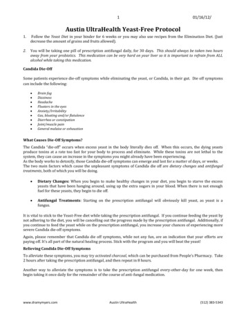
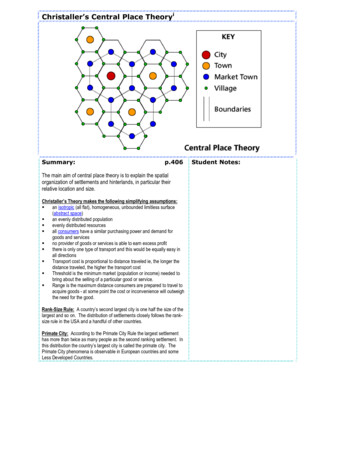
This is a free sample of content from Fission Yeast: A Laboratory Manual.Click here for more information or to buy the book.CHAPTER1Introduction to Fission Yeast as a Model SystemJacqueline Hayles1 and Paul Nurse1Cell Cycle Laboratory, The Francis Crick Research Institute, London WC2A 3LY, United KingdomHere, we briefly outline the history of fission yeast, its life cycle, and aspects of its biology that make it auseful model organism for studying problems of eukaryotic molecular and cell biology.INTRODUCTIONOver the last half century, the fission yeast Schizosaccharomyces pombe has been used increasingly as amodel organism for investigating eukaryotic cellular and molecular processes. This has been drivenmainly by the development of new investigative methodologies and the fact that several biologicalprocesses conserved in S. pombe and in other eukaryotes have been lost or adapted in the modelbudding yeast Saccharomyces cerevisiae. S. pombe has been used as a laboratory organism since the1950s, but until the early 1970s, only approximately 15 laboratories throughout the world worked withfission yeast, publishing approximately 20 papers each year. Now there are more than 300 fission yeastlaboratories worldwide, with more than 400 papers published each year (Fig. 1A). The amount of newinformation about fission yeast has also significantly increased over the past 10 years (as indicated bythe number of annotations per paper; see Fig. 1B and http://www.pombase.org/).In this Introduction, we provide a short history of fission yeast as a model organism, and to give asense of the type of work that is now possible with this model eukaryote, we summarize some of thespecific cellular and molecular processes that have been studied more extensively in fission yeast. Werefer to other contributions in this collection for more detail and for descriptions of protocols coveringdifferent techniques and procedures that can be used to study this organism.HISTORYS. pombe was initially observed and isolated by Saare and colleagues from contaminated millet beer,which had been delayed on the journey from East Africa to Germany. A pure culture of S. pombe wasisolated by Ziedler, under the supervision of Paul Lindner who was the first person to describe it indetail, in the early 1890s. Lindner named it Schizosaccharomyces to distinguish it from budding yeast(because it reproduces vegetatively by means of fission) and pombe after the Swahili word for beer(Lindner 1893). A translation of this report is available at http://www-bcf.usc.edu/ forsburg/. Lindner’s illustrations showed fission yeast to be a single-celled rod-shaped organism that formed ascospores and was able to undergo filamentous growth.S. pombe has proven to be an excellent laboratory model organism. But does it make good beer?Well, the travel writer Tim Cahill (Cahill 1996) has tasted pombe, and this is what he thought about it.1Correspondence: Jacqueline.Hayles@crick.ac.uk; paul.nurse@crick.ac.ukCopyright Cold Spring Harbor Laboratory Press; all rights reservedCite this introduction as Cold Spring Harb Protoc; doi:10.1101/pdb.top0797491 2016 Cold Spring Harbor Laboratory Press. All rights reserved.
This is a free sample of content from Fission Yeast: A Laboratory Manual.Click here for more information or to buy the book.Chapter 5199019952000200520102015YearAverage annotations/publicationB2001501005001970–1974 1975–19791980–19841985–19891990–1994 1995–1999 2000–2004 2005–2009 2010–2014YearFIGURE 1. Numbers of fission yeast publications and annotations in the past 45 yr. (A) Number of fission yeastpublications per year (not cumulative) since 1970. (B) Average number of annotations extracted per paper per 5-yrperiod since 1970 (using the same annotation procedures). An annotation is a piece or unit of information thatconnects a gene to a defined term from an ontology such as Gene Ontology (GO) and that includes supportingevidence attributed to a source (preferably, and usually, a published paper).In central Africa, under the Virunga volcanoes, people make a kind of banana beer they call pombe that is servedin one liter brown glass bottles that once contained beer. Pombe simply means beer in Swahili, but I wascautioned about this banana variety: don’t pour it into a glass, said the brewer himself; you don’t want toactually see it.The pombe is best drunk with a wooden straw. This is because the fermenting bananas leave a thick layer of blacksludge on the glass. I’ve since learned that, in the final brewing process, the beer can be filtered through a finecloth. I’m thinking that my brewer may have found that process superfluous.In the interests of science, Paul Nurse decided to visit a jungle brewery in Uganda, near the Virungavolcanoes, to taste pombe for himself. He trekked into the jungle and located an open-air bananabrewery in a village near the Virunga foothills. He found the beer to be certainly particulate but notblack. The taste was sweet and very alcoholic but definitely drinkable. However, the distilled versionlooked decidedly safer. So draw your own conclusions.The establishment of fission yeast as an experimental laboratory organism came with Urs Leupoldwho, as his PhD project in the 1940s, developed S. pombe for genetic analysis. The strains he derived2Cite this introduction as Cold Spring Harb Protoc; doi:10.1101/pdb.top079749 2016 Cold Spring Harbor Laboratory Press. All rights reserved.
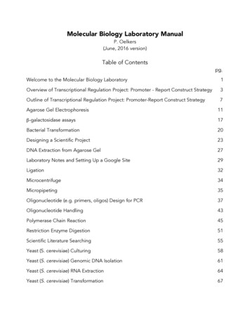
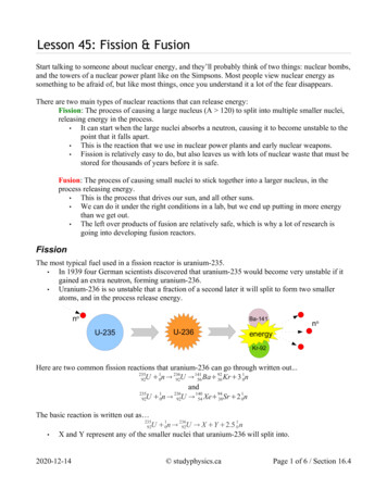
This is a free sample of content from Fission Yeast: A Laboratory Manual.Click here for more information or to buy the book.Fission Yeast as a Model Systemare not from the Lindner strain and are thought to be from a strain deposited in the yeast collection inDelft, the Netherlands in 1924, under the name S. pombe var. liquefaciens. Osterwalder had isolatedthis strain from rancid wine at the Federal Experimental Station of Vini- and Horticulture in Wädenswil, Switzerland.Leupold isolated two homothallic strains, h90 (968) and h40 (which has since been lost), and twoheterothallic strains with opposite mating types, h and h , thus allowing the development of classicalgenetic procedures (Leupold 1950). There are several heterothallic strains with different genomicconfigurations at the mating type locus, but the heterothallic strains commonly used in the laboratoryare h N (975) and h S (972) (Egel 1989). From the 1950s until the 1970s, genetic studies were carriedout in several laboratories including those of Urs Leupold, Jürg Kohli, and Peter Munz in Bern workingmainly on genetics (Munz and Leupold 1970; Kohli et al. 1977) and Murdoch Mitchison, in Edinburgh,studying cell biology, particularly the cell cycle (Mitchison 1957). Mitchison chose fission yeast becauseit was rod-shaped and grew by tip elongation so that the length of the cells reflected cell cycle progression, with the longest cells about to enter mitosis and shortest cells having just septated at the beginningof the cell cycle. The two fields of genetics and cell biology were brought together in Murdoch Mitchison’s laboratory in the mid-1970s, by a group of postdocs and graduate students including Peter Fantes,Kim Nasmyth, Paul Nurse, and Pierre Thuriaux who used fission yeast genetics to study the cell cycle.Other laboratories working early on with fission yeast included Chris Bostock, Colin Clarke, RichardEgel, Herbert Gutz, Brian Johnson, Anwar Nasim, Carl Robinow, and Eva Streiblova. A little later fissionyeast work started in Japan with Chikashi Shimoda, Masayuki Yamamoto, and Mitsuhiro Yanagida.Fission yeast is a member of a small clade currently consisting of only four Schizosaccharomycesspecies, S. pombe, S. japonicus, S. cryophilus, and S. octosporus. All four species have been sequenced, andS. japonicus, in particular, is now being developed as a second model organism (see Furuya and Niki2009 and Chapter 18 Introduction: Schizosaccharomyces japonicus: A Distinct Dimorphic Yeast amongthe Fission Yeast [Aoki et al. 2016]). An understanding of the evolutionary history of Schizosaccharomyces is a current topic of research with comparative studies between these different species (Rhindet al. 2011) and between wild S. pombe strains collected from around the world, particularly fromvineyards and distilleries (Brown et al. 2011; Jeffares et al. 2015). The global dispersal of S. pombe strainsis thought to have started around 340 BCE and was probably associated with the spreading of technologies such as brewing. It reached the New World sometime between 1422 and 1752 CE, which iscoincident with, and likely to be the result of, the European colonization of the Americas (Jeffares et al.2015). Schizosaccharomyces is thought to have diverged from budding yeast hundreds of million yearsago and, at the molecular level, is as closely related to humans as it is to budding yeast (Hedges 2002).LIFE CYCLEFission yeast is a rod-shaped unicellular eukaryote that is 7–14 µm in length and 4 µm wide. Itgrows by tip elongation and divides by medial fission. It has a rapid life cycle with a generation time invegetative growth of 2–4 h at 36 C–25 C in complex and minimal media (see Chapter 2 Introduction:Growth and the Environment of Schizosaccharomyces pombe [Petersen and Russell 2016]). Cellscontinue to undergo mitotic cell cycles until nutrients become limiting and, as growth ceases, theyenter the stationary phase (see Chapter 12 Introduction: Analysis of the Schizosaccharomyces pombeCell Cycle [Hagan et al. 2016]). In appropriate conditions fission yeast cells produce mating pheromones; h cells produce P factor and h cells produce M factor. If nitrogen levels become limiting,cells accumulate in G1 and, in response to mating pheromones, cells of the opposite mating typeconjugate and undergo nuclear fusion to form a diploid zygote. This zygote proceeds to premeioticS phase followed by two successive meiotic nuclear divisions to form four haploid spores (seeChapter 11 Introduction: Analysis of Schizosaccharomyces pombe Meiosis [Yamashita et al. 2016]).If nutrients are restored, these spores will germinate and re-enter the mitotic cell cycle. S. pombe isnormally a haploid organism, however, at the diploid zygote stage, if cells are not committed to themeiotic programme and nitrogen levels are increased, they reenter vegetative growth as diploids(Fig. 2C). Thus S. pombe can be studied either as a haploid or a diploid organism.Cite this introduction as Cold Spring Harb Protoc; doi:10.1101/pdb.top079749 2016 Cold Spring Harbor Laboratory Press. All rights reserved.3
This is a free sample of content from Fission Yeast: A Laboratory Manual.Click here for more information or to buy the book.Chapter 1WHY IS S. pombe A GOOD MODEL EUKARYOTE?S. pombe has excellent classical and molecular genetics, which has enabled the isolation of many mutantsto study specific cell and molecular biological processes. In Chapter 3 Introduction: Genetics Analysis ofSchizosaccharomyces pombe (Ekwall and Thon 2016), Karl Ekwall and Geneviève Thon introduce thebasic classical genetic protocols and describe how these are used to isolate mutants in processes ofinterest, and in Chapter 4 Introduction: Molecular Genetic Tools and Techniques in Fission Yeast(Murray et al. 2016), Johanne Murray and colleagues describe the basic molecular genetic techniquesavailable for research. S. pombe was the fourth eukaryote to be sequenced, and its 14-Mb genomecurrently has 5059 protein-coding genes annotated, of which 67% are conserved in humans (http://www.pombase.org) (Wood et al. 2002). The sequencing and annotation of the S. pombe genome pavedthe way for the development of a range of methodologies for genome-wide analyses in fission yeast (seeChapter 16 Introduction: High-Throughput Quantitative Genetic Interaction Mapping in the FissionYeast Schizosaccharomyces pombe [Roguev et al. 2016], Chapter 7 Introduction: SILAC Technology inFission Yeast [Maček et al. 2016], and Chapter 6 Introduction: Elementary Protein Analysis inSchizosaccharomyces pombe [Grallert and Hagan 2016]).S. pombe has a typical eukaryotic cell cycle, shows a highly polarized growth pattern, and has adefined shape, features that make it very useful for studying processes such as the mitotic and meioticcell cycles, cell shape, and cellular growth. It can also be used for the study of developmental switchesin response to environmental cues such as those that control the transition between the vegetative andsexual life cycles. Although of a similar size to the budding yeast genome, the S. pombe genome consistsof only three chromosomes of 5.6, 4.8, and 3.6 Mb, respectively, for Chromosomes I, II, and IIIcompared with the 16 chromosomes in budding yeast. Genome organization in S. pombe showsseveral features also found in higher eukaryotes: Compared with budding yeast, the three chromosomes show more extensive chromosome condensation during mitosis (Robinow 1977; Hiraoka et al.1984; Petrova et al. 2013); the telomeres, centromeres, and origins of replication are more similar tocomplex eukaryotes than is the case for budding yeast (Wood et al. 2002); and heterochromatin can beformed by both RNAi-dependent and RNAi-independent pathways (see Reyes-Turcu and Grewal(2012) and Chapter 14 Introduction: Analysis of Heterochromatin in Schizosaccharomyces pombe[Cam and Whitehall 2016]). The process of splicing also appears to be more similar to splicing inhuman cells. Nearly 50% of fission yeast genes have at least one intron, and in total there are 5300introns in 2510 protein-coding genes (http://www.pombase.org/status/statistics). By comparison, inbudding yeast there are only 344 genes with a total of 376 introns in the genome as a whole (www.yeastgenome.org/).Mitotic Cell CycleThe S. pombe mitotic cell cycle is separated into the G1, S, G2, and M phases (Fig. 2A; see Chapter 12Introduction: Analysis of the Schizosaccharomyces pombe Cell Cycle [Hagan et al. 2016]). Nucleardivision is normally followed by cell division generated by the formation of a septum to give twoequally sized daughter cells, each containing a complete copy of the genome. There are two majorcontrols regulating progress through the cell cycle, the G1–S transition and the G2–M transition. Thehighly conserved cyclin-dependent serine/threonine protein kinase CDK1 (Cdc2 in fission yeast),which forms a complex with a regulatory cyclin subunit, is required at both these control points forthe timing and size at which cells undergo these transitions. During the fission yeast cell cycle, themajor size control occurs at the G2–M transition, and the G1–S control is cryptic, as newly divided cellsare of greater cell size than required to undergo the G1–S transition. However, mutants that entermitosis and divide at a small cell size to generate newborn cells of smaller than the size requirement forthe G1–S transition have a longer G1 phase before entering S phase (Fig. 2A,B; Nurse and Thuriaux1980).Cdc2 activity appears at the G1–S transition, reaches a peak as cells enter mitosis, and is reduced toa low level during anaphase and G1. Cdc2 forms a complex with a number of different cyclins (Puc1,4Cite this introduction as Cold Spring Harb Protoc; doi:10.1101/pdb.top079749 2016 Cold Spring Harbor Laboratory Press. All rights reserved.
This is a free sample of content from Fission Yeast: A Laboratory Manual.Click here for more information or to buy the book.Fission Yeast as a Model SystemABSG1G1MMSG2G2C1S2G13 nutrientsCommittment to meiotic cell cycleG2MDiploid mitotic cell cycleFIGURE 2. The S. pombe mitotic cell cycle. (A) A wild-type haploid mitotic cell cycle. In wild-type cells, the G1–Stransition control is normally cryptic. Cells are of greater cell size than the size requirement for the G1–S transition aftercell division and thus spend minimal time in G1. Progress through the cell cycle is regulated at the major control pointindicated by the black arrowhead. (B) A wee mutant mitotic cell cycle. Cells that enter mitosis at a small cell size (weemutants) may be below the size requirement for the G1–S transition after cell division. These small cells need to grow toreach the size requirement for the G1–S transition before they can enter S phase; therefore, they spend more time in G1and a shorter time in G2 compared with wild-type cells. Progress through the cell cycle is regulated at the major controlpoint, which is indicated by the black arrowhead. (C ) The formation of diploids. This occurs in three stages: (1) cells ofopposite mating types arrest in G1 under conditions of low nitrogen; (2) cells conjugate in response to matingpheromones; and (3) the nuclei of the mating cells fuse to form a diploid zygotic ascus. If nutrients are provided atthis stage, the diploid zygote returns to the mitotic cell cycle as a diploid h /h cell. If nutrients are not increased andthe mei2 gene is activated, the zygote becomes committed to the meiotic cell cycle.Cig1, Cig2, and Cdc13) at different stages of the cell cycle. Cdc13 (cyclin B) is the only essentialcyclin in fission yeast and cells with only the Cdc2–Cdc13 complex present and lacking the othercyclins can undergo an apparently normal cell cycle, suggesting that, rather than cyclin-dependentsubstrate specificity, a properly regulated pattern of Cdc2 kinase activity is all that is required tobring about normal progression through the cell cycle (Fisher and Nurse 1996; Coudreuse and Nurse2010). The activity of the Cdc2–Cdc13 complex during the cell cycle is inhibited by Cdc2tyrosine15 (Y15) phosphorylation brought about mainly by the Wee1 kinase (Gould and Nurse1989). A low Cdc2 kinase activity is sufficient to bring about S phase, whereas high levels are requiredto undergo mitosis and cell division. At G2–M, Y15 dephosphorylation by the phosphatase Cdc25dramatically increases the kinase activity of Cdc2–Cdc13, and cells enter mitosis (Moser and Russell2000; Morgan 2006).As cells enter mitosis interphase microtubules are disassembled (Hagan and Hyams 1988; Sato andToda 2010), the duplicated spindle pole body (SPB) inserts into the nuclear membrane (Ding et al.1997), and daughter SPBs separate to generate a mitotic spindle. Partially condensed chromosomesbecome aligned on the spindle through attachment at the kinetochore (Hiraoka et al. 1984; SanchezPerez et al. 2005; Tang and Toda 2013), and an actinomyosin band forms over the nucleus defining thefuture site of cytokinesis (Lee et al. 2012). Once correctly aligned, sister chromatids segregate toopposite poles of the elongating spindle, followed by spindle breakdown (Watanabe 2010; Hsu andCite this introduction as Cold Spring Harb Protoc; doi:10.1101/pdb.top079749 2016 Cold Spring Harbor Laboratory Press. All rights reserved.5
This is a free sample of content from Fission Yeast: A Laboratory Manual.Click here for more information or to buy the book.Chapter 1Toda 2011). The nuclear envelope remains intact during mitosis, and the nucleus elongates anddivides to form two daughter nuclei (McCully and Robinow 1971; Ding et al. 1997). The actinomyosinband forms a ring at the cell center between the two daughter nuclei, and as it contracts a septum isformed, which is then degraded to bring about cytokinesis and cell separation (Krapp and Simanis2008; Gupta et al. 2014; Stachowiak et al. 2014). The inactivation of Cdc2 at the metaphase anaphasetransition allows both exit from mitosis and the formation of prereplicative complexes required forthe initiation of DNA replication (Stern and Nurse 1996; Dischinger et al. 2008). As Cdc2 kinaseactivity starts to rise again, cells undergo the G1–S transition and DNA replication is initiated, startingthe cell cycle again.Growth and MorphologyFission yeast cells normally grow until they reach a length of 14 µm before undergoing the G2–Mtransition. The size at which cells enter mitosis is modulated by environmental cues such as nutrientlevels and stress (Fantes and Nurse 1977). In poor nutrient conditions, cells divide at a smaller sizethan in nutrient-rich media. This size modulation occurs through the MAP (mitogen-activatedprotein) kinase and TOR (target of rapamycin) signaling pathways acting on Cdc25 (Hartmuthand Petersen 2009) (see Chapter 2 Introduction: Growth and the Environment of Schizosaccharomycespombe [Petersen and Russell 2016]). Cell size at mitosis is also affected by the Pom1 pathway, whichacts through Wee1, bringing about Cdc2 Y15 phosphorylation to prevent entry into mitosis (Youngand Fantes 1987; Martin and Berthelot-Grosjean 2009; Moseley et al. 2009). There is cross talkbetween these two pathways, and controls acting independently of Cdc2Y15 phosphorylation alsoaffect the timing of entry into mitosis (Navarro and Nurse 2012).Growth at the cell ends is also regulated during the cell cycle. As cells enter mitosis, cell growthceases as actin is delocalized from the growing ends and becomes localized at the cell center (Markset al. 1986). Actin in newly divided daughter cells becomes relocated to the end that was present in themother cell (old end), and growth is initiated here. Later in the cell cycle, actin also becomes localizedat the new end, formed at septation, resulting in initiation of growth at this site; this is known as newend takeoff (NETO) (Mitchison and Nurse 1985). During a normal cell cycle, NETO is dependent onattainment of both a certain cell size and DNA replication. The significance of this growth pattern isunclear but may, for example, be important for linking the DNA replication checkpoint and microtubule-dependent polarized growth (Kume et al. 2011), ensuring both daughter cells obtain onepreviously growing end or to prevent h90 sister cells mating with each other (Merlini et al. 2013).Fission yeast cells mate in G1 when they only have one growth zone at the old end and thus are morelikely to mate with another cell rather than with their sister.Because it is rod shaped and has this highly polarized growth pattern, S. pombe is an excellentmodel for studying cell morphology. The identification of a range of shape mutants and the development of excellent imaging techniques has been instrumental to our understanding of how cell shapeis generated and maintained (see Chapter 8 Introduction: Electron Microscopy of Fission Yeast[McIntosh et al. 2016], Chapter 9 Introduction: Fixed Cell Imaging of Schizosaccharomyces pombe[Hagan and Bagley 2016], and Chapter 10 Introduction: Live Cell Imaging in Fission Yeast [Mulvihill2016]). Many of the shape mutants have defects in microtubule organization and the positioning andsize of growth zones (Chang and Martin 2009; Piel and Tran 2009; Hayles et al. 2013; Mishra et al.2014). The interphase microtubule cytoskeleton consists of three to four bundles of microtubulesorientated with the plus ends toward the cell tips and the minus ends overlapping at the cell center(Foethke et al. 2009). These microtubule bundles are important for transporting factors to the cellends to ensure growth along the long axis of the cell, thus maintaining a rod shape (Brunner and Nurse2000; Terenna et al. 2008; Chang and Martin 2009). Defects in the cell wall structure can also lead tocell shape defects (Calonge et al. 2000), suggesting that the cell wall and the cytoskeleton interact todetermine the overall shape of the cell. For information about the cell wall structure and reliablemethods for rapid analysis of cell wall polymers, see Chapter 15 Introduction: Fission Yeast Cell WallAnalysis (Pérez and Ribas 2016).6Cite this introduction as Cold Spring Harb Protoc; doi:10.1101/pdb.top079749 2016 Cold Spring Harbor Laboratory Press. All rights reserved.
This is a free sample of content from Fission Yeast: A Laboratory Manual.Click here for more information or to buy the book.Fission Yeast as a Model SystemMating and the Meiotic Cell CycleFission yeast is also an excellent model for meiosis, and the transition from mitotic to meiotic cycles isa straightforward example of cellular differentiation in a highly malleable model system. The decisionto enter the mitotic cell cycle is determined at a point of commitment in G1, and once cells entermitotic S phase they cannot normally undergo meiosis until they have completed that mitotic cellcycle and reentered G1 (Nurse and Bissett 1981). During vegetative growth, Mmi1 degrades meioticspecific transcripts and the Pat1 kinase phosphorylates and inactivates Mei2, an RNA-binding proteinand a key component of the switch to the meiotic cell cycle (Yamamoto 2010). In nitrogen-limitingconditions, cells become arrested in G1 and cells of the opposite mating type conjugate to form adiploid zygote, known as a zygotic ascus (Merlini et al. 2013). An h /h diploid cell after nitrogenstarvation can proceed directly to meiosis without conjugation, forming an azygotic ascus. In bothsituations the two Mat1 mating type loci, Mat1-Pc, Pi, and Mat1-Mc, are expressed in the same cell,allowing expression of Mei3, which inhibits Pat1 (see Fig. 1 in Chapter 11 Introduction: Analysis ofSchizosaccharomyces pombe Meiosis [Yamashita et al. 2016]). This allows the accumulation of active,dephosphorylated Mei2, which inhibits Mmi1, thus allowing meiotic-specific transcripts to accumulate, and cells become committed to the meiotic programme (see Chapter 11 Introduction: Analysis ofSchizosaccharomyces pombe Meiosis [Yamashita et al. 2016] and references therein).Many of the genes required for mitosis are also required for meiosis, suggesting that the meioticcell cycle is a modified mitotic cell cycle (Gutiérrez-Escribano 2015). Premeiotic S phase is followed bythe first meiotic division (MI) and during prophase of MI, homologous chromosome pairing andrecombination occur. This stage is characterized by an oscillatory horsetail movement of the chromosomes, led by attachment, via the telomeres, to the SPB, which is the fission yeast equivalent of thecentrosome (Robinow 1977, 1978; Chikashige et al. 2014). The telomere bouquet, as this arrangementis known, is important for promoting chromosome pairing and recombination and for subsequentformation of the meiotic spindle (Tomita et al. 2013). A similar telomere arrangement is also found inother eukaryotes including humans (Shibuya et al. 2014). Like other eukaryotes MI is a reductionaldivision, and there is no intervening S phase before the second meiotic division (MII), which is themore mitotic-like nuclear division (see Fig. 3 in Chapter 11 Introduction: Analysis of Schizosaccharomyces pombe Meiosis [Yamashita et al. 2016]). The meiotic pathway is completed when the fourhaploid daughter nuclei become enclosed in the spore wall to form four haploid spores.Stationary PhaseStationary phase in fission yeast has often been compared with the G0 stage during the cell cycle ofmulticellular eukaryotes. However, there are differences between the two cellular states. Stationaryphase cells (quiescence) have highly reduced metabolic activity (Su et al. 1996), whereas G0 cells arestill metabolically active. S. pombe cells can enter and exit stationary phase from any phase of themitotic cell cycle (e.g., when nitrogen is limiting cells arrest in G1), and if glucose is limiting cells arrestin G2. When the level of nitrogen or glucose is increased, cells exit the stationary phase and reenter thecell cycle in G1 or G2, respectively (Costello et al. 1986; Wei et al. 1993). Genes required for a numberof different processes have been identified that are necessary for both normal proliferation and entryinto stationary phase in response to nitrogen starvation (Sajiki et al. 2009). Recent metabolomicanalyses have identified the early changes that occur in response to limiting nitrogen (see Sajikiet al. 2013). For discussion of techniques for metabolomics analysis in S. pombe, see Chapter 17Introduction: Metabolic Analysis of Schizosaccharmyces pombe: Sample Preparation, Detection, andData Interpretation (Pluskal and Yanagida 2016).SplicingSplicing in S. pombe has a number of similarities to splicing in higher eukaryotes, including thepresence of SR-like proteins, important for alternative splicing (Biamonti et al. 2014) and the requirement for the essential splicing factor U2AF (Sridharan et al. 2011). Around 50% of genes containone or more introns in fission yeast; an average intron is 81-bp long with a mode of 48 bp and theyCite this introduction as Cold Spring Harb Protoc; doi:10.1101/pdb.top079749 2016 Cold Spring Harbor Laboratory Press. All rights reserved.7
This is a free sample of content from Fission Yeast: A Laboratory Manual.Click here for more information or to buy the book.Chapter 1tend to be found at the 5′ end of genes (Wood et al. 2002) with a branch site consensus sequence of[C/T]TAA[T/C] (Bitton et al. 2014). Although no alternative splicing has yet been observed in fissionyeast, because of the high number of genes that contain introns, and the presence of genes encodingsplicing factors such as SR-like proteins, it is possible that alternative splicing could occur in S. pombe(Awan et al. 2013; Bitton et al. 2014). For an overview of methods used to analyze mRNAs, includingmethods to investigate splicing, see Chapter 5 Introduction: Analysis of RNA Metabolism in FissionYeast (Wise and Nielsen 2016).Chromosome StructureTelomeresAs discussed above, S. pombe chromatin has a number of features common to mammalian cells that areabsent or much modified in budding yeast. Telomeric DNA is 300 bp in length and is composed of adouble-stranded stretch of repeats with the consensus sequence TTAC(A)GG(G1–4) followed by a 5′single stranded overhang of the G-rich strand. The sequence-specific proteins that bind to the doubleand single stranded repeats, Taz1 and Pot1, respectively, as well as associated proteins, are conserved inhumans (Nandakumar and Cech 2013). The telomeres of Chromosomes I and II are flanked proximallyby subtelomeric regions of about 20–40 kb and contain three repetitive regions STE1, STE2, and STE3,whereas
Cig1, Cig2, and Cdc13) at different stages of the cell cycle. Cdc13 (cyclin B) is the only essential cyclin in fission yeast and cells with only the Cdc2-Cdc13 complex present and lacking the other