Transcription
Introduction to FlowCytometryAarika MacIntyreSenior Flow Core Technologist
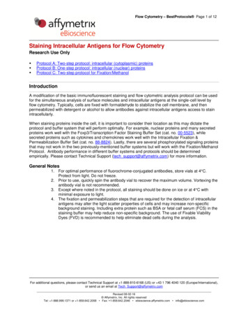
What is Flow Cytometry? Single-cell analysis. Uses monoclonal antibodies to tag markers on/inside the cells. Fluorescent molecules (flurochromes) are bound to the antibodies. Flurochromes are excited by lasers at specific wavelengths. Fluorochromes emit light at a higher wavelength, which is read by thecytometer.
Basics of Flow – FluidicsSheath FluidFlow CLaser
Basics of Flow - Fluidics
Basics of Flow - Fluidics
Basics of FlowSSCMammal whole blood/bone marrowFSC
Basics of FlowIntensityWHATime
Basics of Flow
Fluorochromes
Fluorochromes Many “colors” to choose from. Each fluorochrome has two properties:excitation and emission. Excitation: wavelength at which thefluorochrome absorbs the most energy. Emission: wavelength at which thefluorochrome produces the mostenergy.
Fluorochromes
Fluorochromes Fluorescent proteins GFP, YPF, CFP, RFP Viability Dyes DNA Dyes – DAPI, PI, 7AAD, etc. Fixable Viability Dyes
Compensation
Compensation
Compensation Single-stained controls are required; one for each fluorochrome. Cells Beads Unstained control also necessary.Fully Stained SamplePerCP onlyPE onlyFITC only Software will calculate the percentoverlap for each color.Unstained Cells Cells are ideal
Compensation
Compensation Compensation problems usually caused by poor panel design. In general, if two fluorochromes:a) are excited by the same laser, andb) use the same filter for detection, thenthey cannot be run together on a conventional cytometer.
Compensation
Compensation Sometimes colors that should work together don’t.
Panel Design Spread color choices across available excitation lasers. Choose colors with emissions as far apart as possible. Match brighter colors with dimmer markers. Some colors fluoresce brighter than others. Some markers are less frequently found on the cell surface. Come to Heidi’s presentation to learn more!
Panel Design488nm Blue647nm Red532nm Y/G355nm UV405nm Violet
Sample Prep Single-cell suspension 106 cells/mL, minimum 250µL 12x75 Polystyrene tube Filter at the instrument – bring a pipette!For help with a staining protocol or tissue prep, see Ailing or othermembers of the Flow Core.
Thank you!Dewayne Falkner – Operational Director falkner@pitt.eduAarika MacIntyre – Senior Flow Core Technologist alm253@pitt.eduAiling Liu – Research Specialist ail13@pitt.eduHeidi Gunzelman – Senior Flow Core Tech hmg44@pitt.eduNan Sheng – Flow Core Tech nas260@pitt.edu
What is Flow Cytometry? Single-cell analysis. Uses monoclonal antibodies to tag markers on/inside the cells. Fluorescent molecules (flurochromes) are bound to the antibodies. Flurochromes are excited by lasers at specific wavelengths. Fluorochromes emit light at a higher wavelength, which is read by the cytometer.
