Transcription
(2022) 23:82Pereira et al. BMC Open AccessRESEARCHProgesterone differentially affectsthe transcriptomic profiles of cow endometrialcell typesGonçalo Pereira1†, Yongzhi Guo2†, Elisabete Silva1, Claudia Bevilacqua3, Gilles Charpigny4,Luís Lopes‑da‑Costa1*† and Patrice Humblot2†AbstractBackground: The endometrium is a heterogeneous tissue composed of luminal epithelial (LE), glandular epithelial(GE), and stromal cells (ST), experiencing progesterone regulated dynamic changes during the estrous cycle. In thecow, this regulation at the transcriptomic level was only evaluated in the whole tissue. This study describes specificgene expression in the three types of cells isolated from endometrial biopsies following laser capture microdissectionand the transcriptome changes induced by progesterone in GE and ST cells.Results: Endometrial LE, GE, and ST cells show specific transcriptomic profiles. Most of the differentially expressedgenes (DEGs) in response to progesterone are cell type-specific (96%). Genes involved in cell cycle and nuclear divi‑sion are under-expressed in the presence of progesterone in GE, highlighting the anti-proliferative action of pro‑gesterone in epithelial cells. Elevated progesterone concentrations are also associated with the under-expression ofestrogen receptor 1 (ESR1) in GE and oxytocin receptor (OXTR) in GE and ST cells. In ST cells, transcription factors suchas SOX17 and FOXA2, known to regulate uterine epithelial-stromal cross-talk conveying to endometrial receptivity, areover-expressed under progesterone influence.Conclusions: The results from this study show that progesterone regulates endometrial function in a cell type-spe‑cific way, which is independent of the expression of its main receptor PGR. These novel insights into uterine physiol‑ogy present the cell compartment as the physiological unit rather than the whole tissue.Keywords: Transcriptome, Progesterone, Endometrium, LCM, RNA-seqBackgroundThe bovine endometrium is composed of different cellcompartments, comprising luminal epithelial (LE), glandular epithelial (GE), and stromal cells (ST), which is submitted to intense tissue remodelling during the estrous*Correspondence: lcosta@fmv.ulisboa.pt†Gonçalo Pereira and Yongzhi Guo shared first authorship.†Luís Lopes-da-Costa and Patrice Humblot shared senior authors.1CIISA‑Centro de Investigação Interdisciplinar em Sanidade Animal,Faculdade de Medicina Veterinária, Universidade de Lisboa, Avenida daUniversidade Técnica, 1300‑477 Lisbon, PortugalFull list of author information is available at the end of the articlecycle, embryo implantation and puerperal involution[1, 2]. Progesterone (P4) released by the corpus luteum(CL) plays a key role regulating endometrial function andremodelling [3, 4]. At the transcriptomic level, this P4regulation has been evaluated so far, only from the wholeendometrial tissue, following luteolysis [5], comparingdifferent oestrus cycle stages [6] or status (diestrus versus anoestrus) and type of ovulation (single versus multiple ovulation) [7]. However, as in other heterogeneoustissues, quantification of gene expression from the wholeendometrium may not reflect the specific transcriptionof the cell compartments. Laser capture microdissection The Author(s) 2022. Open Access This article is licensed under a Creative Commons Attribution 4.0 International License, whichpermits use, sharing, adaptation, distribution and reproduction in any medium or format, as long as you give appropriate credit to theoriginal author(s) and the source, provide a link to the Creative Commons licence, and indicate if changes were made. The images orother third party material in this article are included in the article’s Creative Commons licence, unless indicated otherwise in a credit lineto the material. If material is not included in the article’s Creative Commons licence and your intended use is not permitted by statutoryregulation or exceeds the permitted use, you will need to obtain permission directly from the copyright holder. To view a copy of thislicence, visit http:// creat iveco mmons. org/ licen ses/ by/4. 0/. The Creative Commons Public Domain Dedication waiver (http:// creat iveco mmons. org/ publi cdoma in/ zero/1. 0/) applies to the data made available in this article, unless otherwise stated in a credit line to the data.
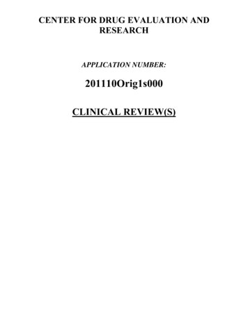
Pereira et al. BMC Genomics(2022) 23:82(LCM) emerged as a research tool to isolate cell populations for molecular analyses [8]. This method has beenused to study the interactions between the endometrial epithelial and stromal compartments with seminalplasma in a murine model [9] and the conceptus inducedregulation of endometrial function in the porcine [10],ovine [11] and equine [12] species. In the cow, recentstudies based on this approach described the specificmolecular signatures of endometrial stromal, glandularand luminal epithelial cells, as well as the effect of negative energy balance on the transcriptomic profiles ofendometrial compartments in the mid-luteal phase [13,14]. The above experiments revealed that the differentendometrial cell types show distinct transcription patterns. The main objective of this study was to evaluatethe effects of P4 on the transcription patterns of the threemain bovine endometrial cell types, which, to our knowledge, have not been documented so far.ResultsThis study was performed initially while considering themain effects of cell type, P4 concentrations, and theirinteraction, on the transcriptomic profile of endometrialcells of postpartum cows.Due to the fact that only one sample from LE cellswas associated with high progesterone, transcriptomicchanges induced by P4 and their interaction with celltype were studied only from GE and ST cells. Fromthe full list of DEGs (GE plus ST cells), where the maineffects was significant (FDR adjusted P-value 0.05),the interaction was only significant for 1% of these (6 /591). Furthermore, for most of these genes, significancewas mainly associated with a low level of expression. Forthree genes, the interaction resulted from progesteronebeing associated with an effect in one cell type but notin the other cell type. For three other genes, (ERP27, GKand ENSBTAG00000003408) the significant interactionresulted from opposite effects of P4 in GE and ST cells.Due to the very low number of genes for which a significant interaction was detected, the results presented anddiscussed below address principally the main effects ofcell type and P4 concentrations.Overall gene expression and differentially expressed genesbetween the three endometrial cell typesThe total number of genes with more than 10 transcriptsper million (TPM) was 15,420, 15,555, and 15,308 for LE,GE, and ST cells, respectively. From these, 274 (1.78%)were LE specific, 280 (1.80%) were GE specific and 346(2.26%) were ST specific (Fig. 1A). Among genes specifically transcribed by LE cells, TM4SF4, C29H11orf86,CSF2, CAPN14, SERPINB10, CA1, SLC5A5, SLC6A12,and two uncharacterized genes (ENSBTAG00000036102,Page 2 of 19ENSBTAG00000055111) had the highest level of averagetranscription. Among GE-specific genes, BTG4, IBSP,TMEM212, C4BPA, DPP6, ANKS4B, PROC, CFAP58,LDLRAD1 and GAS2L2 were the most transcribed,whereas, among the ST-specific genes, the most transcribed were CHRNA2, KLK5, SST, DMRT2, GABRA4,NFASC, CDH9, DLK1, and two uncharacterized genes(ENSBTAG00000054090, ENSBTAG00000045630). Thefull lists of genes specifically transcribed by each cell typewith respective transcription levels are provided in Supplementary File 1.Although a very high proportion of genes was expressedover 10 TPM in all three cell types (14,509/16337; 88.8%;Fig. 1A), their expression level was not the same. Thisis shown by the PCA analysis revealing a clear separation of the samples from the three cell types. The twofirst dimensions explain 70% of the variability (Fig. 1B),the first allowing the distinction of epithelial from stromal cells, whereas the second differentiates GE from LEcells. The genes which explain the most differences inthe first dimension (levels of expression the most associated with GE and LE cells) were TMEM125, ESRP1,ELMO3, KDF1, C3H1orf210, RHPN2, AP1M2, CYB561,TJP3, and SLC44A4, whereas these genes were WT1,SFRP1, TAGLN, HOXA10, ACTA2, C15H11orf96, CCN3,CNN1, TPM2, and STRA6 for ST cells. When analyzing differences between GE and LE cells (second dimension), PPP1R1B, MYOC, AGT , MGP, CPN1, SNAP91,MPTX, PRSS35, CYBRD1 and CCDC146 were the geneswhich were the most specific of GE cells, whereas BCHE,SMOC2, GM2A, DES, ENPP5, TNMD, ZBTB16, CHODL,BPIFB1 and PDZK1 were the most associated with LEcells. The lists of genes most correlated (P 0.01) witheach dimension are provided in Supplementary File 2.The DESeq2 analysis revealed 4045 DEGs between GEand LE cells, 7974 between GE and ST cells, and 7839between LE and ST cells.Gene ontology enrichment analysis of cell‑specific genesEnriched GO terms in each cell-specific genes list werevisualized using the REVIGO algorithm to reduce termredundancy and identified 64, 30, and 79 GO terms clusters in LE, GE, and ST cells, respectively (SupplementaryFig. 1). The most significant over-represented terms inLE cells included transport processes, immune systemprocess, cytokine production and regulation, response tostimulus and cell surface receptor signalling, whereas forGE cells, these terms included cilium movement, regulation of triglyceride biosynthetic process, regulation ofpeptide secretion and transport, regulation of glucosetransmembrane transport, and complement activation.For ST cells, the most significant over-represented termsincluded signalling, cell communication, multiple
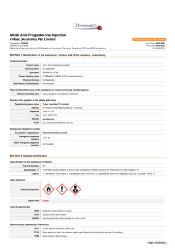
Pereira et al. BMC Genomics(2022) 23:82Page 3 of 19Fig. 1 Transcriptome of endometrial cell types and effect of different progesterone concentrations of cows from which samples were issued. AVenn diagram with genes expressed ( 10 transcripts per million (TPM)) in endometrial cells (stromal cells (ST), glandular epithelium (GE), andluminal epithelium (LE) (numbers of identified genes are indicated). B Principal component analysis (PCA) of cell types (ST, GE and LE) among cowgroups (High progesterone, P4high and low progesterone, P4low)developmental and biological regulation processes. Thecomplete list of over-represented GO terms is providedin Supplementary File 3.Differentially expressed genes between elevated and lowprogesterone cowsFollowing microdissection, only 1 high RNA quality LEsample collected from cows with high progesteroneconcentrations was available. Therefore, differences inthe transcriptomic profiles in response to progesteronewill be presented and further discussed here from listsof DEGs obtained for GE and ST cells (Supplementaryfile 4). However, given its potential interest for futurestudies, the list of putative DEGs from LE samples is alsopresent in Supplementary file 4. The number of DEGsfound between samples collected from cows with elevated or low P4 concentrations were 386 and 205 in GEand ST cells, respectively. From these DEGs, 365 (95%)
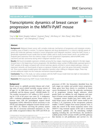
Pereira et al. BMC Genomics(2022) 23:82Page 4 of 19Fig. 2 A Venn diagram from differentially expressed genes (DEGs) between samples issued from cows of the high and low progesterone groupsin glandular epithelial (GE) and stromal (ST) endometrial cells. (Numbers and percentage of DEGs are indicated). B) Linear regression analysis of thefold changes of the 21 common DEGs from ST (x-axis) and GE (y-axis) cellsand 184 (90%) were cell type-specific (Fig. 2A). Twentyone DEGs were common to GE and ST cells (TNC,ADAMTS18, P4HA2, APEX1, PNPLA2, SOSTDC1,TUBB, FBLN7, MAPK4, EEF1G, TNFRSF13B, ENSBTAG00000015493, B9D1, RACK1, OXTR, RPL8, P1, ENSBTAG00000040367). When consideringthe above 21 DEGs, the regression analysis of the log2fold change in response to progesterone from the two celltypes showed a similar effect in the two cell types (regression slope 0.72; adjusted R-squared 0.975; Fig. 2B). Theslope coefficient lower than 1 indicates that the magnitude of response was only slightly higher in ST than inGE cells which is consistent with the average log2 foldchange for all DEGs observed in ST and GE cells, 3.81and 2.95, respectively. Moreover, elevated P4 was most
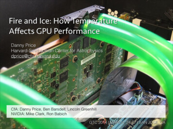
Pereira et al. BMC Genomics(2022) 23:82often associated with under-expression of genes in GE(280/386; 73%) and over-expression in ST (118/205; 58%)(Fig. 3).Gene ontology enrichment analysis of samples from cowswith elevated and low progesterone concentrationsEnriched GO terms in the DEGs lists of elevated andlow P4 cows are presented in Fig. 4, and the corresponding lists of genes in Supplementary File 5. In GE cells,the over-expressed and under-expressed genes underthe effect of P4 relate to 42 and 62 enriched GO terms,respectively. Over-expressed genes are mostly categorised in cellular metabolic processes (GO:0044237)(n 49), response to organic substances (GO:0010033)(n 19) and regulation of diverse biological processeslike response to stimulus (GO:0048583) (n 20), cellcommunication (GO:0010646) (n 18) and signalling(GO:0023051) (n 18). The under-expressed genes areoverrepresented in cell cycle (GO:0007049) (n 31),nuclear division (GO:0000280) (n 14), nuclear chromosome segregation (GO:0098813) (n 14), mitotic cellcycle (GO:0000278) (n 20), localisation (GO:0051179)(n 58), response to stimulus (GO:0050896) (n 7),response to stress (GO:0006950) (n 36) and cell-cellsignalling (GO:0007267) (n 23).In ST, the analysis revealed 22 and 2 enriched GOterms corresponding to over- and under-expressedgenes, respectively. The over-expressed genes relate tocell motility (GO:0048870) (n 14), movement of cell orsubcellular component (GO:0006928) (n 18), locomotion (GO:0040011) (n 18), animal organ morphogenesis (GO:0009887) (n 15), localization (GO:0051179)(n 37) and regulation of localization (GO:0032879)(n 18), whereas the under-expressed genes are associated to anatomical structure development (GO:0048856)(n 21) and developmental processes (GO:0032502)(n 22).GeneCards and protein‑protein interaction (PPI) networkanalysisThe comparison between the genes of the GeneCardsdatabase (http:// www. genec ards. org) corresponding to“hormonal regulation”, “uterine receptivity” and “pregnancy”, and the DEGs identified in the present study inrelation with the presence of P4 is shown in Fig. 5. ForGE and ST cells, a very large proportion of DEGs areinvolved in hormonal regulation ( 60%) and pregnancy( 40%), and all DEGs participating in uterine receptivityalso relates to the above terms. A more thorough analysisof the present DEGs involved in “hormonal regulation”,“pregnancy” and “uterine receptivity” revealed that someof the genes important for these processes are differentially expressed under progesterone in both cell typesPage 5 of 19(Fig. 6). For instance, ESR1 is under-expressed under theeffect of P4 in GE, OXTR is under-expressed under theeffect of P4 in GE and ST, and transcription factors suchas SOX17 and FOXA2 as well as interferon related genes,which are known to regulate uterine epithelial-stromalcross-talk conveying to endometrial receptivity, are overexpressed under the effect of P4 in ST whereas no changeis observed in GE cells.In high P4 cows, the STRING-generated protein interaction network obtained from GE DEGs revealed 11clusters of under-expressed genes including one of avery large size (Fig. 7A) and 5 clusters of over-expressedgenes (Fig. 7B), whereas, in ST cells, the analysis revealed6 clusters of under-expressed (Fig. 7C) and 5 clusters ofover-expressed (Fig. 7D) genes.DiscussionThis study combined LCM to isolate endometrial cellcompartments from uterine biopsies and RNAseq toanalyze their full transcriptomes, identifying changesinduced by P4. This approach provides novel information regarding cell-specific gene expression that remainsundetected when analyzing whole tissue samples. This isparticularly relevant for epithelial cells for which the celltype’s representability is low, as specific gene expressiondata is diluted in the average of the whole tissue. Furthermore, as observed here and recently also reportedin another model [14], genes may only be differentiallyexpressed in a specific cell type and remain unaffected inthe others.This study confirms the difficulty in capturing sampleswith good quality RNA from LE. As in Chankeaw et al.[13, 14], despite preparation of a high number of slidesfrom biopsies, the number of sequenced samples waslower for LE than for GE and ST cells.Of the three different endometrial samples (LE, GE,and ST), ST samples are the most heterogeneous, comprising the stratum compactum and stratum spongiosum,and including blood vessel and migrating immune cells.By carefully selecting the capture areas, contaminationwith blood vessel cells was avoided, and contaminationwith migrating immune cells was minimized by includingonly healthy cows, as assessed by endometrial cytologyand histology. Variation regarding the different strata thatcompose endometrial ST was addressed by capturingsamples from the most superficial layers (stratum compactum) as these may induce paracrine interactions withthe neighbouring epithelial cells.The design model here applied revealed strong maineffects of cell type and P4 concentrations, but showedlack of significant interaction for most of the genes influenced by progesterone. The fact that ERP27, GK andENSBTAG00000003408) exhibit opposite changes in
Pereira et al. BMC Genomics(2022) 23:82Page 6 of 19Fig. 3 Volcano plots with the distribution of differentially expressed genes between high and low progesterone cows for glandular epithelial (GE)and stromal (ST) endometrial cells
Pereira et al. BMC Genomics(2022) 23:82Page 7 of 19Fig. 4 Scatterplot representation of enriched GO terms in semantic space using REVIGO [15], from lists of over-expressed and under-expressedgenes in glandular epithelial (GE) and stromal (ST) endometrial cells, between high and low progesterone cows. Circle size represents the frequencyof the GO term in the underlying GOA database (bubbles of more general terms are larger) and colour indicates the uniqueness valueFig. 5 Comparison of differentially expressed genes in glandular epithelial (A) and stromal (B) cells to GeneCards database. Numbers and senseof variation (arrows) of DEGs participating in hormonal regulation, pregnancy, and uterine receptivity. From the 386 DEGs identified in GE, 237participate in hormonal regulation, 184 in pregnancy and 26 in uterine receptivity. From the 205 DEGs that emerged in ST, 130 participate inhormonal regulation, 85 in pregnancy and 15 in uterine receptivity
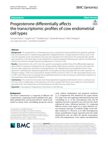
Pereira et al. BMC Genomics(2022) 23:82Page 8 of 19Fig. 6 Log normalized transcripts per million (TPM) of ESR1, FOXA2 and SOX17 genes for glandular epithelial (GE) and stromal (ST) endometrial cellsissued from high (red) and low (blue) progesterone cows. Horizontal black lines indicate median; boxes extend from the 25th to the 75th percentileand vertical lines indicate values within 1.5 interquartile range of the 25th and 75th percentiles. Dots indicate outliersexpression under P4 in GE and ST cells, would deservefurther investigations in relation with endometrialfunction.Transcriptome of endometrial cell typesThe PCA from full RNA-seq data corroborates previouswork [13] documenting that LE, GE, and ST cells of thebovine endometrium exhibit different molecular signatures. The full description of the characteristics of thetranscriptome of the different cell types is beyond thescope of this article (lists are set in Supplementary File 4).A low percentage (around 2%) of cell type-specificgenes were identified, which is in agreement with datafrom the porcine endometrium [10]. These cell-specificexpressed genes encode proteins that putatively supportspecialized functions of each cell type, which is supported by the overrepresentation of different enrichedGO terms in the cell-specific gene lists (SupplementaryFig. 1). Discussion of the compared function of these proteins is beyond the scope of this article, but some relevantexamples are summarized below.LE specific genes encode proteins involved in processesof transport and uptake across epithelial surfaces (solutecarrier family 5 member 5, solute carrier family 6 member 12, transmembrane 4 L six family member 4). SLC6has been shown to be differentially transcribed throughdifferent phases of the human oestrus cycle [16] andstimulated by INF-Tau in the cow [17]. These comprisealso an enzyme responsible for maintaining acid-basehomeostasis (carbonic anhydrase 1), a serine peptidaseinhibitor (serpin family B member 10), an embryokine(colony stimulating factor 2), and a member of the calpain family (calpain 14).Carbonic anhydrase 1 is a member of the large family ofzinc metalloenzymes that catalyze the reversible hydration of carbon dioxide and plays a pivotal role maintaining acid-base homeostasis [18] possibly impacting inthe endometrium, sperm fertilization capacity, embryotransport, development and implantation [19].Colony stimulating factor 2 (CSF2) is among the moststudied embryokines, being secreted into the bovineuterine lumen [20]. This cytokine is involved in therecruitment, differentiation and function of neutrophils,when secreted by mouse uterine epithelial cells, following stimulation with TLR agonists [21]. CFS2 treatmentduring the preimplantation period improved the development and survival of bovine embryos [22].In postpartum dairy cows, LE cells are a primary lineof defence against bacteria, and an important component of the innate immune system [2]. This is here evidenced with the over-representation of genes related toimmune response and interleukin production, especiallyinterleukin-23 which was shown to be involved in humanendometrial immune regulation [23]. Also, the secretoryrole of LE cells is evidenced by the overrepresentation ofprocesses regarding multiple transport processes (ion,chloride, oxalate, bicarbonate) (Supplementary File 3).Overall, gene data from LE cells highlight their putative
Pereira et al. BMC Genomics(2022) 23:82Page 9 of 19Fig. 7 STRING-generated protein-protein networks from differentially expressed genes of glandular (GE), and stromal (ST) endometrial cellsbetween high and low progesterone cows. A Under-expressed in GE; B Over-expressed in GE; C Under-expressed in ST; D) Over-expressed in ST.Arrows pointed to target nodesspecialized functions, such as the regulation of uterinefluid composition, providing favourable microenvironments for sperm and embryos, and the immune responseagainst potential pathogens.GE specific genes encode proteins involved in cellcycle regulation (BTG anti-proliferation factor 4), adhesion processes (integrin binding sialoprotein), immuneresponse (complement component 4 binding proteinalpha, protein C), localized in brush border (ankyrinrepeat and sterile alpha motif domain containing 4B, ciliaand flagella associated protein 58, low density lipoprotein receptor class A domain containing 1, growth arrestspecific 2 like 2) and transmembrane proteins associatedwith voltage-gated potassium channels (Dipeptidyl peptidase-like protein 6).The expression of genes encoding proteins related tomicrovilli adhesion and assembly support that bovineendometrial GE cells form a cluster of tightly packed
Pereira et al. BMC Genomics(2022) 23:82microvilli, as observed in rat endometrial GE cells [24].As example, low density lipoprotein receptor class Adomain containing 1 is a membrane receptor alreadyidentified in GE cells in previous work [13] and alsoexpressed by mature ciliated cells in airway epithelium[25]. Cow endometrial GE cells have specialized functions regarding synthesis, transport and secretion of substances into the uterine lumen [26], here illustrated bythe overrepresentation of biological processes of regulation of peptide secretion and cilium movement, the latter believed to be essential for moving secretory productsacross the surface of GE cells into the uterine lumen [27].In addition, the role of GE cells in innate and adaptiveimmune responses is highlighted by the overrepresentation of genes involved in complement activation, asobserved in woman’s endometrial GE cells [28]. Complement component 4 binding protein alpha is a protein thatcontrols the activation of the complement cascade and isupregulated in the bovine endometrium following exposure to seminal plasma components, likely involved inthe regulation of peri-implantation events [29]. ProteinC is a potent anticoagulant and anti-inflammatory molecule which regulates the functions of different epithelialbarriers by controlling inflammation [30, 31]. Dipeptidylpeptidase-like protein 6 is a transmembrane protein thatbinds to voltage-gated potassium channels from the Kv4family [32], associated with uterine capacity for pregnancy and fertility in beef heifers [33], and relevant foruterine function [34].The overrepresented processes in ST cells are morenumerous and diverse than in GE and LE cells. Most ofthem relate to regulatory processes, which is consistentwith the ST regulatory role exerted on adjacent epithelial cells in co-culture systems [35], or within the cowendometrium [13]. This regulatory role of ST cells alsoemerges from the overrepresentation of cell communication and signalling processes, which are paramountfor the coordination of cellular responses. ST specificgenes encode, among others, extracellular proteases (Kallikrein related peptidase 5) and cell adhesion molecules.In women, kallikrein related peptidase 5 is an extracellular protease expressed in endometrial GE cells, suggested to play a role in host defense [36]. This protein isalso involved in remodelling and repair of epithelial barriers [37] and able to generate plasmin indicating a rolein wound healing [38]. Different collagens were describedas substrates for kallikrein-related peptidase 5, hinting arole in extracellular matrix remodelling and cell migration [38].The specific roles of some of the most correlated genesto each PCA dimension, which were previously foundto be involved in endometrial function or associated topathologies, are also explored below. In the first PCAPage 10 of 19dimension, which separates stromal from epithelial cells,the most specific ST cells’ gene was the Wilms’ tumorsuppressor gene 1 (WT1), previously reported as specifically expressed in woman endometrial ST cells [39]. Thiswas also the case of smooth muscle cell markers (TAGLN;ACTA2; CNN1; TPM2), also specifically detected in theendometrial stromal compartment of healthy women[40]. Additionally, STRA6 and SFRP1, encoding a receptor for retinol uptake and a soluble modulator of Wnt signalling, respectively, were highly expressed in ST cells, aspreviously described in woman’s endometrium [41, 42].The most specific GE and LE genes were those encoding an epithelial splicing regulatory protein (ESRP1),which is a regulator of FGFR2 splicing [43], the keratinocyte differentiation factor (KDF1), strongly expressed inthe dental epithelium of mouse embryos [44], and theMsh homeobox 1 (MSX1), with strong nuclear localization in GE and LE cells of fertile woman’s endometrium[45]. In addition, a large set of genes involved in epithelial cell differentiation, epithelium development, and celladhesion (CLDN3, SYNE4, LRP2, F2RL1,DLX6, ELF3,SPINT1, PHGDH, OVOL1, TACSTD2, ST14, EHF, MSX1,EPCAM, ST14, KDF1, IRF6, TJP3, SLC44A4, RAB25,DSP, MCOLN3) were identified, also previously correlated with epithelial cells (GE LE) in the endometriumof dairy cows [13].In the second PCA dimension, which separates GEcells from LE cells, the most specific GE cells’ genes werePPP1RB, which encodes DARPP-32, a phosphoproteinexpressed in ciliary epithelia [46], and CCDC146, a ciliated cell marker [47]. Although both LE and GE containciliated cells [48], the number of these cells is expectedto be lower in LE than in GE, as documented in women[49]. Also as in the human endometrium [50], the angiotensinogen coding gene (AGT ), was highly expressedin GE cells. A set of genes involved in axonemal dyneincomplex assembly and cilium movement processes(CCDC65, DRC1, DRC3, DAW1, CFAP45, DNAH5,DNAH9) was associated to GE, as previously reported[13].LE cells were correlated with SMOC2, encoding anextracellular glycoprotein recognized as an endometrialcancer stem cell signature [51]. Stem cells were identifiedin the epithelial and stromal compartments of humanendometrium, where they are said to be responsible forits remarkable regenerative capacity [52]. The endometrium of postpartum dairy cows experiences intensetissue remodelling and re-epithelialization, suggesting SMOC2 as a putative uterine stem cell maker. Thedesmin coding gene (DES) was also specific of LE cells,despite being identified as a smooth muscle cell marker[53]. However, desmin has also been used to distinguish epithelial cells undergoing epithelial-mesenchymal
Pereira et al. BMC Genomics(2022) 23:82transition (EMT) [54], and there is evidence of EMT participation in endometrial regeneration and re-epithelialization [55, 56]. This indicates desmin expression to be aputative EMT marker in LE cells. BPIFB1, a gene encoding an innate defence protein identified in other epithelialbarriers, such as human airways [57] was also specific ofLE cells.Impact of progesterone on the transcription profileof endometrial cell typesFor GE and ST cells, the PCA analysis did not identifyoutliers and individual samples clustered nicely, showing similar gene expression profiles within each P4 group.Overall, data on number of DEGs and overrepresentation of biological processes indicate that the response toelevated P4 was more significant in GE than in ST cells.As documented in the methods section, the average log2fold change of DEGs in
(GE), and stromal cells (ST), experiencing progesterone regulated dynamic changes during the estrous cycle. In the cow, this regulation at the transcriptomic level was only evaluated in the whole tissue. This study describes specic . cic way, which is independent of the expression of its main receptor PGR. These novel insights into uterine .