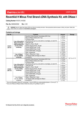
Transcription
Synthesis and Evaluation of a Thermoresponsive DegradableChitosan-Grafted PNIPAAm Hydrogel as a “Smart” Gene DeliverySystemZiminska, M., Wilson, J. J., McErlean, E., Dunne, N., & McCarthy, H. O. (2020). Synthesis and Evaluation of aThermoresponsive Degradable Chitosan-Grafted PNIPAAm Hydrogel as a “Smart” Gene Delivery System.Materials, 13(11), [2530]. https://doi.org/10.3390/ma13112530Published in:MaterialsDocument Version:Publisher's PDF, also known as Version of recordQueen's University Belfast - Research Portal:Link to publication record in Queen's University Belfast Research PortalPublisher rightsCopyright 2020 the authors.This is an open access article published under a Creative Commons Attribution License ch permits unrestricted use, distribution and reproduction in any medium, provided the author and source are cited.General rightsCopyright for the publications made accessible via the Queen's University Belfast Research Portal is retained by the author(s) and / or othercopyright owners and it is a condition of accessing these publications that users recognise and abide by the legal requirements associatedwith these rights.Take down policyThe Research Portal is Queen's institutional repository that provides access to Queen's research output. Every effort has been made toensure that content in the Research Portal does not infringe any person's rights, or applicable UK laws. If you discover content in theResearch Portal that you believe breaches copyright or violates any law, please contact openaccess@qub.ac.uk.Download date:08. Jul. 2022
materialsArticleSynthesis and Evaluation of a ThermoresponsiveDegradable Chitosan-Grafted PNIPAAm Hydrogel asa “Smart” Gene Delivery SystemMonika Ziminska 1 , Jordan J. Wilson 1,2 , Emma McErlean 1 , Nicholas Dunne 1,3,4,5,6,7,8,9, * andHelen O. McCarthy 1,10, *12345678910*School of Pharmacy, Queen’s University of Belfast, 97 Lisburn Road, Belfast BT9 7BL, UK;m.ziminska@qub.ac.uk (M.Z.); jwilson88@qub.ac.uk (J.J.W.); E.McErlean@qub.ac.uk (E.M.)School of Chemistry and Chemical Engineering, Queen’s University of Belfast, Belfast BT9 5AG, UKSchool of Mechanical and Manufacturing Engineering, Dublin City University, Dublin 9, IrelandCentre for Medical Engineering Research, School of Mechanical and Manufacturing Engineering, DublinCity University, Dublin 9, IrelandDepartment of Mechanical and Manufacturing Engineering, School of Engineering, Trinity College Dublin,Dublin 2, IrelandAdvanced Manufacturing Research Centre (I-Form), School of Mechanical and Manufacturing Engineering,Dublin City University, Glasnevin, Dublin 9, IrelandAdvanced Materials and Bioengineering Research Centre (AMBER), Trinity College Dublin, Dublin 9, IrelandAdvanced Processing Technology Research Centre, Dublin City University, Dublin 9, IrelandTrinity Centre for Biomedical Engineering, Trinity Biomedical Sciences Institute, Trinity College Dublin,Dublin 2, IrelandSchool of Chemical Sciences, Dublin City University, Dublin 9, IrelandCorrespondence: nicholas.dunne@dcu.ie (N.D.); h.mccarthy@qub.ac.uk (H.O.M.);Tel.: 353-(0)1-7005712 (N.D.); 44-(0)28-90972149/1993 (H.O.M.) Received: 18 May 2020; Accepted: 29 May 2020; Published: 2 June 2020Abstract: Thermoresponsive hydrogels demonstrate tremendous potential as sustained drug deliverysystems. However, progress has been limited as formulation of a stable biodegradable thermosensitivehydrogel remains a significant challenge. In this study, free radical polymerization was exploited toformulate a biodegradable thermosensitive hydrogel characterized by sustained drug release. Highlydeacetylated chitosan and N-isopropylacrylamide with distinctive physical properties were employedto achieve a stable, hydrogel network at body temperature. The percentage of chitosan was alteredwithin the copolymer formulations and the subsequent physical properties were characterized using1 H-NMR, FTIR, and TGA. Viscoelastic, swelling, and degradation properties were also interrogated.The thermoresponsive hydrogels were loaded with RALA/pEGFP-N1 nanoparticles and release wasexamined. There was sustained release of nanoparticles over three weeks and, more importantly,the nucleic acid cargo remained functional and this was confirmed by successful transfection of theNCTC-929 fibroblast cell line. This tailored thermoresponsive hydrogel offers an option for sustaineddelivery of macromolecules over a prolonged considerable period.Keywords: hydrogel; thermoresponsive; hydroxyapatite; nanoparticles; drug delivery1. IntroductionHydrogels are three-dimensional, crosslinked polymeric networks capable of absorbing largeamounts of water or biological fluids [1]. This group of synthetic biomaterials encompasses a widerange of chemical compositions and bulk physical properties, since they can be prepared from avast library of available monomers and crosslinkers [2]. Hydrogels can encapsulate and protectMaterials 2020, 13, 2530; ls
Materials 2020, 13, 25302 of 22hydrophilic drugs from hostile environments with a resultant slow and controlled release by diffusionor erosion [3–5]. Multiple studies have demonstrated the applicability of hydrogels for the release of abroad range of therapeutic agents including growth factors, DNA, small interfering ribonucleic acid(siRNA), and anti-cancer drugs [6–10].Hydrogel that can be administered through minimally invasive delivery is an attractive propositionthat can enhance patient convenience, ease surgical administration, and reduce costs associatedwith surgery [11]. Injectable hydrogels can readily mold into the shape of the surgical cavity andadhere to the surrounding tissues during hydrogel formation, which facilitates treatment of anynon-standard geometry [3]. The therapeutic cargo dissolved within the structure of hydrogel isshielded from injection-associated shear forces and can be released with complex dynamics ina controlled manner [12]. These injectable hydrogels can be prepared using non-toxic chemicalcrosslinkers, enzymes, or physical interactions, such as hydrophobic and ionic interactions, and canrespond to external stimuli (e.g., temperature, pH, and light), conferring “smart” or “intelligent”systems [5,13]. The thermosensitive hydrogel (i.e., responsive to temperature change) is one of themost popular and widely analyzed systems for drug delivery, facilitating an in situ gelation [3].Poly(N-isopropylacrylamide) (PNIPAAm) is one such example these stimuli-responsive polymers.PNIPAAm undergoes a sharp and reversible coil-to-globule phase transition above 32 C (the lowcritical solution temperature, LCST) [9,14]. Below this temperature, the hydrogel is hydrophilic as thehydrogen bonding between isopropyl side groups on the polymer and water molecules is dominant.The polymer chains are thus excessively hydrated and the expanded structures form a hydrogel [15].Above the LCST, PNIPAAm displays a sharp transition phase with a shrinking conformation as a resultof interactions between the hydrophobic segments with a concurrent reduction in the polymer-waterhydrogen bonding. At this point, the hydrogel is hydrophobic and the chains rapidly dehydrateand aggregate to form a viscous gel [16]. This temperature-dependent reversible reaction with theLCST close to body temperature has resulted in the application of PNIPAAm hydrogels for drugdelivery [17,18]. Grafting PNIPAAm chains onto the backbone of natural polymers (e.g., chitosan,collagen, alginate, and hyaluronic acid) is a strategy that ensures biodegradation, flexibility of gellingtemperature, and manipulation of the mechanical properties in hydrogels [19,20].Chitosan (Cs) can be employed as a drug delivery copolymer and has many advantages, includingbiocompatibility, non-toxicity, bioactivity, biodegradability, low cost, and ease of modification throughhydroxyl and amino groups [19]. Cs and derivatives have been utilized as delivery vehiclesin various forms, such as film, hydrogel, and microsphere for delivery of vaccines, DNA, andinsulin [21–24]. The hydrogel and nanoparticles composed of Cs backbone grafted with PNIPAAm(Cs-g-PNIPAAm) have been reported as non-cytotoxic with improved drug loading efficiency comparedto PNIPAAm alone [16,18,25,26]. Chuang et al. fabricated Cs-g-PNIPAM porous nanoparticles for theencapsulation of a hydrophilic drug (doxycycline hyclate) and reported continuous in vitro release over10 days [18]. In addition, Cs-g-PNIPAAm copolymers polymerized by free radical copolymerizationusing ammonium persulfate (APS) and N,N-methylenebisacrylamide resulted in a dual responsivetargeted drug delivery system of magnetic nanoparticles with drug loading of 86% followed by a70% drug release profile over 10 min [25]. PNIPAAm-based hydrogels can also be utilized as carriers fornucleic acid therapies as the affinity of PNIPAAm copolymers to DNA and the transfection efficiencyof delivered DNA can be controlled via temperature change [27]. Trimethyl Cs (TMCs)-g-PNIPAAmhas been demonstrated as an effective thermoresponsive gene carrier with 80% transfection efficiencyand minimal cytotoxicity in human embryonic kidney cells, HEK293 [16]. However, nucleic acidsoften require vectors to protect from degradation in the physiological environment and to augmenttransport across the plasma membrane and facilitate endosomal escape. A 30-mer cationic amphipathicpeptide known as RALA (i.e., WEARLARALARALARHLARALARALRACEA) has been successfullyemployed as a non-viral system for successful gene delivery [28]. RALA condenses nucleic acidsthrough spontaneous electrostatic self-assembly to form cationic nano-sized complexes with thephysiochemical properties required for cellular delivery [29]. The rise in utilizing copolymers as
Materials 2020, 13, 25303 of 22delivery platforms for therapeutic nanoparticles has led to the need for a greater understanding ofstructure-property relationships between hydrogel and therapeutic cargo. This study was designedto characterize the structure-property relationship of Cs-g-PNIPAAm hydrogels where dissolutionmedia and Cs components are considered in the context of how the physiochemical properties relate tosuccessful localized gene delivery.Simple free radical polymerization was employed to conduct the grafting of PNIPAAm onto a Csbackbone as reported by Pentlavalli et al. [14]. Radical polymerization is a swift and effective wayto produce grafted copolymers with unsaturated monomers such as NIPAAm monomers. It negatesthe requirement for regioselective activation and/or protection of functional groups of the Cs aswould be required for condensation methods of polymerization. Ammonium persulfate (APS) andtetramethyl-ethylenediamine (TEMED) initialize the formation of primary radicals, as previouslydescribed by Feng [30]. The free radical polymerization avoids multistep synthesis and associatedtoxicity issues offering a simple, cyto-compatible, scalable, and cost-effective copolymer fabrication [14].In this study, the structure, morphology, thermoresponsive properties, and in vitro drug releaseof electrostatic self-assembled nanoparticles were examined. The effect of differing the ratio of Cs inrelation to PNIPAAm on the properties of copolymer was also interrogated. Cytotoxicity, swellingbehavior, and drug release profiles in different media (i.e., water and Tris-ethylenediaminetetraaceticacid (EDTA) buffer) was also measured. To explore tunable swelling and release properties of thehydrogel, the Cs-g-PNIPAAm copolymer was crosslinked with genipin, a water soluble, non-toxiccrosslinker that can undergo self-polymerization [5].2. Materials and Methods2.1. MaterialsN-Isopropylacrylamide (NIPAAm) with purity 99%, low molecular weight Cs with a degreeof acetylation of 75–85%, and molecular weight (MW) of 50-190 kDa based on viscosity, APS,TEMED, and genipin were purchased from Sigma-Aldrich, UK. Double distilled water (DDW)was used for copolymer preparation, and the hydrogels were purified by using the membranefilter, Spectra/Por molecular porous membrane tubing with cut-off 10,000 Da, purchased fromSpectrum Laboratories, Inc. (Rancho Dominguez, CA, USA). The RALA peptide (peptide sequenceWEARLARALARALARHLARALARALRACEA) was supplied as a lyophilized powder from BiomatikCorporation (Wilmington, DE, USA). The powder was stored at 20 C and reconstituted inDNase/RNase free water (Life Technologies, Paisley, UK) for experimentation. A plasmid encoding thegreen fluorescent protein (GFP), pEGFP-N1, Clontech, Mountain View, CA, USA was propagated inMAX Efficiency DH5α Competent Cells (Life Technologies, Paisley, UK), purified using PureLink HiPure Plasmid Filter Maxiprep Kit (Life Technologies, Paisley, UK), and quantified by UV absorptionat 260 nm. NCTC-929 murine fibroblast cell lines were obtained from ATCC, USA. Cells werecultured and maintained in Minimum Essential Medium (MEM) (Gibco, Life Technologies, Paisley, UK)supplemented with 10% horse serum (HS) (Life Technologies, Paisley, UK) in a humidified incubator at37 C and 5% CO2 . Prior to use, cells were trypsinized using trypsin/EDTA in phosphate buffer solution(PBS) (2X) (Life Technologies, Paisley, UK), centrifuged, and resuspended in MEM. Cytotoxicity of thedegraded hydrogel product was measured using CellTiter 96 Aqueous One Solution Cell ProliferationAssay (MTS) (Promega, Southampton, UK). Tris-EDTA buffer (1X) was purchased from Thermo Fisher,Abingdon, UK. Quant-iT PicoGreen Reagent was purchased from Life Technologies, Paisley, UK.2.2. Preparation of Cs-g-PNIPAAmCs-g-PNIPAAm, with 10, 20, and 30 wt% of Cs in relation to PNIPAAm was synthesized throughfree radical polymerization: NIPAAm was added to 1% weight/volume (w/v) Cs in acetic acid. Solutionwas purged with dry nitrogen for 60 min prior to polymerization to remove any oxygen in order to avoida reaction with free radicals, followed by addition of 0.131 mmol initiator APS and 0.2 mmol accelerator
2.2. Preparation of Cs-g-PNIPAAmCs-g-PNIPAAm, with 10, 20, and 30 wt% of Cs in relation to PNIPAAm was synthesized throughfree radical polymerization: NIPAAm was added to 1% weight/volume (w/v) Cs in acetic acid.Materials 2020, 13, 25304 of 22Solution was purged with dry nitrogen for 60 min prior to polymerization to remove any oxygen inorder to avoid a reaction with free radicals, followed by addition of 0.131 mmol initiator APS and 0.2TEMED. Copolymerizationwas achieved12 hwithinunder12ambientThe copolymermmol acceleratorTEMED. Copolymerizationwaswithinachievedh underconditions.ambient conditions.was dialyzedfor 5 daysatDDW20 Catto20removeany unreactedmonomersand recoveredbyThe copolymerwas dialyzedforin5 DDWdays in C to removeany unreactedmonomersand freeze-dryingat 50 atC 50(Figure1a). Lyophilizedcopolymerwas thanby mechanicalrecoveredby freeze-drying C (Figure1a). Lyophilizedcopolymerwas solubilizedthan solubilizedbyagitationin DDW,Tris-EDTAbuffer,or PBSat a 5%ratioratiofor 18thethecrosslinkedsamples,mechanicalagitationin DDW,Tris-EDTAbuffer,or PBSat a w/v5% w/vforh.18Forh. ples, genipin was dissolved in 60% ethanol to 1 mg/mL solution and added to the reconstituted1%atw/v1b). Genipinreactsreactswith Csproducinga blue-coloredfluorescentsample,as shown inhydrogel1%(Figurew/v (Figure1b). GenipinwithCs producinga blue-coloredfluorescentsample,Figure1b [31].as shownin Figure1b [31].1. (a) Schematicof Chitosan-grafted-Poly(NIPAAm)synthesis.synthesis. (b)(b) CrosslinkingCs-g-PNIPAAmFigure Figure1. (a) Schematicof ithgenipin(GE).PNIPAAm with genipin (GE).2.3. Synthesis of Nano-Complexes2.3. Synthesis of Nano-ComplexesThe RALA/pEGFP-N1 nanoparticles (NPs) were prepared at a N:P ratio of 10 (the molar ratio ofThe RALA/pEGFP-N1 nanoparticles (NPs) were prepared at a N:P ratio of 10 (the molar ratio ofpositively charged nitrogen atoms in the peptide to negatively charged phosphates in the pEGFP-N1positively charged nitrogen atoms in the peptide to negatively charged phosphates in the pEGFP-N1backbone) by adding appropriate volumes of RALA peptide solution to 1 µg pEGFP-N1. The complexesbackbone) by adding appropriate volumes of RALA peptide solution to 1 μg pEGFP-N1. Thewere incubated at ambient temperature for 30 min to allow the formation of the NPs.complexes were incubated at ambient temperature for 30 min to allow the formation of the NPs.To visualize the incorporation of the NPs into the hydrogel matrix using scanning electronTo visualize the incorporation of the NPs into the hydrogel matrix using scanning electronmicroscopy [32], representative RALA/hydroxyapatite (HA) NPs were used as the nano-sized HA canmicroscopy [32], representative RALA/hydroxyapatite (HA) NPs were used as the nano-sized HAbe easily observed using scanning electron microscopy [33]. The nano-complexes were formulated at acan be easily observed using scanning electron microscopy [33]. The nano-complexes weremass ratio 7:1 via electrostatic attraction as previously described [34]. Briefly, negatively charged HAformulated at a mass ratio 7:1 via electrostatic attraction as previously described[34]. Briefly,was incubated with the positively charged RALA peptide for 30 min at 20 C. The resultant NPs hadnegatively charged HA was incubated with the positively charged RALA peptide for 30 min at 20 C.an average diameter of 153.7 18.9 nm and ζ-potential of 24.8 5.4 mV.The resultant NPs had an average diameter of 153.7 18.9 nm and ζ-potential of 24.8 5.4 mV.All NPs were lyophilized with 5% w/v trehalose as a cryoprotectant using VirTis Wizard 2.0 (SPAll NPs were lyophilized with 5% w/v trehalose as a cryoprotectant using VirTis Wizard 2.0 (SPScientific, Ulster, NY, USA) and stored at ambient temperature.Scientific, Ulster, NY, USA) and stored at ambient temperature.2.4. Chemical Characterization of Cs-g-PNIPAAm Hydrogel2.4. Chemical Characterization of Cs-g-PNIPAAm Hydrogel(1) Nuclear magnetic resonance analysis(1) Nuclear magnetic resonance analysismagneticresonance(1 H-NMR)were recordedon a Bruker400 -NMR)spectraspectrawere recordedon a Bruker400 plusspectrometerLtd., Durham,in deuteriumoxide(Da2 O)at a frequencyof 400 MHz.spectrometer(Bruker(BrukerLtd., Durham,UK) inUK)deuteriumoxide (D2O) atfrequencyof 400 MHz.(2) Fourier-transforminfraredspectroscopy(2) Fourier-transforminfraredspectroscopyFourier transform infrared spectra were obtained using Jasco FT/IR 4100 (Jasco Ltd., Dunmow,UK) to determine the copolymer formation. Samples were scanned in a spectral range of 400–4000 cm 1at 256 scans per spectrum and a resolution of 4 cm 1 .(3) Thermogravimetric analysis
Materials 2020, 13, 25305 of 22Thermogravimetric Analysis (TGA) of Cs-g-PNIPAAm copolymers was performed using a TGAQ500 analyzer (TA Instruments, New Castle, DE, USA). Lyophilized samples weighing approximately8–10 mg of were placed on a platinum pan and heated at a rate of 10 C min 1 from 20 to 600 C ina nitrogen atmosphere at a flow rate of 40 mL/min for the balance and 60 mL/min for the sample.The loss of copolymer weight was measured and the onset temperature was calculated.The grafting efficiency was calculated using Equation (1):Grafting efficiency (%) mass of graft copolymer 100%.mass of NIPAAm mass of chitosan(1)(4) Zeta potential analysisCs-g-PNIPAAm with 10, 20, and 30% of Cs component was reconstituted in DDW, Tris-EDTAbuffer, or PBS at a concentration of 2.5% w/v. Fifty µL of each copolymer and Cs in 1% acetic acid weremixed with 950 µL of ultrapure H2 O and the ζ-potential was analyzed using a Nano ZS Zetasizer andDTS software (version 7.11, Malvern Instruments, UK).2.5. Rheological AnalysisRheological properties of 5% w/v hydrogels in DDW, Tris-EDTA buffer, or PBS were tested usinga stress-controlled rheometer model AR-2000ex (TA Instruments, New Castle, DE, USA) equippedwith a parallel plate geometry (40 mm diameter). Then, 1.5 mL of the solubilized hydrogel was placedon the rheometer base and analyzed through oscillation mode. Measurements were performed inthe linear viscoelasticity region of each sample. The system was equilibrated for 120 s before eachmeasurement and a water trap was used during data acquisition to minimize evaporation.A time-dependent experiment was carried out at a frequency of 1 Hz for 20 min at 37 C, measuringthe storage modulus (G0 ) and loss modulus (G”) to determine stability of the hydrogel. Additionally,the viscosity was measured in a flow mode at 20 C through continuous ramp with shear rate from0 to 100 s 1 for 10 min. The G0 and G” were measured through the temperature sweep procedureat a frequency of 1 Hz over the temperature from 20 to 50 C at a rate of 3 C/min. The LCST wasdetermined as the temperature at which tan δ 1, where tan δ G”/G0 .2.6. Scanning Electron Microscopy (SEM)Lyophilized copolymers were individually solubilized in RALA-HA NPs (HA concentration50 µg/mL) solution or HA-only solution (50 µg/mL) to a concentration of 5% w/v under agitationfor 18 h at 4 C. The hydrogels NPs were lyophilized prior to analysis to remove water content.The analysis was conducted using a JEOL 6500 FEG SEM (Advanced Microbeam Inc., Vienna, OH,USA) at an operational voltage of 3.0 kV. Samples were fixed onto aluminum discs and sputter-coatedwith gold to avoid electrical charging.2.7. Swelling Ratio of the Cs-g-PNIPAAm HydrogelsSwelling behavior was measured by solubilizing 25 mg of each lyophilized copolymer with 1 mLDDW, Tris-EDTA buffer, or PBS at 4 C for 18 h. Hydrogels were then solidified at 37 C for 1 h. After adefined time interval, excess DDW, TE, or PBS was removed carefully from each well to ensure theincrease in weight was due to the fluid absorption, and mass of swollen hydrogel was measured.The equilibrium swelling ratio was calculated according to Equation (2).Swelling ratio (%) (Wt Wo) 100%Wo(2)where Wo was the weight of initial lyophilized copolymer and Wt was the weight of swollenhydrogel sample.
Materials 2020, 13, 25306 of 222.8. Injectability AnalysisSolubilized hydrogels (5% w/v in DDW) were injected into the air through a 25 gauge (25-G)cannulated needle for a distance of 20 mm at a displacement rate of 1 mm/s. The injection force wasrecorded using a TA.XT plus Tensile Analyzer using Exponent software (Version 6, Godalming, UK).2.9. Indirect Cytotoxicity AssessmentMouse fibroblast NCTC-929 cells were plated in 96-well plates at 10 103 cells/well and incubatedfor 18 h at 37 C. Then, 100 mg of the lyophilized Cs-g-PNIPAAm hydrogel was sterilized in an UVA-10Ultraviolet Transilluminator (Ultra-Lum, Claremont, CA, USA) at a wavenumber of 365 nm for 16 h atmaximum intensity. Sterilized copolymer was incubated in 2 mL of MEM cell culture media for 48 h at37 C. Supernatant was withdrawn and stored at 4 C and the extract concentration was calculated as1.2 mg/mL.NCTC-929 cells were transfected with 100 µL of residual product (120 µg/well) for 4 h, followedby PBS wash and replenishment with normal culture medium. Extract from high-density polyethylene(HDPE) sheet (10 mm 20 mm) was incubated in 2 mL of MEM cell culture media at 37 C for 48 h and10% DMSO in MEM was used as negative and positive control as per ISO 10993-5 [35]. Cell viabilitywas assessed 24 h post-transfection with CellTiter 96 Aqueous One Solution Cell Proliferation Assayusing manufacturer’s protocol, and absorbance was measured at 490 nm using a FLUOstar OmegaMicroplate Reader (BMG Labtech, Aylesbury, UK). Cell viability of each treatment was normalized toan untreated control group.2.10. RALA/pEGFP-N1 NP Delivery from Cs-g-PNIPAAm HydrogelThe lyophilized Cs-g-PNIPAAm copolymers were solubilized to 5% w/v concentration withRALA/pEGFP-N1 NPs’ suspension for 18 h at 4 C (pEGFP-N1 concentration in hydrogel wasequivalent to 33.3 µg/mL). Then, 300 µL of each hydrogel containing 10 µg of pEGFP-N1 were formedinto discs by incubating at 37 C for one hour in individual wells of 24-well plate. Then, 1.5 mLof DDW was added to each well containing hydrogel disc and stored at 37 C. At defined timeintervals, three supernatant samples of 50 µL were withdrawn and stored at ambient temperature.To measure pEGFP-N1 release in supernatants at each time point, supernatant samples were analyzedvia Quant-iT Picogreen assay (Life Technologies, Paisley, UK). Percentage DNA release was calculatedusing a standard curve between concentrations of 0.1 and 10 µg/mL. Then, 50 µL of each sample wasplaced in individual wells of a black, 96-well plate. To dissociate complexed RALA/ pEGFP-N1 NPs,50 µL of 1 µg/µL Proteinase K (Sigma Aldrich, Dorset, UK) was added to the NPs’ suspension andincubated for 120 min at 37 C. Finally, 50 µL of diluted Picogreen reagent was added to each well.Samples were excited at a wavelength of 480 nm and the fluorescence emission was measured at520 nm using a FLUOstar Omega Microplate Reader (BMG Labtech, Aylesbury, UK).To assess the DNA transfection efficiency following release from the hydrogel, the supernatantcontaining released NPs was collected at a 24-h time point. Then, 15 103 NCTC-929 cells wereplated into a 96-well plate and incubated for 18 h at 37 C, followed by incubation in Opti-MEMmedia for 2 h at 37 C. The supernatant containing RALA/pEGFP-N1 NPs released from the hydrogelwas transferred to a well plate for a direct contact with cells. The plate was incubated for 6 h at37 C followed by replacing the supernatant with complete MEM media. Images were taken 48 hpost-transfection, using an EVOS FL fluorescent microscope (Life Technologies, Paisley UK). Sampleswere prepared for measurement of transfection efficiency using fluorescence-activated cell sorting(FACS) flow cytometry. The percentage of GFP-expressing cells was detected by FACS Calibur system(BDBiosciences , Berkshire, UK), fixing fluorescence gating at 1.5%.
Materials 2020, 13, 25308 of 22stated. A statistically significant difference was considered when p-value was 0.05 or less, (*) p 0.05,(**) p 0.01, (***) p 0.001).Materials2020, 13, 25303.Results7 of 22Cs-g-PNIPAAm copolymers were formulated with 10, 20, or 30% of Cs in relation to NIPAAmmonomers.TheAnalysis10% Cs-g-PNIPAAm composition was assessed via 1H-NMR (Figure 2a). Spectra of2.11. StatisticalNIPAAm monomer, PNIPAAm, and 10% Cs-g-PNIPAAm exhibited peaks at 1 ppm originated fromThe difference between the groups was performed using one-way or two-way ANOVA (analysisthe methyl group (–CH3) and at 3.7 ppm attributed to isopropyl (–CH) signal. The intensity of the twoof variance) with Bonferroni post hoc test using GraphPad Prism version 8 (GraphPad, San Diego,broad peaks of ethyl groups (–CH2–CH) at 1.4 and 1.9 ppm observed in PNIPAAm and 10% Cs-gCA, USA). Results represent the mean of three independent repeats (n 3) SEM, unless otherwisePNIPAAm were enhanced due to the presence of the methyl group of PNIPAAm component [36].stated. A statistically significant difference was considered when p-value was 0.05 or less, (*) p 0.05,The peak at 4.7 ppm originated from the D2O solvent [9]. The double-bond peaks of the NIPAAm(**) p 0.01, (***) p 0.001).monomer observed at 5.7 ppm and 6.1 ppm were not present in PNIPAAm and 10% Cs-g-PNIPAAm,indicating3. Results successful polymerization of NIPAAm [37]. The 1H-NMR analysis confirmed the presenceof both PNIPAAm and Cs in the molecular structure. At higher magnification (Figure 2b) peaksCs-g-PNIPAAm copolymers were formulated with 10, 20, or 30% of Cs in relation to NIPAAmbetween 3 and 4 ppm were assignable to protons of C-3, C-4, C-5, andC-6 of the Cs pyranose ringmonomers. The 10% Cs-g-PNIPAAm composition was assessed via 1 H-NMR (Figure 2a). Spectra[38,39]. Two reactive groups in the Cs backbone were used for grafting, the amine groups of theof NIPAAm monomer, PNIPAAm, and 10% Cs-g-PNIPAAm exhibited peaks at 1 ppm originateddeacetylated units on C-2 carbons and C-3 and C-6 hydroxyl groups of acetylated or deacetylatedfrom the methyl group (–CH3 ) and at 3.7 ppm attributed to isopropyl (–CH) signal. The intensity ofunits of Cs component [40]. Dueto the higher reactivity of C-2 compared to hydroxyl groups at C-3the two broad peaks of ethyl groups (–CH2 –CH) at 1.4 and 1.9 ppm observed in PNIPAAm and 10%and C-6 on the chitosan backbone, most grafting reactions occurred at C-2 [39]. In agreement withCs-g-PNIPAAm were enhanced due to the presence of the methyl group of PNIPAAm component [36].other studies [41], the lack of 1H-NMR spectra associated with C-2 in the pyranose ring of Cs indicatedThe peak at 4.7 ppm originated from the D2 O solvent [9]. The double-bond peaks of the NIPAAmthat this site was used for grafting in the 10% Cs-g-PNIPAAm.monomer observed at 5.7 ppm and 6.1 ppm were not present in PNIPAAm and 10% Cs-g-PNIPAAm,Cs in 1% acetic acid exhibited a high positive ζ-potential of 50.6 mV 8.6. The 10% Cs-gindicating successful polymerization of NIPAAm [37]. The 1 H-NMR analysis confirmed the presence ofPNIPAAm was close to neutral for DDW, Tris-EDTA buffer, and PBS. Increasing the Cs componentboth PNIPAAm and Cs in the molecular structure. At higher magnification (Figure 2b) peaks betweento20% in relation to NIPAAm increased the ζ-potential in all media. Increasing the Cs content to 30%3 and 4 ppm were assignable to protons of C-3, C-4, C-5, and C-6 of the Cs pyranose ring [38,39].resultedin a surge when the hydrogel was reconstituted in Tris-EDTA buffer and PBS, but no increaseTwo observedreactive groupsthe Csbackbonewere usedthe aminegroups of [42],the deacetylatedwaswhen inDDWwasused (Figure2c). forCs grafting,is a cationicpolyelectrolytewhich etylatedordeacetylatedunitsof Cscon
Synthesis and Evaluation of a Thermoresponsive Degradable Chitosan-Grafted PNIPAAm Hydrogel as a "Smart" Gene Delivery System Monika Ziminska 1, Jordan J. Wilson 1,2, Emma McErlean 1, Nicholas Dunne 1,3,4,5,6,7,8,9,* and Helen O. McCarthy 1,10,* 1 School of Pharmacy, Queen's University of Belfast, 97 Lisburn Road, Belfast BT9 7BL, UK;