Transcription
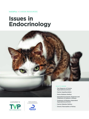
VetEdPlusE-BOOK RESOURCESIssues inEndocrinologyWHAT’S INSIDE The Diagnosis of CanineHyperadrenocorticism Canine Hypothyroidism Feline Diabetes Mellitus Hypoadrenocorticism: Diagnosis andTreatment of Addison’s DiseaseA SUPPLEMENT TOMade possible byan educational grant: Treatment of Pituitary-dependentHyperadrenocorticism Canine Diabetes Mellitus Chronic Pancreatitis in Felines
E-BOOKPEER REVIEWEDThe Diagnosis ofCanine HyperadrenocorticismAudrey Cook, BVM&S, MSc VetEd, MRCVS, DACVIM-SAIM, DECVIM-CA, DABVP (Feline)Department of Small Animal Clinical Sciences,Texas A&M College of Veterinary Medicine and Biomedical SciencesCollege Station, TexasThere are occasional reports of dogs with HACdue to an aberrant response to a digestive hormone(i.e., food-dependent HAC) or from ectopicACTH secretion, but these are extremely rare.CLINICAL PRESENTATIONSpontaneous HAC is usually diagnosed in olderdogs, particularly Boston terriers, dachshunds,miniature poodles, and beagles.1 It is uncommonin dogs younger than 5 years of age. Onset isoften insidious, and owners frequently attributechanges to aging. A strong understanding of theclinical manifestations of HAC is essentialbecause it helps us identify suitable candidatesfor further testing. A dog with Cushing’s2todaysveterinarypractice.comsyndrome must have some (usually many) of theclassic signs (BOX 1).More than 95% of dogs are polyuric/polydipsic;a normal water intake makes HAC less likely.Additionally, most manifest dermatologicchanges;2 in my experience, a good hair coatBOX 1 Clinical Signs CommonlyAssociated With Canine HAC Polyuria and polydipsia Polyphagia Panting Abdominal distention Hepatomegaly Muscle weakness Dermatologic changes Symmetric truncal alopecia Hyperpigmentation Comedones Thin skin (FIGURE 2) Poor hair regrowthEkaterina Brusnika/shutterstock.comHyperadrenocorticism (HAC or Cushing’ssyndrome) describes the clinical manifestationsof chronic exposure to excessive glucocorticoids.Spontaneous HAC is often caused byinappropriate secretion of adrenocorticotropichormone (ACTH) by a pituitary tumor (i.e.,pituitary-dependent HAC [PDH]) or may reflectthe autonomous production of cortisol by anadrenal tumor (AT).1
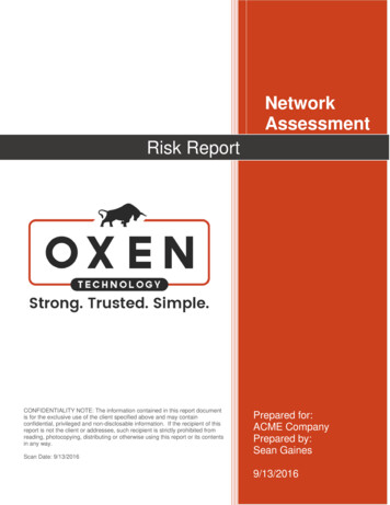
E-BOOKPEER REVIEWEDBOX 2 Indications to PursueDiagnostics for HAC Client concern (you hear something) Increased thirst or urination Hunger, food stealing Panting Difficulty jumping Physical examination (you see something)expected, along with poor forelimb and hindlimbmusculature (FIGURE 1). A small number of dogs withPDH will present with signs referable to a largepituitary tumor, such as personality change, vision loss,and poor appetite.3 Retinal changes may be noted indogs with concurrent hypertension.A careful history is always important in a cushingoidpatient because exogenous steroid administration mayotherwise be overlooked. Endocrine alopecia Thin skin, comedonesDIAGNOSTIC PROCESS Hepatomegaly/abdominal distentionHAC can often be considered an “exam room”diagnosis, meaning that the owner’s concern or thedog’s physical appearance suggests HAC. Lessfrequently, a comorbid condition suggests thepossibility (BOX 2). I do not advise pursuing adiagnostic workup for HAC without a strong clinicalindex of suspicion. Chasing a diagnosis based onbiochemical changes alone often results in substantialfrustration for both client and veterinarian. There areessentially 3 steps to the HAC diagnostic workup(BOX 3). Comorbid condition (you are fighting something) Recurrent urinary tract infection Frequent otitis externa; pyoderma Hypertension(particularly if shaved hair grows back promptly)essentially rules out HAC. Although calcinosis cutis ispathognomonic for HAC, it is uncommon. A “potbellied” appearance with palpable hepatomegaly isStep 1: Scrutinize RoutineLaboratory FindingsMost dogs manifest many (or all) of the expectedpatterns on routine laboratory tests.1Urine is usually dilute, with specific gravity less than1.020. Hyposthenuria may be documented: In fact,HAC is one of the most common causes of a urinespecific gravity less than 1.008. Proteinuria may beFIGURE 1. Dog with PDH. Note the abdominal distentionand hepatomegaly.3todaysveterinarypractice.comFIGURE 2. Dog with PDH. Note the thin skin with alopeciaand loss of elasticity.
E-BOOKPEER REVIEWEDnoted and should be quantified with a urine protein-tocreatinine ratio. Urinary tract infection is also commonand may not be accompanied by significant pyuria orovert clinical signs. A urine culture is oftenrecommended in dogs with evidence of HAC.BOX 3 Stepwise Diagnostic Approach for HACStep 1: Scrutinize routine laboratory findings forevidence of HAC On the biochemistry panel, the most consistent findingis increased alkaline phosphatase (ALP) activity. This isoften substantially elevated ( 1000 U/L; referencerange, 24 to 147 U/L) and routinely accompanied byan increase in γ-glutamyl transferase activity. However,increased ALP activity is common in older and obesedogs, dogs with various physiologic stressors, and dogswith primary hepatobiliary disease. High ALP activityin the absence of clinical signs of Cushing’s syndromeshould not prompt a hunt for HAC. Cholesterol andtriglyceride concentrations are consistently elevated; anormal value is unusual in a cushingoid dog. Othercommon biochemical changes are an increasedphosphorus concentration (seen in 50% of cases butstill a useful marker4), mild hyperglycemia, and(variably) a modest decrease in blood urea nitrogen.Some dogs have mild increases in alanineaminotransferase activity, but this is usually less than 3times the upper limit of normal.The complete blood count shows a stress leukogram(neutrophilia, lymphopenia, monocytosis, andeosinopenia). The hematocrit should be robustlynormal or even mildly high ( 50% is not uncommon);anemia is not consistent with HAC and should promptfurther investigation. Platelets are often increased andmay cause spurious hyperkalemia. Urinalysis Specific gravity 1.020 Proteinuria Chemistry panel Increased ALP activity and cholesterol /- Increased phosphorus /- Hyperglycemia (mild; 150 mg/dL) /- Low BUN /- Increased alanine aminotransferase orγ-glutamyl transferase activity Complete blood count Stress leukogram Robust hematocrit /- Increased platelet countStep 2: Screen for HAC (see text for details) LDDST ACTH stimulation test Urine cortisol-to-creatinine ratio (low specificity; apositive result is not adequate evidence to establishthis diagnosis)Step 3: Differentiate PDH from AT (necessary only ifadrenalectomy is an option) Abdominal ultrasonography Endogenous ACTH measurement H DDST (limited reliability; failure to suppress does notdifferentiate PDH from AT)Step 2: Screen for HACThere is limited consensus about the “best” screeningtest, although sensitivity and specificity data supportthe routine use of the low-dose dexamethsonesuppression test (LDDST).1 However, a clinician’sconfidence in a positive (or negative) result isdetermined by population characteristics, meaning thata result supporting HAC is inherently more believablein a geriatric dog with polydipsia and truncal alopeciathan in an apparently normal juvenile (same test,different patient population).Serum/plasma assays for “cortisol” cross-react withmany synthetic glucocorticoids (apart fromdexamethasone); exogenous steroids should thereforebe withheld for at least 72 hours before adrenalfunction tests are performed. In addition, on of exogenous steroids of any type willsuppress adrenal gland function after 2 to 3 weeks.LDDSTThe LDDST is an elegant way to interrogate thepituitary-adrenal axis, with reported sensitivities of85% to 100%.1 In addition, it is more reliable in dogswith AT than the ACTH stimulation test.5First, collect a baseline cortisol sample; serum isgenerally preferred, although some laboratories willaccept heparinized plasma. Next, administer 0.01 mg/kg of dexamethasone IV. This dose is slightly
E-BOOKPEER REVIEWEDAbout 85% of dogswith HAC have PDH,particularly small dogs.supraphysiologic and will suppress the release of ACTH(and therefore cortisol) in a normal dog for more than12 hours. If the sodium phosphate formulation is used(“Dex SP”), the dose should be adjusted to reflect that1.3 mg of this formulation contains 1.0 mg ofdexamethasone.1 The product used should be dilutedfor accurate dosing in small dogs (BOX 4). Subsequentblood samples are collected at 4 and 8 hours.Cortisol concentrations indicating “suppression” vary,with some laboratories using a value of less than 0.7mcg/dL and others defining suppression as anythingless than 1.4 mcg/dL.1 Lower values increase testsensitivity but may produce more false-positive results.Nonadrenal issues, such as fear and pain, affect cortisolrelease and cause false-positive results. Chronic illnessmay also affect results because long-term endogenoushypercortisolemia may shorten the half-life ofexogenous glucocorticoids;6 the suppressive effects ofdexamethasone therefore abate within 8 hours.One advantage of the LDDST over other screeningtests is its ability to differentiate PDH from AT. PDHcan be diagnosed with confidence if the 4-hour cortisollevel is less than 50% of baseline or below the levelestablished by the laboratory. The LDDST can causeconfusion, so always look at the 8-hour result first andsee if this value indicates HAC. If the answer is “yes,”then look at the 4-hour result; suppression at this pointBOX 4 Protocols for the Dilution ofDexamethasone for LDDSTD examethasone 2 mg/mL:0.5 mL 9.5 mL of saline dexamethasone 0.1 mg/mLAdminister 0.1 mL/kg ( 0.01 mg/kg of dexamethasone) D examethasone sodium phosphate 4 mg/mL:0.32 mL 9.68 mL of saline dexamethasone 0.1 mg/mLAdminister 0.1 mL/kg ( 0.01 mg/kg of dexamethasone) 5todaysveterinarypractice.comindicates PDH. Failure to suppress at the 4-hour markis not diagnostically useful; it is seen in many dogs withPDH and all those with AT.ACTH Stimulation TestThis test is convenient—it takes just over an hour.First, collect a baseline cortisol sample. Then injectcosyntropin (5 mcg/kg IM or IV; maximum, 250 mcgper dog) and collect a second serum sample 1 hourlater. Compounded ACTH gel products are bestavoided because their biological effect is uncertain. Tosave costs, reconstituted cosyntropin can be dividedinto aliquots in plastic syringes and kept frozen for upto 6 months.7This test is based on the premise that the response toexogenous ACTH is proportional to functionaladrenocortical tissue; this is increased in dogs withPDH or AT. Most labs use a cutoff serumconcentration of 17 to 20 mcg/dL; a serumconcentration above this level supports HAC.1 Thedisadvantages to this test include poor specificity indogs with chronic disease, meaning that false-positiveresults are likely. It also has poor sensitivity in dogswith AT; many dogs with this condition have normalresults or findings suggesting hypoadrenocorticism,8likely due to limited ACTH receptor expression byneoplastic cortical cells. A “flat-line” or addisonianresponse is also seen in dogs with iatrogenic HAC;chronic exposure to exogenous glucocorticoids resultsin adrenocortical atrophy and a blunted response toexogenous ACTH.Urine Cortisol-to-Creatinine RatioThis test is overly sensitive and not very specific,meaning that false-positive results are common,particularly in dogs with other polyuric disorders.9However, a negative result is highly reliable andcan be used to exclude HAC when there is a lowindex of suspicion. Test reliability is optimizedby having the owner collect morning urine athome. Stress or fear can quickly increase cortisolconcentrations in normal dogs, so it is unwise tocollect this sample in the clinic environment.Step 3: Differentiate PDH From ATAbout 85% of dogs with HAC have PDH,particularly small dogs. HAC due to AT is most oftenreported in dogs that weigh more than 20 kg.
E-BOOKPEER REVIEWEDDifferentiating the 2 forms is important because thediagnosis affects therapy and prognosis, but it is notnecessary if the dog is a poor candidate for surgery orthe owner is unwilling to consider adrenalectomy (thetreatment of choice for AT).Abdominal ultrasonography can readily differentiatePHD from AT but is operator and machine dependent.Normal adrenal gland width (at the caudal pole) is 3 to5 mm, although it is not unusual to find 7-mmadrenals in large dogs with nonadrenal illness.10Bilateral, symmetric adrenomegaly indicates PDH in adog with a positive result on a confirmatory test forHAC. A solitary mass in 1 gland suggests AT;contralateral atrophy ( 4 to 5 mm) is expected. Dogscan develop PDH and have a concurrentpheochromocytoma or adrenal tumor.11Abdominal radiography is a backup option ifultrasonography is not available. About 50% of ATsbecome calcified and can be seen on a plain lateralabdominal study. Calcification does not indicatemalignancy.Many reference laboratories offer highly sensitive assaysfor endogenous ACTH concentrations, which can beused to reliably differentiate the 2 forms of HAC. Dogswith AT have essentially undetectable levels, whereasthose with PDH have normal or increased levels.12Endogenous ACTH is a fragile hormone, so carefulsample handling is essential. Contact your laboratory toclarify requirements for sample submission.The high-dose dexamethasone suppression test (whichuses 0.1 mg/kg IV of dexamethasone) is rarelyperformed these days because its role has beensupplanted by ultrasonography and measurement ofendogenous ACTH. Suppression of cortisol productionfor the 8-hour test period is diagnostic for PDH, but alack of suppression is inconclusive.1SUMMARY Theclinical recognition of dogs with HACis a key part of the diagnostic process. Do not chase this diagnosis without overtclinical manifestations of HAC. The confirmatory tests have limitations; morethan 1 may be needed to establish a diagnosis. If adrenalectomy is not an option, clientresources may be directed toward treatmentrather than differentiating PDH from AT.However, life expectancy for dogs with ATwill be affected by the biological behavior ofthe tumor (benign vs malignant).References1.Behrend EN, Kooistra KS, Nelson R, et al. Diagnosis of spontaneouscanine hyperadrenocorticism: 2012 ACVIM consensus statement (smallanimal). J Vet Intern Med 2013;27:1292-1304.2. Zur G, White SD. Hyperadrenocorticism in 10 dogs with skin lesions asthe only presenting clinical signs. JAAHA 2011;47:419-427.3. Nelson RW, Ihle SL, Feldman EC. Pituitary macroadenomas andmacroadenocarcinomas in dogs treated with mitotane for pituitarydependent hyperadrenocorticism: 13 cases (1981-1986). JAVMA1989;194:1612-1617.4. Tebb AJ, Arteaga A, Evans H, Ramsey IK. Caninehyperadrenocorticism: effects of trilostane on parathyroid hormone,calcium and phosphate concentrations. J Small Anim Pract2005;46:537-542.5. Reusch CE, Feldman EC. Canine hyperadrenocorticism due toadrenocortical neoplasia. Pretreatment evaluation of 41 dogs. J VetIntern Med 1991;5:3-10.6. Stokes PE, Stoll PM, Schluger JH, Lasley B. Hypercortisolemiadecreases dexamethasone half-life in rabbit. J Psychiatr Res2002;36:423-428.7.Frank LA, Oliver J. Comparison of serum cortisol concentrations inclinically normal dogs after administration of freshly reconstitutedversus reconstituted and stored frozen cosyntropin. JAVMA1998;212:1569-1571.8. Normal EJ, Thompson H, Mooney CT. Dynamic adrenal functiontesting in eight dogs with hyperadrenocorticism associated withadrenocortical neoplasia. Vet Rec 1999;144:551-554.9. Smiley LE, Peterson ME. Evaluation of a urine cortisol:creatinine ratioas a screening test for hyperadrenocorticism in dogs. J Vet Intern Med1993;7:163-168.Audrey K. CookAudrey K. Cook is a Diplomate of both ACVIM andECVIM and is Board Certified in Feline Practice(ABVP). She is currently an associate professorof small animal internal medicine at TexasA&M, with particular interests in endocrinology,gastroenterology, feline medicine and endoscopy.Dr. Cook routinely speaks at national meetingsand is a recipient of the Texas A&M UniversityDistinguished Achievement Award in Teaching.6todaysveterinarypractice.com10. Benchekroun G. Ultrasonography criteria for differentiatingACTH dependency from ACTH independency in 47 dogs withhyperadrenocorticism and equivocal adrenal asymmetry. J Vet InternMed 2010;24:1077-1085.11. Greco DS, Peterson ME, Davidson AP, et al. Concurrent pituitary andadrenal tumors in dogs with hyperadrenocorticism: 17 cases (19871995). JAVMA 1999;214:1349-1353.12. Rodriguez Pineiro MI, Benchekroun G, de Fornel-Thibaud P,et al. Accuracy of an adrenocorticotropic hormone (ACTH)immunoluminometric assay for differentiating ACTH-dependent fromACTH-independent hyperadrenocorticism in dogs. J Vet Intern Med2009;23:850-855.
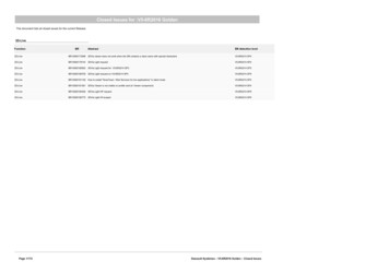
E-BOOKPEER REVIEWEDCanine Hypothyroidism:Diagnosis and TreatmentHypothyroidism is a common endocrine diseaseof dogs. It occurs when the thyroid glands fail toproduce adequate amounts of the hormonesthyroxine (T4) and triiodothyronine (T3).Primary hypothyroidism resulting fromidiopathic thyroid gland atrophy or immunemediated lymphocytic thyroiditis is the mostcommon diagnosis. Uncommon causes of caninehypothyroidism include congenital diseaseresulting from dyshormonogenesis of thyroidhormone, abnormal thyroid-stimulatinghormone (TSH) production, or abnormalthyroid gland development.1Clinical signs (BOX 1) may be nonspecific, whichcan result in overdiagnosis of this disorder;lethargy and weight gain are common.2,3 Clinicalsigns may have an insidious onset and may notbe noticed by the owner. Hypothyroidism oftencauses hair coat changes, including bilaterallysymmetric, nonpruritic alopecia over the trunkor areas of wear, post-clipping alopecia, and adull, lusterless hair coat (FIGURE 1). Skinchanges may include scaling, seborrhea,hyperpigmentation, and recurrent infections(pyoderma or otitis externa).2,3Thyroid hormones are involved in a wide varietyof metabolic processes, and low thyroid hormonelevels result in a constellation of clinical signsand laboratory abnormalities that characterizehypothyroidism. Multiple hormone tests arerequired to make a diagnosis. The diagnosisshould never be based on low T4 concentrationas a sole finding.CLINICAL PRESENTATIONHypothyroidism typically affects middle-ageddogs, although it has been reported in youngerand older dogs. Any breed can be affected.7todaysveterinarypractice.comFIGURE 1. Dog with hypothyroidism—note the excessivebody weight, dull hair coat, and scaling.From the top: taro911 Photographer/shutterstock.com. Opposite: Courtesy of DavidPanciera, Virginia-Maryland College of Veterinary Medicine, Blacksburg, VA.Johanna Heseltine, DVM, MS, DACVIM (Small Animal Internal Medicine), Clinical Assistant ProfessorTexas A&M University College of Veterinary Medicine & Biomedical Sciences, College Station, Texas
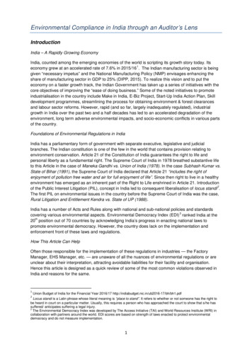
E-BOOKPEER REVIEWEDRare clinical signs and syndromes that have beenassociated with hypothyroidism includemegaesophagus, vestibular dysfunction, facial nerveparalysis, and atherosclerosis.4,5,8BOX 1 Clinical Signs CommonlyAssociated With CanineHypothyroidism2–7 Signs related to decreased metabolic rate Lethargy or dull mentation Inactivity or unwillingness to exercise Weight gain Cold intolerance or heat seeking Dermatologic changes Symmetric, nonpruritic hair loss Post-clipping alopecia Dry, dull hair coat Scaling Hyperpigmentation Recurrent pyoderma or otitis externaUncommon Incoordination Ocular signs Peripheral nervous system signs Facial nerve paralysis Laryngeal paralysis Polyneuropathy Other Vestibular signs Megaesophagus or esophageal dysmotility C ardiovascular abnormalities B radycardia E xacerbation of other cardiac signs Atherosclerosis Reproductive effects Periparturient mortality Lower birth weights Myxedema coma 8 Lipid corneal deposits Depressed mental status Altered thermoregulation B radycardia Hypoventilation Thickened skintodaysveterinarypractice.comDIAGNOSTIC PROCESSDogs should be tested for hypothyroidism only whenthe disease is strongly suspected based on the patient’shistory and physical examination findings (BOX 1).Complete blood count and serum biochemistry panelresults may heighten clinical suspicion forhypothyroidism. Hypothyroidism can be misdiagnosedwhen testing is performed only because a dog isoverweight or because a T4 concentration is includedwith a standard biochemistry panel.A stepwise approach is helpful in accurately diagnosingcanine hypothyroidism (FIGURE 2).Step 1: Evaluate Minimum DatabaseResults for Supportive FindingsResults from a complete blood count, serumbiochemistry panel, and urinalysis are helpful to ruleout concurrent disorders that could affect thyroid testresults. However, none of the abnormal results thatmay be seen on these tests are specific forhypothyroidism.Approximately 75% of hypothyroid dogs have elevatedcholesterol levels.2,3 While mild hypercholesterolemiaalone should not prompt testing for hypothyroidism, itsupports a suspicion of hypothyroidism. Liver enzymesmay be mildly elevated. A mild, nonregenerativeanemia is present in about 30% to 40% of hypothyroiddogs.2,3 The urinalysis typically shows no abnormalities.Dilute urine, if present, should prompt investigationfor concurrent illness or another cause of clinical signs.Step 2: Screen With aT4 ConcentrationTotal T4 concentration is a useful screening test forhypothyroidism. The sensitivity of this test for thediagnosis of canine hypothyroidism is reported to be89% to 100%.9-12 If the T4 concentration is well withinreference range, it is very likely the dog is euthyroid andfurther thyroid testing is not required. Free T4 (fT4) andthyroid-stimulating hormone (TSH) are evaluated onlyif the T4 concentration is low (FIGURE 2). CombinedT4, fT4, and TSH testing is not recommended at thisstage and may add unnecessary expense since a normalT4 concentration effectively rules out hypothyroidism.However, a T4 concentration below reference range isnot diagnostic for hypothyroidism. In addition tonormal daily fluctuations, several medications have
E-BOOKPEER REVIEWEDBOX 3 Euthyroid Sick SyndromeBOX 2 Drugs That Alter CanineThyroid Hormone Functionor Test Results13–20 Prednisone (high dose) Phenobarbital Trimethoprim–sulfamethoxazole A spirin (high dose) Clomipramine Thyroxine supplementationThis syndrome refers to a condition inwhich nonthyroidal illness suppressesthe concentration of circulating thyroidhormones. The mechanism is complexand likely involves changes in hormonedistribution and metabolism and alteredbinding of hormones to proteins.been demonstrated to lower the serum T4concentration of dogs (BOX 2), and some also affectfT4 and TSH concentrations. Certain drugs, such astrimethoprim–sulfamethoxazole, can have direct effectson the pituitary–thyroid axis and result inhypothyroidism.13 Furthermore, nonthyroidal illnessescan alter thyroid hormone metabolism and result in theeuthyroid sick syndrome (BOX 3). Concentrations offT4 are less likely to be affected by concurrent illness,but if the illness is severe enough, fT4 can also below.21,22 Therefore, thyroid testing should not beperformed in dogs that are systemically ill. If a dogwith a concurrent illness is tested for hypothyroidism,test results should be interpreted with caution.Because diagnosing hypothyroidism is not anemergency, sending samples out to a referencelaboratory is advisable. Since additional confirmatorytests are required, it is helpful to collect and hold extraserum when collecting for the T4 test.It is important to remember that “normal” referenceranges for T4 do not apply to sighthounds, as healthydogs of these breeds have lower T4 concentrations thanother breeds.23,24FIGURE 2. Approach to canine thyroid testing.Clinical suspicion based on history, physical exam, and minimum databaseT4 NormalT4 LowTSHHypothyroidism unlikely.Consider other causes forpatient’s signsHIGHConsistent NORMALHypothyroidismpossible.Evaluate fT4LOWConsistent sider othercauses forpatient’s signs
E-BOOKPEER REVIEWEDTABLE 1 Test Results for Diagnosisof Canine HypothyroidismaHORMONECONCENTRATIONTotal T4LowFree T4LowTSHHigh or normalTo confirm the diagnosis, all 3 results must be obtained.aInitiating TherapyStudies have shown that most dogs can be regulatedwith once-daily levothyroxine,26,27 usually initiated at0.02 mg/kg PO q24h. Some clinicians begin withtwice-daily administration of levothyroxine (0.02 mg/kg PO q12h) and attempt to reduce the dosing to oncedaily, once clinical signs are well controlled. Lethargyoften improves after a few weeks. Most clinical signsimprove within 4 to 6 weeks, although dermatologicchanges take months to resolve.Step 3: Confirm With an fT4or TSH ConcentrationMonitoring and Adjusting TherapyWhen a dog suspected to have hypothyroidism has alow total T4 concentration, fT4 and/or TSHconcentrations must be evaluated to help confirm orrefute the diagnosis (TABLE 1). If the TSH concentrationis high, hypothyroidism can be diagnosed. However,13% to 38% of hypothyroid dogs have normal TSHconcentrations,10-12,25 so a normal TSH concentrationdoes not exclude the diagnosis. Because of thislimitation, it is often helpful to evaluate fT4 and TSHsimultaneously as confirmatory tests. If the fT4 is low,a diagnosis of hypothyroidism can be made.After 4 weeks of therapy, blood is collected 4 to 6 hourspost-pill for T4 measurement. (T4 can be measured asearly as 2 weeks after starting or adjusting therapy,26but waiting until 4 weeks allows for assessment ofimprovement in clinical signs at the same visit.) Thepost-pill T4 concentration should be at the upper endof the reference range or slightly above ( 6 mcg/dL).Laboratory reference ranges for “initial” T4concentrations and “post-pill” concentrations may bedifferent, so careful sample labeling and interpretationare important.If T4 is low and fT4 is within reference range,hypothyroidism cannot be diagnosed, and the clinicianshould consider other differentials for the dog’sclinical signs.If the post-pill T4 concentration is below the targetconcentration, the dose of levothyroxine should beincreased by 25%. The T4 concentration is thenrechecked in 2 to 4 weeks. The dose is graduallyincreased until the post-pill T4 concentration is withinthe target range. Similarly, if the post-pill T4concentration is too high, the dose should be decreasedby 25% and the concentration rechecked. Once aneffective dose has been established, the interval betweenmonitoring visits is increased to every 6 months.T3 concentrations vary widely and are notdiagnostically useful.Approximately 75% ofhypothyroid dogs haveelevated cholesterollevels. 2,3 While mildhypercholesterolemia aloneshould not prompt testing forhypothyroidism, it supports asuspicion of .comTreatment Failure andAdverse EffectsTreatment failure is uncommon. Possible reasons forfailure to achieve the targeted T4 concentration includeowner noncompliance in administering medication orpatient refusal to swallow the pills. Variablegastrointestinal absorption of levothyroxine is alsoconsidered to be a possible cause.28 If a target post-pillT4 concentration has been achieved and clinical signsare not controlled, the dosing frequency should beincreased to twice daily. Additionally, the diagnosis ofhypothyroidism should be reconsidered. If thediagnosis of hypothyroidism is definitive and the dog’s
E-BOOKPEER REVIEWEDT4 concentration is well controlled, consider whether aconcurrent disorder could be causing clinical signs.Dogs are generally resistant to the effects of excessivelevothyroxine supplementation. However, clinical signssuch as polyuria/polydipsia and hyperactivity maydevelop.26SUMMARYThyroid testing should be carried out only whenpatients are suspected of having thyroid disease.Measurement of T4 concentration is helpful to rule outhypothyroidism but should not be solely relied on toconfirm the diagnosis. Combined testing that includesthe serum T4 concentration along with fT4 and/orTSH levels is needed for definitive diagnosis and willhelp decrease the possibility for misdiagnosis ofhypothyroidism. Since lifelong therapy is required, it isappropriate to achieve a definitive diagnosis beforestarting therapy.References1.Graham PA, Refsal KR, Nachreiner RF. Etiopathologic findingsin canine hypothyroidism. Vet Clin North Am Small Anim Pract2007;37(4):617-631.2. Panciera DL. Hypothyroidism in dogs: 66 cases (1987-1992). JAVMA1994;204(5):761-767.3. Dixon RM, Reid SW, Mooney CT. Epidemiological, clinical,haematological and biochemical characteristics of caninehypothyroidism. Vet Rec 1999;145(17):481-487.4. Jaggy A, Oliver JE, Ferguson DC, et al. Neurological manifestationsof hypothyroidism: a retrospective study of 29 dogs. J Vet Intern Med1994;8(5):328-336.5. Higgins MA, Rossmeisl JH, Panceria DL. Hypothyroid-associatedcentral vestibular disease in 10 dogs: 1999-2005. J Vet Intern Med2008;20(6):1363-1369.6. Panciera DL, Purswell BJ, Kolster KA, et al. Reproductive effects ofprolonged experimentally induced hypothyroidism in bitches. J VetIntern Med 2012;26(2):326-333.7.Finora K, Greco D. Hypothyroidism and myxedema coma. Comp ContEduc Pract Vet 2007;29(1):19-32.8. Hess RS, Kass PH, Van Winkle TJ. Association between diabetesmellitus, hypothyroidism or hyperadrenocorticism, and atherosclerosisin dogs. J Vet Intern Med 2003;17(4):489-494.9. Nelson RW, Ihle SL, Feldman EC, et al. Serum free thyroxineconcentration in healthy dogs, dogs with hypothyroidism, andeuthyroid dogs with concurrent illness. JAVMA 1991;198(8)1401-1407.10. Peterson ME, Melian C, Nichols R. Measurement of serum totalthyroxine, triiodothyronine, free thyroxine, and thyrotropinconcentration for diagnosis of hypothyroidism in dogs. JAVMA1997;211(11):1
Endocrinology VetEdPlus E-BOOK RESOURCES A SUPPLEMENT TO Made possible by an educational grant: WHAT’S INSIDE The Diagnosis of Canine Hyperadrenocorticism Canine Hypothyroidism Feline Diabetes Mellitus Hypoadrenocorticism: Diagnosis and Treatment of Addison’s Dise