Transcription
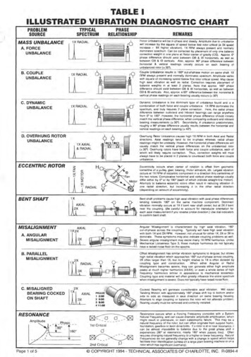
CANINE CARDIAC DIAGNOSTIC SCHEMEABCDs OF MYXOMATOUS MITRAL VALVE DISEASEUPDATED JANUARY 2021
2021 Cardiac Education GroupAcardiaceducationgroup.orgSTAGES OF MYXOMATOUS MITRAL VALVE DISEASE (MMVD)Dogs with no structural disease but at high risk fordeveloping MMVD.DIAGNOSTICS Patient history Yearly auscultation Screening programs for select breedsCEG RECOMMENDATIONS No treatment Client education Annual re-evaluationKEY:Red text: High priorityBlack text: Lower priority
2021 Cardiac Education GroupBcardiaceducationgroup.orgSTAGES OF MYXOMATOUS MITRAL VALVE DISEASE (MMVD)Dogs with structural heart disease that have not yet developedclinical signs of heart failure.Stage B valve disease can be divided into Stage B1 and Stage B2disease.Dogs with Stage B MMVD have no clinical signs of heart failure. Thisstage includes: Stage B1: Dogs with no radiographic or echocardiographic evidence ofcardiac remodeling (heart enlargement) or remodeling that is not severeenough to meet current clinical trial criteria used to determine initiationof treatment. Stage B2: Dogs with remodeling that is severe enough to supportinitiation of treatment.Diagnostics are required to differentiate B1 from B2. Echocardiography is the test of choice to differentiate Stage B1 MMVDfrom Stage B2 MMVD. Echocardiographic enlargement indicativeof Stage B2 includes both LA:Ao 1.6 and LVIDDN 1.71. Details ofdiagnostic approach can be found here. Radiographic criteria may be used to help identify MMVD patientslikely to meet echocardiographic criteria for Stage B2 whenechocardiographic examination is not possible. In dogs with left apical systolic heart murmurs grade 3/6, radiographiccriteria to identify likely Stage B2 dogs includes vertebral heart size(VHS) 11.5 or vertebral left atrial size (VLAS) 3 measured on a lateralradiograph. In cases where an echocardiogram cannot be obtained for staging,serial radiography (with consecutive examinations separated by 6 to 12months) can offer a practical substitute2.FOOTNOTES1. Cornell CC et al. Allometric scaling of M-modecardiac measurements in normal adult dogs. JVet Intern Med 2004;18:311-321.2. Both VHS and VLAS are measured using thesame (right) projection for each exam. A VHSrate of change (“velocity”) is calculated as thechange in VHS in vertebral bodies (VB) dividedby the number of months between the twoexaminations. Values 0.1 VB/month signalsa higher risk for developing CHF.
2021 Cardiac Education GroupBcardiaceducationgroup.orgSTAGES OF MYXOMATOUS MITRAL VALVE DISEASE (MMVD)Dogs with structural heart disease that have not yet developedclinical signs of heart failure.DIAGNOSTICS: STAGE B1 & B2 MMVD Patient history3Cardiac and pulmonary auscultation4Echocardiography5, 6Thoracic radiographs7, 8- Measure Vertebral Heart Size (VHS)- Measure Vertebral Left Atrial Size (VLAS) Blood pressure Electrocardiogram (ECG) when cardiac arrhythmia is evident duringclinical examination. NT-proBNP9 increases over time are associated with progression ofMMVD. Clinical lab tests: serum biochemistries, PCV/TS (or CBC) and urinalysis toestablish baseline values in older patients.KEY:Red text: High priorityBlack text: Lower priorityFOOTNOTES3.4.The history should include: questions about exercisetolerance, resting or sleeping respiratory rate,appetite, interaction with family, suggestive clinicalsigns (cough, collapse), and overall assessmentof quality of life. Hacking unproductive coughwithout respiratory distress may be a sign ofclinically significant cardiomegaly in dogs withMMVD. Evaluation for the presence of Stage B2 isrecommended.Left apical systolic heart murmurs are consistent withmitral regurgitation due to MMVD. Echocardiographyis strongly recommended if murmur grade is 3/6 toidentify Stage B2 dogs.5.Echocardiography is the diagnostic test of choice toestablish the diagnosis of MMVD in dogs.6.Echocardiographic enlargement indicative of StageB2 includes LA:Ao 1.6 and LVIDDN 1.7; reviewflow chart found in The EPIC Trial: Pimobendan inPreclinical Myxomatous Mitral Valve Disease inResources page at cardiaceducationgroup.org.7.Thoracic radiographs may be helpful as part of thediagnostic evaluation of dogs with possible MMVD.If echocardiography is not available, radiographicmeasurements of VHS and VLAS may help identifydogs likely to meet Stage B2 echocardiographiccriteria. A second opinion from a radiologist orcardiologist may be helpful.8.See cardiaceducationgroup.org for more informationabout radiographs and the Vertebral Heart Size (VHS)and Vertebral Left Atrial Size (VLAS).9.NT-proBNP testing may help differentiate coughingdue to respiratory disease from coughing due tocardiac enlargement. See NT-proBNP Testing in theDog in Resources page at cardiaceducationgroup.org.
2021 Cardiac Education GroupBcardiaceducationgroup.orgSTAGES OF MYXOMATOUS MITRAL VALVE DISEASE (MMVD)Dogs with structural heart disease that have not yet developedclinical signs of heart failure.CEG RECOMMENDATIONS - STAGE B MMVDStage B2 MMVD Dogs: Pimobendan treatment should be initiated in dogs with B2 MMVD10. See The EPIC Trial: Pimobendan in Preclinical Myxomatous MitralValve Disease Avoidance of high salt dog treats and human food is stronglyrecommended.Stage B2 MMVD:Left atrial enlargementAoLAAll Stage B MMVD Dogs: Mild dietary sodium restriction and feeding of a diet with adequate proteinand calories for maintaining optimal body condition is recommended.Severe sodium restriction is not recommended. WSAVA global nutritioncommittee recommendations for selecting a pet food manufacturer canbe found here. Manage systemic hypertension and concurrent systemic diseases, ifpresent. Re-evaluation in 6-12 months unless clinical signs develop. Client education: Discuss typical disease progression and signs to watch for.Stage B2 MMVD:Left ventricular dilationLV Tips for Diagnosing Heart Failure Counsel/ train owners to monitor resting respiratory rateStage B2 MMVD:Left ventricular dilation Using Resting Home Respiration Rate Client handout can be provided to aid owners in RRR recording.See Monitoring Your Pet’s Respiratory (Breathing) Rate (Client handout)LV Discuss nutritional information Nutritional Management of Heart Disease Implementing an Optimal Nutrition Plan for Your Cardiovascular Patient Encourage exercise in B1 MMVD patients as tolerated. Stage B2patients can exercise as tolerated, avoiding strenuous exerciseduring periods of high heat or humidity.KEY:Red text: High priorityFOOTNOTES10. The use of other therapies (ACEi andspironolactone) in Stage B2 MMVD iscontroversial. Consultation with a cardiologistmay be helpful.Black text: Lower priority
2021 Cardiac Education GroupCcardiaceducationgroup.orgSTAGES OF MYXOMATOUS MITRAL VALVE DISEASE (MMVD)Dogs with past or current clinical signs of heart failure.Common clinical signs of MMVD with congestive heart failure (CHF) includeincreased resting or sleeping respiratory rate or effort, cough associatedwith changes in respiratory rate/effort, abdominal effusion (ascites), exerciseintolerance, collapse or syncope11.DIAGNOSTICS: STAGE C MMVDStage C MMVD:CHF: active Stage C MMVD:Resolved CHF: stable onmedications KEY:Patient historyCardiac and pulmonary auscultationThoracic radiographs12Blood pressureEchocardiography for definitive diagnosis of underlying heart diseaseand screening for complications, including pulmonary hypertension, leftatrial rupture and ruptured chordae tendineae, as soon as it can be safelyperformed.Electrocardiogram (ECG) when cardiac arrhythmia is evident duringclinical examination.Point of care ultrasound to identify pulmonary infiltrates and cavitaryeffusions may be helpful but are not a substitute for thoracic radiographsto diagnose the presence of congestive heart failureClinical lab tests: serum biochemistries, PCV/TS (or CBC) and urinalysis toestablish baseline values prior to institution of any therapy.NT-proBNP might help discriminate between dogs with respiratory causesof clinical signs or congestive heart failure.24 hour ambulatory electrocardiography (using Holter ECG monitor) forassessment of heart rhythm disturbances or ambulatory event monitor forassessment of syncope.Red text: High priorityFOOTNOTES11. The presence of some respiratory conditionsmay complicate classification of dogs withheart disease. This is especially problematicin distinguishing stage B2 from stage C dogs.Collapsing trachea, mainstem bronchialcompression due to left atrial enlargement,chronic bronchitis or pulmonary hypertensionmay cause symptoms similar to those ofBlack text: Lower prioritycongestive heart failure. These includecoughing, tachypnea or symptoms relatedto airway obstruction. These patients mightbenefit from additional diagnostic testing,referral to a specialist or therapeutic trials.Coughing in Dogs: Is It Heart Failure?12. Confirm cardiomegaly (VHS, VLAS) andpulmonary infiltrates consistent withpulmonary edema.
2021 Cardiac Education GroupCcardiaceducationgroup.orgSTAGES OF MYXOMATOUS MITRAL VALVE DISEASE (MMVD)Dogs with past or current clinical signs of heart failure.CEG RECOMMENDATIONS – STAGE C MMVDInitial Therapy of Life Threatening Congestive Heart Failure Initial treatment of acute CHF should include injectable furosemide,oxygen and butorphanol sedation if needed. Administer pimobendan if the patient can tolerate oral medication. These patients require 24-hour care and may benefit from specialtyreferral and additional individualized therapy. Stabilize the patientbefore transport is considered.Therapy of chronic CHF due to MMVD Standard therapy for congestive heart failure: furosemide13,pimobendan, ACEi, spironolactone. If atrial fibrillation is present, diltiazem therapy is recommended14. Avoiding high-salt snacks and treats is strongly recommended.Nutritional Management of Heart Disease (Part 1) Exercise should be limited in acute phase of any episode of congestiveheart failure. When pulmonary edema has resolved, gentle exercise isencouraged as tolerated, avoiding prolonged strenuous activity. Continuation of or gradual introduction of moderate dietary sodiumrestriction is recommended if tolerated by the patient. A highlypalatable diet with adequate protein and calories will help maintainoptimal muscle condition.KEY:Red text: High priorityFOOTNOTES13. Furosemide and pimobendan are typicallythe first medications to be initiated; ACEiand spironolactone can be added to chronictherapy within the first 1-2 weeks as tolerated.Black text: Lower priority14. Addition of digoxin to diltiazem therapymay add heart rate control benefit. Relativecontraindications for digoxin therapy include:impaired renal function, pre-existing ventricularectopy or conduction disease of the sinusnode or atrioventricular node (AV block).
2021 Cardiac Education GroupDcardiaceducationgroup.orgSTAGES OF MYXOMATOUS MITRAL VALVE DISEASE (MMVD)Dogs with end-stage disease and clinical signs of heart failurerefractory to standard therapy.Persistent clinical signs of end-stage MMVD and congestive heart failure despiteuse of standard doses of recommended medications (Canine Formulary, MMVDConsensus Statement). Signs may include persistent increased resting or sleepingrespiratory rate or effort, cough associated with changes in respiratory rate/effort,abdominal effusion (ascites), poor apetite, weight loss or muscle wasting, exerciseintolerance, collapse or syncope.DIAGNOSTICS 15 Patient history, with special attention to assessment of client complianceand monitoring of clinical signs16, 17. Full physical examination with careful cardiac and pulmonaryauscultation Careful screening for concurrent systemic disease to identify possiblenon-cardiac causes of clinical signs. Thoracic radiographs Blood pressure Echocardiography for definitive diagnosis of underlying heart diseaseand screening for complications, including pulmonary hypertension, leftatrial rupture and ruptured chordae tendineae as soon as it can be safelyperformed. Electrocardiogram (ECG) when cardiac arrhythmia is evident duringclinical examination. Clinical lab tests: serum biochemistries, PCV/TS (or CBC), thyroidassessment if appropriate. 24 hour ambulatory electrocardiography (using Holter ECG monitor) forassessment of heart rhythm disturbances or ambulatory event monitor forassessment of syncope.KEY:Red text: High priorityFOOTNOTES15. The order of diagnostic tests should betailored to the patient’s current clinical signs.16. Assess for medication compliance, check forrecent changes in diet, environment and bodyweight.17. The presence of some respiratory conditionsmay complicate assessment of dogs withBlack text: Lower priorityheart disease. Collapsing trachea, mainstembronchial compression due to cardiomegaly,chronic bronchitis or pulmonary hypertensionmay cause symptoms similar to those ofcongestive heart failure. These includecoughing, tachypnea or symptoms related toairway obstruction. Stage D MMVD patientswith these types of clinical signs may benefitfrom additional diagnostic testing, referral to aspecialist or therapeutic trials.
2021 Cardiac Education GroupDcardiaceducationgroup.orgSTAGES OF MYXOMATOUS MITRAL VALVE DISEASE (MMVD)Dogs with end-stage disease and clinical signs of heart failurerefractory to standard therapy.CEG RECOMMENDATIONS Standard therapy for congestive heart failure: furosemide, pimobendan,ACEi, spironolactone18. Torsemide may be considered in place of furosemide if high doses offurosemide are not effective for recurrent CHF. If atrial fibrillation is present, diltiazem therapy with or without digoxin isrecommended. Other therapies may be helpful, see MMVD consensus statement;consultation with a cardiologist is strongly recommended. Avoiding high-salt snacks and treats is strongly recommended. Exercise should be limited in acute phase of each episode of congestiveheart failure. When pulmonary edema and cavitary effusions haveresolved, gentle exercise is encouraged as tolerated, avoiding prolongedstrenuous activity. Continuation of or gradual introduction of moderate dietary sodiumrestriction is recommended if tolerated by the patient. A highly palatablediet with adequate protein and calories will help maintain optimal musclecondition. Appetite stimulants may be useful to support oral intake (CanineFormulary).KEY:Red text: High priorityFOOTNOTES18. Doses of recommended medications can beuptitrated to the high end of the dosing rangein patients with difficult to treat heart failure.Black text: Lower priority
cardiaceducationgroup.orgABOUT THE CARDIAC EDUCATION GROUP (CEG)Founded in 2009, the Cardiac Education Group is a registered not-forprofit organization of board-certified veterinary cardiologists from bothacademia and private practice that offers independent recommendationsfor the evaluation and treatment of canine and feline heart disease. TheCEG mission is to improve the lives of dogs and cats with heart diseaseby providing resources and information in order to promote detection,diagnosis and therapy of heart disease and heart failure with greateraccuracy and confidence.John D. Bonagura, DVM, MS, Diplomate ACVIMAdjunct Professor, North Carolina State UniversityRebecca L. Stepien, DVM, MS, Diplomate ACVIMClinical Professor of Cardiology,University of Wisconsin-MadisonBrian A. Scansen, DVM, MS, Diplomate ACVIMAssociate Professor of Cardiology,Service Head, Cardiology & Cardiac Surgery,Colorado State UniversityBarret J. Bulmer, DVM, Diplomate ACVIMTufts Veterinary Emergency Treatment & Specialties,Walpole, MAWhit M. Church, DVM, Diplomate ACVIMDesert Veterinary Medical Specialists, Phoenix, AZAlan W. Spier, DVM, PhD, Diplomate ACVIMBluePearl Veterinary Partners, Tampa, FLSonya G. Gordon, DVM, DVSc, Diplomate ACVIMProfessor of Cardiology,Eugene Ch’en Chair of Cardiology,Texas A&M UniversityTO LEARN MORE OR SIGN UPFOR OUR NEWSLETTER, VISITcardiaceducationgroup.org . 2021 Cardiac Education GroupThe CEG is sponsored by educational grants fromBoehringer Ingelheim Vetmedica, Inc. and IDEXX Laboratories.
Dogs with end-stage disease and clinical signs of heart failure refractory to standard therapy. Persistent clinical signs of end-stage MMVD and congestive heart failure despite use of standard doses of recommended medications (Canine Formulary, MMVD Consensus Statement). Signs may include persistent increased resting or sleeping