Transcription
CLINICAL ARTICLEJ Neurosurg 126:796–804, 2017Traumatic hemorrhagic brain injury: impact of locationand resorption on cognitive outcomeRyan M. Martin, MD,1,2 Matthew J. Wright, PhD,3 Evan S. Lutkenhoff, PhD,4Benjamin M. Ellingson, PhD, MS,5 John D. Van Horn, PhD,6 Meral Tubi, BA,1 Jeffry R. Alger, PhD,2David L. McArthur, PhD, MPH,1 and Paul M. Vespa, MD1,2Departments of 1Neurosurgery, 2Neurology, 3Pyschiatry & Biobehavioral Sciences, 4Psychology, and 5Radiological Sciences,University of California, Los Angeles; and 6Department of Neurology, University of Southern California, Los Angeles, CaliforniaOBJECTIVE Hemorrhagic contusions are often the most visible lesions following traumatic brain injury. However, theincidence, location, and natural history of traumatic parenchymal hemorrhage and its impact on neurological outcomehave been understudied. The authors sought to examine the location and longitudinal evolution of traumatic parenchymalhemorrhage and its association with cognitive outcome.METHODS Sixteen patients with hemorrhagic contusions due to acceleration-deceleration injuries underwent MRI inthe acute (mean 6.3 days postinjury) and chronic (mean 192.9 days postinjury) phases. ImageJ was used to generateGRE and FLAIR volumes. To account for the effect of head-size variability across individuals, the authors calculatedeach patient’s total brain tissue volume using SIENAX. GRE and FLAIR volumes were normalized to the total braintissue volume, and values for absolute and percent lesion volume and total brain volume change were generated.Spearman’s rank correlations were computed to determine associations between neuroimaging and 6-month postinjuryneuropsychological testing of attention (Symbol Digit Modalities Test [SDMT], oral [O] and written [W] versions), memory(Selective Reminding Test, total learning and delayed recall), and executive function (Trail Making Test Part B [TMT-B]).RESULTS The patients’ mean age was 31.4 14.0 years and their mean Glasgow Coma Scale score at admission was 7.9 2.8. Lesions were predominantly localized to the frontal (11 lesions) and temporal (9 lesions) lobes. The average percentreductions in GRE and FLAIR volumes were 44.2% 46.1% and 80.5% 26.3%, respectively. While total brain and frontallesion volumes did not correlate with brain atrophy, larger temporal lobe GRE and FLAIR volumes were associated with largervolumes of atrophy (GRE: acute, -0.87, p 0.01, chronic, -0.78, p 0.01; FLAIR: acute, -0.81, p 0.01, chronic, -0.88, p 0.01). Total percent volume change of GRE lesions correlated with TMT-B (0.53, p 0.05) and SDMT-O (0.62, p 0.05) scores.Frontal lobe lesion volume did not correlate with neuropsychological outcome. However, robust relationships were seen in thetemporal lobe, with larger acute temporal lobe GRE volumes were associated with worse scores on both oral and written versions of the SDMT (SDMT-W, -0.85, p 0.01; SDMT-O, -0.73, p 0.05). Larger absolute change in temporal GRE volumewas strongly associated with worse SDMT scores (SDMT-W, 0.88, p 0.01; SDMT-O, 0.75, p 0.05). The same relationshipswere also seen between temporal FLAIR lesion volumes and neuropsychological outcome.CONCLUSIONS Traumatic parenchymal hemorrhages are largely clustered in the frontal and temporal lobes, and significant residual blood products are present at 6 months postinjury, a potential source of ongoing secondary brain injury.Neuropsychological outcome is closely tied to lesion volume size, particularly in the temporal lobe, where larger GREand FLAIR volumes are associated with more brain atrophy and worse SDMT scores. Interestingly, larger volumes ofhemorrhage resorption were associated with worse SDMT and TMT-B scores, suggesting that the initial tissue damagehad a lasting impact on attention and executive .JNS151781KEY WORDS MRI; neuropsychological; traumatic brain injury; hemorrhage; brain; traumaABBREVIATIONS DAI diffuse axonal injury; FLAIR fluid-attenuated inversion recovery; GCS Glasgow Coma Scale; GOS Glasgow Outcome Scale; GOSE GOSextended; GRE gradient recalled echo; ICH intracerebral hemorrhage; ICP intracranial pressure; MNI Montreal Neurological Institute; MP-RAGE magnetizationprepared gradient echo; ROI region of interest; SDMT Symbol Digit Modalities Test; SDMT-O SDMT-Oral; SDMT-W SDMT-Written; SRT-6 6-trial version of theSelective Reminding Test; TBI traumatic brain injury; TMT-B part B of the Trail Making Test.SUBMITTED July 30, 2015. ACCEPTED March 24, 2016.INCLUDE WHEN CITING Published online May 27, 2016; DOI: 10.3171/2016.3.JNS151781.796J Neurosurg Volume 126 March 2017 AANS, 2017Unauthenticated Downloaded 07/06/22 10:01 AM UTC
SCognitive outcome following traumatic ICHtraumatic brain injury (TBI) is a devastatinginjury leading to significant morbidity and mortalitythat results in an annual hospitalization rate of 85 per100,000 persons in the United States.22 TBI is a dynamicprocess involving immediate injury to some structures ofthe brain and delayed injury to more remote areas in theensuing hours, days, and weeks. Although TBI is associated with heterogeneous pathological injuries (i.e., brainhemorrhage, edema, and ischemia), most visible lesionson MRI and CT tend to be hemorrhagic. The influence ofbrain hemorrhage on mortality and overall neurologicaloutcome has been studied in TBI, and important lessonshave been learned about the subtypes of brain hemorrhage(i.e., epidural vs subdural). However, the incidence, location, natural history, and biological impact of traumaticparenchymal hemorrhage on neurological outcome hasheretofore been understudied. In this study, we sought toexamine the location and longitudinal evolution of traumatic parenchymal hemorrhage and its association withlong-term cognitive outcome.Intraparenchymal blood is known to be toxic and maycontribute to early secondary brain injury through masseffect and edema formation within the first 24 hours after trauma.42 In addition, the components of blood, including thrombin, heme, and iron activate harmful molecular cascades that may contribute to neurotoxicity andworse neurological outcomes.41 Despite the known toxiceffects of intracerebral blood products, there is conflicting data about the influence of brain hemorrhage volumeon long-term outcome. Recently, Iwamura et al.14 showedthat multiple and large hemorrhages associated with diffuse axonal injury (DAI) measured 1-month post injurywas associated with poorer outcome as measured by theGlasgow Outcome Scale (GOS). In contrast, Kurth et al.16showed no relationship between the number and volumeof petechial hemorrhagic contusions with neuropsychological outcome. Animal models of TBI have suggestedthat initial hemorrhage volume can be used as a predictorof long-term outcome.13,20 However, there has been littlestudied on the evolution of traumatic parenchymal hemorrhage and its relationship with outcome in a longitudinalmanner.Gradient recalled echo (GRE) MRI is the ideal modality for monitoring brain hemorrhage longitudinallybecause of its inherent sensitivity to paramagnetic bloodproducts,25,28,40 making it quite sensitive for picking uphemorrhages not visible on CT or other MRI modalities.24,27,40 In addition, GRE signal intensity and volumechange over time, making it a suitable biomarker to follow longitudinally in the post-TBI patient. Wardlaw andStatham40 showed that 10% of hemorrhages seen on admission CT scans following TBI do not have a GRE signal over 10 months out from injury, and Messori et al.24showed that hemosiderin signal on GRE attenuates after 6months following TBI. Even more recently, Moen et al.27showed that a small, but statistically significant number ofGRE lesions related to traumatic axonal injury disappearentirely with time, as does total hemorrhage volume. Howthis change in GRE volume over time affects cognitiveoutcome has not been elucidated.Neuropsychological profiles following severe TBI haveeverelargely been characterized as “frontal”34,37,41 and/or “frontal-temporal”19,41 in nature, as primary deficits in attention,memory, and executive function are common followingTBI. While the presence of focal lesions is known to predict cognitive outcome1,3,18,39 and may even have a moreprofound effect than DAI,39 it is unclear how change incontusion volume over time relates to neuropsychologicaloutcome.In this study, we measured hemorrhage volumes onGRE images obtained within the first 2 weeks of injuryand in corresponding images obtained 6 months after injury in patients with hemorrhagic contusions from nonpenetrating TBI. This longitudinal design allowed us todefine whether all blood products in particular brain regions resolved during the first 6 months following TBI.We then defined how hemorrhage features are associatedwith long-term neuropsychological outcome. We hypothesized that 1) larger acute hemorrhage volumes, 2) largerchronic hemorrhage volumes, and 3) smaller amounts ofhemorrhage resolution would be associated with poorerneuropsychological performance. The last goal was to determine a relationship between traumatic hemorrhage andbrain atrophy.MethodsParticipantsThe current study was approved by the medical institutional review board of the University of California,Los Angeles, and all participants consented directly orby proxy to voluntary participation between January 20,2002, and December 15, 2007. The current study included16 patients with focal traumatic hemorrhages due to acceleration-deceleration injuries (Table 1).The acute care of patients with TBI at our institutionis discussed in detail elsewhere.38 Briefly, all participantswere admitted to a neurointensive care unit following surgery or initial stabilization in the emergency room. Craniotomies or craniectomies, at the discretion of the treatingprovider, were performed for evacuation of intracranialmass lesions. Intracranial pressure (ICP) was measuredwith an external ventricular drainage (EVD) system, witha goal of maintaining ICP less than 20 mm Hg. Our standardized treatment protocol for ICP management includedelevation of the head of the bed up to 30 , mild hyperventilation (PaCO2 30–35 mm Hg), external ventriculostomy with cerebrospinal fluid drainage, moderate sedationwith low doses of propofol or versed, and maintenance ofnormoglycemia (100–140 mg/dl) and mild hypernatremia (sodium 140–145 mmol/L). Refractory ICP elevationwas managed by pentobarbital-induced burst suppressioncoma. Per standard of care, cerebral perfusion pressureabove 60 mm Hg was maintained with volume repletionand vasopressors. Jugular venous oxygen saturation wasmonitored continuously and kept at 60%–70% via adjustments in cerebral perfusion pressure. Possible seizure activity and barbiturate effects were assessed via continuouselectroencephalographic monitoring.Imaging Protocol and Volumetric AnalysesAcute (mean 6.3 days postinjury) and follow-up (meanJ Neurosurg Volume 126 March 2017797Unauthenticated Downloaded 07/06/22 10:01 AM UTC
R. M. Martin et alTABLE 1. Clinical and demographic characteristics of the study cohortInterval Btwn Injury & MRI (days)Case No. Age (yrs), Sex1234567891011121314151644, M44, M41, M18, M42, M53, F16, M22, M43, M18, F20, M25, M18, M50, M51, M18, MInitial GCS*Mechanism of InjuryHemorrhage LocationGOSE†Acute1477311NA576814436154Ped vs MVPed vs MVMotorcycle vs MVPed vs MVFell down stairsPed vs MVMVAMVAPed vs MVMVAMVAMotorcycleMVABicyclePed vs MVSkateboardFrontalFrontal, temporalTemporal, basal ganglia, spleniumFrontalFrontalFrontal, temporalFrontal, temporal, parietalFrontal, temporalFrontalFrontal, temporalParietalTemporal, basal ganglia, spleniumBasal gangliaFrontal, temporalFrontal, temporalFrontal, 00MV motor vehicle; MVA motor vehicle accident; NA not available (not determined); ped pedestrian; splenium splenium of the corpus callosum.* GCS score at time of admission.† GOSE score at 6 months’ follow-up.192.9 days postinjury) structural imaging studies wereconducted using a Siemens Sonata 1.5-T MRI scanner(Siemens Healthcare GmbH). Both acquisitions includedvolumetric T2*-weighted GRE (TR 1500 msec, TE 7msec, FOV 512 384, 3 mm slice thickness), volumetricT1-weighted magnetization-prepared gradient echo (MPRAGE; TR 1900 msec, TE 3.5 msec, FOV 256 256, 3mm slice thickness), and an axial fluid-attenuated inversion recovery (FLAIR; TR 9590 msec, TE 70 msec, FOV512 384, slice thickness 3 mm).Digital Imaging and Communication in Medicine(DICOM) images of both the GRE and FLAIR studieswere imported into ImageJ software (NIH) 1.37v (http://rsb.info.nih.gov). Using a home-written plug-in, we outlined regions of interest (ROIs) around hypointensities inthe GRE sequences and hyperintensities in the FLAIR.Lesion volumes from these ROIs could then be calculatedusing the ImageJ plug-in. For purposes of analysis andcomparisons with neuropsychological data, lesions wereanalyzed in their respective lobar or subcortical compartment. When hemorrhage or FLAIR lesions extendedacross 2 or more lobar regions, the lesion was separatedinto 2 or more ROIs based on lobar location. Lobar boundaries were defined using a standard approach10 and ananatomical atlas 9.htm).Using the acute and chronic volumes, we calculatedthe absolute change in volume size for both GRE andFLAIR lesions as the follow-up volume minus the acutevolume. Negative numbers represented a decrease in volume over the 6-month follow-up period. Percent volumechange (calculated as the absolute change in volume divided by the acute volume, multiplied by 100) was used to798normalize the data to the acute volume size. For purposesof comparison with results of neuropsychological testingand to account for the effect of head size variability acrossindividuals, we calculated each subject’s total brain tissuevolume, from a single image, normalized for skull size using SIENAX.32,33 We also calculated frontal and temporallobar volumes. First, each cortical lobe was identified andmasked using the Montreal Neurological Institute (MNI)structural atlas.6,23 Next, a nonlinear registration was performed using ANTs (Advanced Normalization Tools)2 toalign each individual subject’s T1 whole-head structuralMRI to the MNI atlas standard space. To ensure an accurate alignment, the results of these transforms were visually inspected. The nonlinear transform was then invertedto calculate the registration from the MNI atlas to eachindividual subject’s space. Since the cortical lobe maskswere delineated in MNI standard space, the inverted nonlinear transform was applied to the masks to register themto each individual subject’s space. The transformed lobarmasks were again visually inspected in individual subjectspace and the volume of each mask (cortical lobe) was estimated using FSL utilities.15 Percent total brain atrophyand lobar brain atrophy were then calculated by dividingthe absolute change by the acute volume and multiplyingby 100.Neuropsychological TestingThe participants (n 16) completed a modified version of a neuropsychological test battery designed for TBIclinical trials5 at 6 months postinjury. The test battery included the Symbol Digit Modalities Test (SDMT),31 the6-trial version of the Selective Reminding Test (SRT-6),9and part B of the Trail Making Test (TMT-B).36 A clini-J Neurosurg Volume 126 March 2017Unauthenticated Downloaded 07/06/22 10:01 AM UTC
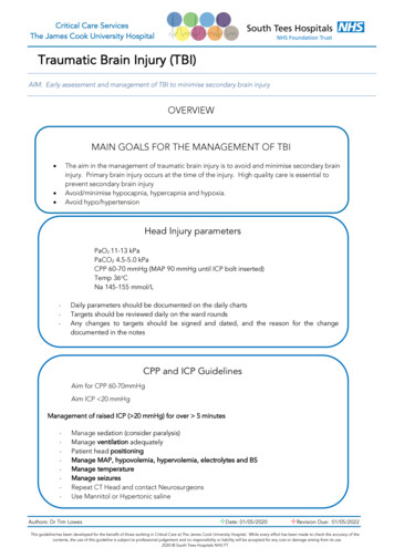
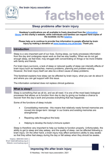
Cognitive outcome following traumatic ICHcal neuropsychologist administered the test battery, whichtook about 40 minutes to complete. Measures such as theSDMT, SRT-6, and the TMT-B have been shown to correlate with general cortical integrity, particularly in theintactness of the frontal and temporal lobes.25 We normedthe aforementioned tests (SDMT31 and SRT-617,26) andused T-scores for the current analysis. The Glasgow Outcomes Scale–extended (GOSE) score35 was determinedconcomitantly with the neuropsychological testing. TheGOSE score was not obtained for 1 patient at the followup visit.Statistical AnalysesA threshold of p 0.05 was set for statistical significance for all analyses. Nonparametric statistics were usedin the current study due to our sample size and non-normal distribution of some of the neuropsychological data.Spearman’s rank correlations were computed to determineassociations between neuroimaging and 6-month postinjury neuropsychological test values. Prior to analysis, theneuropsychological test values were normed, and T-scoreswere used for the current analysis. The Yuen-Welch testwas used to analyze for significant change in lesion vol-ume between the acute and follow-up lesion volumes.Spearman’s rank correlations were analyzed to determineassociations between acute GRE and FLAIR volumeswith frontal lobe, temporal lobe, and total brain atrophy.Data are reported as the mean standard deviation.ResultsPatient CharacteristicsThe participants’ demographic and baseline clinicalcharacteristics (including initial GCS scores and mechanism of injury) can be found in Table 1. In summary,16 patients were evaluated, with an average age of 31.4 14.0 years and with a mean educational level of 12.5 3.72 years. Most (87.5%) of the participants were maleand 62.5% were white. All had experienced accelerationdeceleration traumatic brain injuries; the mean GCS scoreat admission was 7.9 2.8. Two patients (Cases 11 and 16)underwent decompressive hemicraniectomy for refractoryelevated ICP, 2 patients underwent craniotomy for evacuation of a subdural hematoma (Case 2) or an epidural hematoma (Case 14), and 2 patients underwent evacuation ofparenchymal hemorrhage (Cases 6 and 15).FIG. 1. Montage of representative slices from the acute and follow-up GRE images from each patient in the study cohort (labeledto match the case numbers in Table 1). Hypointensities within the brain parenchyma represent blood products. The patient in Case1 also had a left frontal subdural hematoma. The patient in Case 13 had a right frontal epidural hematoma.J Neurosurg Volume 126 March 2017799Unauthenticated Downloaded 07/06/22 10:01 AM UTC
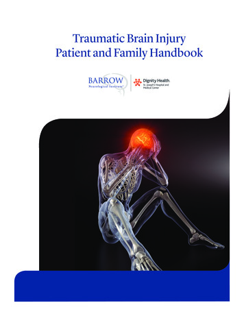
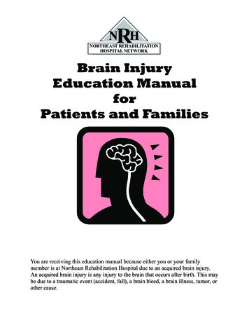
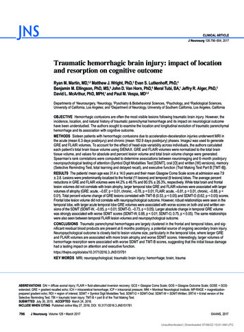
R. M. Martin et alThe mean interval between injury and the acute MRIof the brain was 6.3 5.0 days (range 1–16); the mean interval between injury and follow-up MRI was 189.4 12.2days (range 170–211).GRE VolumesRepresentative lesions can be seen in Fig. 1. GRE lesions were predominantly seen in the frontal and temporallobes and were less frequent elsewhere (Table 1). The totalGRE volume decreased from the acute (10.6 14.5 cm3)to chronic (3.9 4.8 cm3) time points, with a trend towardlarger volumes in the frontal lobes (acute 6.4 5.5 cm3,chronic 2.7 2.6 cm3) compared with the temporal lobes(acute 3.0 4.8 cm3, chronic 1.0 0.6 cm3). Over the entirebrain, the percent GRE lesion volume reduction from theacute to chronic images was 44.2% 46.1%. (Table 2).Larger acute and chronic GRE volumes were associatedwith larger absolute change (rho 0.990, p 0.005, Fig.2, upper), but not percent change (rho 0.317, p 0.23,Fig. 2, lower). This relationship was the same in the frontal(absolute change, rho 0.926, p 0.005; percent change,rho 0.359, p 0.252) and temporal (absolute change, rho 0.984, p 0.005; percent change, rho 0.568, p 0.087)lobes as well.Adjacent FLAIR Hyperintensity VolumesThe FLAIR hyperintensity volumes were substantiallylarger than the GRE volumes, with total acute and chronicFLAIR volumes of 43.4 44.5 cm3 and 5.0 7.4 cm3 (p 0.02), respectively. This corresponded to a mean percentvolume reduction of 80.5% 26.3% (Table 2). As was seenfor GRE lesions, mean frontal lobe volumes (acute, 25.2 14.1 cm3; chronic, 6.5 9.6 cm3) were larger than meantemporal lobe volumes (acute, 20.1 26.1 cm3; chronic,5.0 7.4 cm3).Brain AtrophyThe mean value for total brain volume decreased by125.5 74.3 cm3 or by 1.8% 1.9% over the 6-monthfollow-up period. Frontal lobe lesions were associatedwith a 2.9% 5.1% decline in frontal lobe volumes, whiletemporal lobe lesions were associated with a 6.5% 4.6%reduction in temporal lobe volumes. Total hemorrhagevolumes and frontal hemorrhage volumes did not correlate with the percent volume change (for correlation withtotal hemorrhage volume, rho ranged from -0.65 to 0.45,TABLE 2. Mean change in total GRE, FLAIR, and total brainvolume after a 6-month follow-up period*ParameterAbsolute Change in6 Mos (cm3)% Change in6 Mosp ValueGRE volFLAIR volTotal brain vol 6.7 10.0 34.7 41.1 125.5 74.3 44.2 46.1 80.5 26.3 1.8 1.90.020.03 0.01* Data are presented as mean SD. Absolute change is calculated as chronicvolume acute volume; percent change as (absolute change/acute volume) 100. Negative values indicate an overall decrease in volume; p values are forcomparison of acute to chronic volumes, which are not shown.800FIG. 2. Scatter plots showing that larger total acute hemorrhage volumes (hemorrhage cm3 per total brain cm3) were associated with largerabsolute hemorrhage change (upper), but not larger percent hemorrhage change (lower) over a 6-month follow-up period. rho Spearman’s rank correlation coefficient. Figure is available in color online only.with p values ranging from 0.07 to 0.54; for correlationwith frontal hemorrhage volume, rho ranged from -0.39to 0.09, with p values ranging from 0.21 to 0.98). However,larger acute and chronic hemorrhage volumes in the temporal lobe were strongly associated with more temporallobe atrophy (acute, rho 0.88, p 0.002; chronic, rho 0.78, p 0.012; Fig. 3A and B). Larger absolute volumesof hemorrhage change in the temporal lobe (rho 0.83, p 0.003, Fig. 3C), but not percent change in hemorrhagevolume (rho -0.44, p 0.203, Fig. 3D), were also associated with more temporal lobe atrophy. The same association was seen with FLAIR volumes (acute, rho 0.87, p 0.003; chronic, rho 0.83, p 0.003).Neuropsychological OutcomeGiven the sample size and the small number of associated lesions, associations with neuropsychological testingperformance were only made with total lesion volume,frontal lobe lesion volume, and temporal lobe lesion volume. The mean GOSE score was 4.7 1.2 at 6-monthfollow-up.Correlations between total lesion volume and neuropsychological testing results revealed that larger percentchange in total GRE volumes was associated with worsescores on the SDMT-O (rho -0.62, p 0.020) and TMT-J Neurosurg Volume 126 March 2017Unauthenticated Downloaded 07/06/22 10:01 AM UTC
Cognitive outcome following traumatic ICHFIG. 3. Scatter plots showing that larger acute (A) and chronic (B) temporal hemorrhage volumes (hemorrhage cm3 per total braincm3) are associated with larger volumes of percent temporal lobe atrophy over a 6-month follow-up period. Larger absolute changein temporal lobe hemorrhage volume (C), but not percent change in temporal lobe volume (D), was associated with larger volumesof percent temporal lobe atrophy. Figure is available in color online only.B (rho -0.53, p 0.040). No other test scores were foundto correlate with GRE volumes (rho ranged from -0.49to 0.36, with p values ranging from 0.06 to 0.84). TotalFLAIR volumes did not significantly correlate with neuropsychological test scores (rho ranged from -0.37 to 0.50,with p values ranging from 0.06 to 0.73).Associations between frontal lobe lesion volumes andneuropsychological testing showed no significant trend(rho ranged from -0.49 to 0.53, with p values ranging from0.06 to 0.98).Associations between temporal lobe lesion injury andneuropsychological testing can be found in Fig. 4. Largeracute temporal lobe GRE volumes were strongly associated with worse scores on both the SDMT-W (rho -0.85,p 0.004, Fig. 4A) and SDMT-O (rho -0.73, p 0.025,Fig. 4B) components. Similarly, absolute volume changein the temporal GRE volumes was also associated withworse scores on both versions of the SDMT (SDMT-W,rho -0.88, p 0.002, Fig. 4C; SDMT-O, rho -0.75, p 0.020, Fig. 4D). Lastly, larger temporal FLAIR volumesacutely (SDMT-W, rho -0.833, p 0.005; SDMT-O, rho -0.783, p 0.013), chronically (SDMT-W, rho -0.763,p 0.017; SDMT-O, rho -0.763, p 0.017), and absolutechange (SDMT-W, rho -0.750, p 0.020; SDMT-O, rho -0.700, p 0.036) were strongly associated with worseSDMT scores. However, larger percent change in temporalFLAIR volumes was associated with higher SDMT scores(SDMT-W, rho 0.686, p 0.041; SDMT-O, rho 0.686,p 0.041).DiscussionOur main objective was to characterize the distributionand evolution of hemorrhagic contusions following TBIand their relationship with neuropsychological outcome.We found that hemorrhagic contusions in our patient serieswere largely confined to the frontal and temporal lobesand that a significant amount of residual blood productswere present 6 months after injury. There was considerablepatient-to-patient variability, but on average, over 50% ofthe initial intracerebral blood volume was present at 6months’ follow-up. This is an important finding as hemorrhage and iron deposition are thought to play an importantrole in secondary brain injury and comorbidities.Mechanisms of hemorrhage-induced secondary braininjury have largely been reported in stroke literature inthe context of models of spontaneous intracerebral hemorrhage (ICH). Initial hemorrhage-related injury is due toJ Neurosurg Volume 126 March 2017801Unauthenticated Downloaded 07/06/22 10:01 AM UTC
R. M. Martin et alFIG. 4. Scatter plots showing that larger acute temporal hemorrhage volumes are associated with worse scores on a test of attention, Symbol Digit Modalities Test (SDMT), in both written (A) and oral (B) modalities. Higher test scores on the SDMT reflectbetter performance. Larger absolute change in temporal lobe hemorrhage was also associated with worse SDMT scores (C andD). Figure is available in color online only.edema formation and mass effect, possibly contributingto alterations in cerebral blood flow and metabolic disturbance.42 Thrombin is activated immediately after hemorrhage and has been shown in experimental models to leadto worsening edema, apoptosis of neurons and astrocytes,potentiation of glutamate toxicity, and activation of inflammatory microglia.11,29,42 Erythrocyte lysis can occur within24 hours of injury, resulting in worsening edema and therelease of heme into the extracellular space. Heme is thendegraded into iron, ferritin, carbon monoxide, and biliverdin, all of which lead to neuronal dysfunction and deathand hemosiderin deposition.4,11,29,42 Interestingly, higher serum levels of ferritin on admission for ICH have been associated with worse outcome, suggesting that body storesof iron can contribute to neuronal toxicity.7 Iron depositionmay also play a role in seizure formation, with models ofcortical iron injections causing focal epileptiform paroxysmal discharges.12,28,42 The potential role of iron in secondary brain injury is further suggested in animal modelsof ICH and TBI in which deferoxamine, an iron chelator,attenuates the effects of brain injury on both neuronal celldeath and behavioral outcomes.8,21,43 However, despite allthis work, the role of traumatic ICH in patient outcomeshas not been well documented.Inconsistent findings are the norm when correlating802neuropsychological outcomes with acute and chronicstructural brain imaging, with the vast majority of studies looking at DAI and nonfocal contusions.14,16,30 In ourstudy, the strongest associations between GRE volumesand neuropsychological outcome were those related to thetemporal lobes, such that larger acute volumes were associated with worse SDMT scores, indicating poorer attention, as well as higher rates of temporal lobe atrophy.Intuitively, it makes sense that the brain would be more“overwhelmed” by larger hemorrhage volumes and lesslikely to recover. Residual hemosiderin then leads to brainatrophy, which is associated with poorer attention. In addition, larger volumes of hemorrhage resorption tended to beassociated with lower SDMT scores, a result related to thefact that larger hemorrhage volumes were associated withlarger volumes of resorption. Contrary to our expectationthough, percent change in GRE signal did not correlatewith cognitive outcome, suggesting that the initial tissuedamage had a lasting impact on these cognitive abilities.FLAIR volumes in the temporal lobes had the strongest association with outcome, such that larger acute andchronic FLAIR volumes in the temporal lobe were associated with lower SDMT scores. This is consistent withMoen et al.27 who showed that total FLAIR volume inwhite matter predicted long-term outcome, as measuredJ Neurosurg Volume 126 March 2017Unauthenticated Downloaded 07/06/22 10:01 AM UTC
Cognitive outcome following traumatic ICHby the GOSE. In addition, the relationship between attention (i.e., SDMT scores) and FLAIR resorption ratesseems, at first glance, to be contradictory when looking atthe absolute change versus the percent change. However,like hemorrhage volumes, larger acute FLAIR volumescan have larger absolute change without a correspondinglyhigher percent change. In this regard, the relationships between temporal lobe FLAIR percent change and neuropsychological data are likely more clinically relevant, andhigher relative resorption volumes in the temporal lobe areassociated with better outcomes.LimitationsWhile our study shows a relationship between hemorrhage resolution and neuropsychiatric outcome, ourstatistical power was limited by the small sample size ofpatients who underwent neuropsychological testing, andthus, relationships may be under- or over-appreciated. Inaddition, the small sample size precluded detailed regionalanalysis, such as hemispheric differences or a
(Selective Reminding Test, total learning and delayed recall), and executive function (Trail Making Test Part B [TMT-B]). RESULTS The patients' mean age was 31.4 14.0 years and their mean Glasgow Coma Scale score at admission was 7.9 2.8. Lesions were predominantly localized to the frontal (11 lesions) and temporal (9 lesions) lobes.