Transcription
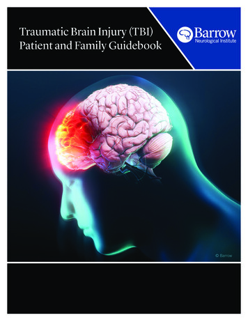
Traumatic Brain InjuryPatient and Family Handbook
Table of ContentsTreatment Information . . . . . . . . . . . . . . . . . . . . . . . . . . . . . . . . . . . . . . . . . . . . . . . . . . . . . . . . . . . . .1The Brain and Traumatic Brain Injury . . . . . . . . . . . . . . . . . . . . . . . . . . . . . . . . . . . . . . . . . . . . . . .3Behavior Management after a TBI . . . . . . . . . . . . . . . . . . . . . . . . . . . . . . . . . . . . . . . . . . . . . . . . . .7Treatment for a TBI . . . . . . . . . . . . . . . . . . . . . . . . . . . . . . . . . . . . . . . . . . . . . . . . . . . . . . . . . . . . . . . .13Recovery and Rehabilitation . . . . . . . . . . . . . . . . . . . . . . . . . . . . . . . . . . . . . . . . . . . . . . . . . . . . . . .17Resources . . . . . . . . . . . . . . . . . . . . . . . . . . . . . . . . . . . . . . . . . . . . . . . . . . . . . . . . . . . . . . . . . . . . . . . . .25Locations and Map . . . . . . . . . . . . . . . . . . . . . . . . . . . . . . . . . . . . . . . . . . . . . . . .Inside Back Cover
Treatment InformationDiagnosis:Treatment TeamStaff Member NameSpecialtyProceduresDateProcedurePurpose1
The Brain and Traumatic Brain InjuryTraumatic Brain InjuryTraumatic brain injury (TBI) is an injury to the brain from an external force. A TBI can cause achange in brain function.Symptoms of a TBI can be mild, moderate, or severe depending on the extent of the damage to thebrain.Mild TBI is not always associated with loss of consciousness, but mild TBI can causeunconsciousness for a few seconds or minutes. Other symptoms of mild TBI include: headacheconfusionlightheadednessdizzinessblurred vision or tired eyesringing in the earsbad taste in the mouthfatigue or lethargya change in sleep patternsbehavioral or mood changestrouble with memory, concentration, attention, or thinkingModerate or severe TBI can include all or some symptoms of a mild TBI, along with: headache that gets worse or does not go awaynausea or repeated vomitingconvulsions or seizuresinability to awaken from sleepdilation of one or both pupilsslurred speechweakness or numbness in the extremitiesloss of coordinationincreased confusion, restlessness, or agitationloss of consciousnessAbout The BrainThe brain controls important functions like movement, digestion, breathing, and sensoryperception (for example, sight, touch, and taste). It is also controls higher functions like thinking,learning, and emotions. To work properly, the brain needs to be protected from infections, trauma,and receive an adequate supply of oxygenated blood.3
Although the brain gets some nutrition from cerebrospinal fluid (CSF), it is mainly nourished byoxygenated and nutrient-rich blood that is supplied by the left and right carotid arteries andvertebral arteries. Blood delivers essential substances that brain cells need to function.Because of its importance to the body and its fragile, gelatin-like consistency, there are several layersof protection around the brain. The skull is the hard, bony covering that forms the outermost layer of protection for the brain. The dura mater is a tough, skin-like covering that is located between the skull and brain. Cerebrospinal fluid (CSF) is a clear, watery fluid that is produced in the ventricles, or hollowspaces in the center of the brain. CSF circulates outward from the ventricles, nourishing thebrain and spinal cord. It also serves as a shock absorber that dissipates forces applied to theskull before they can be transferred to the brain.Types of TBISkull fractures can occur anywhere in the skull and can be open or closed. A closed fracture is a break in the bone of the skull without a break in the skin or scalp. An open fracture is a break both in the bone of the skull and break in the skin or scalp. Openfractures exposing the brain to the external environment and can lead to infections of thecentral nervous system like meningitis if not treated. Bone fragments from an open fracturealso can be pushed into the brain, resulting in a depressed fracture. Depressed fractures candamage brain tissue and may require surgery.Contusion is bruising of the brain that occurs from the brain hitting the skull during trauma. Thereare two types of brain contusions:1. Coup contusions occur at the site of directimpact to the skull.2. Contrecoup contusions occur opposite from thesite of direct impact and are the result of forcetransfer from the site of the injury to the oppositeside of the brain.Diffuse axonal injury is damage to the white matter(axons) of the brain. Violent force to the brain maystretch, tear, and twist the axons. Axons connect areas ofthe brain and spinal cord to each other and to the rest thebody. When they are damaged, messages from the brainor spinal cord to the rest of the body are slowed or lost.Swelling of the brain tissue may also occur, worsening thedamage. The brain is not able to repair damaged axons,and there is no medical treatment that can restore their function. Therefore, treatment concentrateson limiting the damage by reducing swelling.Concussion is an injury deep in the brain that can cause impaired consciousness. Concussion isconsidered a mild form of diffuse axonal injury and is sometimes called mild TBI.4
Anoxic brain injury results from trauma—such as cardiac arrest or blood loss—that preventsoxygenated blood from reaching the brain. After approximately five minutes of oxygen deprivation,brain cells begin to die and anoxic brain injury occurs. Once brain cells die, they do not regenerate,although sometimes other surviving neurons can adapt and take over the functions formerly carriedout by dead neurons. However, if this does not happen the neurological impairments caused byneuronal death are permanent.Penetrating head injury describes gunshot wounds and other trauma originating from a projectilestriking the skull. Higher speed projectiles tend to cause more damage to the brain because theypenetrate deeper and deliver more energy.Hematomas, also known as blood clots, result when a blood vessel is broken during an injury. Iflarge enough, a hematoma can compress or shift the brain, and requires surgery.Locations of Hematomas and HemorrhagesIntracerebral hemorrhages or hematomasoccur within brain tissue (Intra within, thecerebrum).Intraventricular hemorrhages or hematomasoccur within the ventricles of the brain(Intra within, the ventricle).Epidural hematomas (EDH) are blood clotslocated between the skull and dura mater (Epi onor upon, the dura). This blood clot may growquickly and endanger the brain by putting pressureon it. Emergency surgery may be required toremove the blood clot.Subdural hematomas (SDH) are blood clots that form between the dura and the brain(Sub below or underneath, the dura). SDH can occur acutely—at the time of the trauma—or theycan form slowly over weeks to months (chronic SDH). Surgery may be required depending on theseverity of the symptoms and the increase in intracranial pressure caused by the SDH.Subarachnoid hemorrhage (SAH) is a clot that forms in the space between the brain and itsarachnoid membrane (Sub- below or underneath, the arachnoid). The arachnoid membrane is adelicate surface that is attached to the underside of the dura mater.The Delayed Effects of TBIBrain damage occurs immediately after TBI. However, injury to the brain can also occur as a result ofswelling or bleeding in and around the brain following the initial injury.Increased intracranial pressure (ICP) is an increase in the pressure within the skull. The brainand its tissues, along with blood and cerebrospinal fluid (CSF), take up a given amount of spacewithin the skull. Brain swelling, blockages in CSF circulation, and blood clots can all cause increasedintracranial pressure, leading to further brain injury.5
Brain anoxia or hypoxia is complete (anoxia) or partial (hypoxia) loss of oxygen to part or all of thebrain. Anoxia or hypoxia can occur when blood flow to the brain stops or is reduced because ofinjury to the brain. Injuries to other parts of the body, especially the heart and lungs, can also causehypoxia and anoxia of the brain.Brain edema describes swelling of the brain, causing it to push against other contents in the skull.This is a major cause of brain injury, and can cause death if not treated.Hydrocephalus is enlargement of the cerebral ventricles due to a blockage of CSF flow. Injuriesthat cause bleeding or swelling can distort and block the ducts that carry and circulate CSF withinthe central nervous system. When this happens, the cerebral ventricles can increase in size toaccommodate the larger volume of CSF trapped within the skull. This can also lead to an increase inintracranial pressure (see above).Brain herniation is when some structures of the brain move or are pushed across or through otherstructures of the skull due to very high ICP. Brain herniation is life threatening and can result inpermanent neurological damage and disability.Posttraumatic Amnesia (PTA)Posttraumatic amnesia (PTA) is a period of confusion , memory impairment, or both, after an injury.Long term memory (such as address, date of birth, or significant historical events) often remainsintact after TBI. There are two types of amnesia after TBI: retrograde and anterograde amnesia. Retrograde amnesia is partial or total loss of memory for the time period immediately beforean injury. The span of retrograde amnesia can be as short as the few seconds before an injury,or as much as a month or more before the injury. Anterograde amnesia is reduced ability to store new memories after TBI. To determine ifsomeons is experiencing PTA, the care team may ask the patient to recall information abouttheir identity, location, and the date and time of accident or injury. In most rehabilitationprograms, PTA is measured daily until the patient consistently and correctly answer questionswithout cues. It is important to avoid signing important documents and making criticaldecisions during the period of anterograde amnesia, as the patient may not be able toremember making that decision minutes or hours later.The length of PTA is associated with severity of injury and long term recovery.6
Behavior Management after a TBIA patient’s behavior may change after a TBI in ways that may cause difficulty for them and thosecaring for the patient. Behavioral problems after TBI can interfere with recovery, so it is important toreduce or eliminate these behaviors so that the patient can get the most out of rehabilitation.Types of problematic behaviors seen after TBI include: Impulsivity—acting before thinking about the consequences of an action. Disinhibition—the loss of socially appropriate inhibitions. Agitation—strong emotional reaction to overstimulation (from too much noise, light,thought, or social interaction), frustration, confusion, or irritation. Perseveration—getting stuck on a certain thought, idea, or movement. Confabulation—the replacement of a gap in memory with false information that thepatientbelieves is true. Misperception—holding a false, sometimes paranoid belief about the reality of events.It is important to remember that: TBI patients do not behave in this way intentionally. Changes in behavior are a result of damage to the brain, most commonly the frontal andtemporal lobes. People with frontal lobe injuries can become frustrated or act impulsively,exhibiting agitated or aggressive behaviors like yelling and cursing. They also may be overlysuspicious, have problems with short term memory, and be easily disoriented. The team of therapists, doctors, and nurses will work together to create an appropriatebehavior management program. All parties—patient and supporting cast—must work togetherto generate consistent reponses and attitudes toward problem behaviors. Some tips for dealing with problem behaviors:º Take frequency into account. Episodes may be tolerable if they don’t happen frequently,but may be difficult to cope with as frequency increases.º Consider the severity of the behavior. Mild arguments and episodes of frustration canbe benign. If they escalate, try to identify the cause.º Frequent and severe behaviors need to be addressed. Continuing negative behaviorcan hinder recovery.º Set up an environment that fosters success. The environment should not be toostimulating (for example, not too many visitors at once, turn the television or music offwhen socializing or doing tasks) because overstimulation can increase confusion oragitation.º Give direct and immediate feedback. Cues and feedback should be simple and direct.Feedback can be positive (giving praise for doing something well) or negative (stopping aproblem behavior). Disorientation, short-term memory loss, and difficulty with abstractthinking can complicate feedback, and people with brain injury may act out to avoid adifficult task or therapy.º Remember age. Addressing a patient in a way that is not appropriate for their age can7
cause frustration and hostility.º Praise and encourage. These interactions tend to help more than punishment.º Set realistic goals. Break big, long-term goals into small, attainable goals.º Modify. Change interactions as behavior improves and recovery progresses, but be asconsistent as possible.º Encourage. When working to eliminate or change a behavior, the behavior may get worsebefore it gets better. Encouragement can help speed this process.º Consult a doctor. If negative behavior continues and disrupts the recovery plan,medications can be combined with the behavioral plan to jumpstart recovery.º Restrain. Though unpleasant, these may be necessary if there is a threat of harm to self orothers. Our staff can provide information about the least-restrictive options.The Rancho Levels of Cognitive FunctioningThe Rancho Levels of Cognitive Functioning is an evaluation tool used to identify patterns ofbehavior after a traumatic brain injury and help caregivers and all who interact with the patientunderstand what to make of this behavior and how to respond. It is a way to monitor how anindividual is recovering from brain injury.The scale identifies eight levels or stages that describe behavioral and cognitive deficits, allowing thetreatment team to develop the most appropriate rehabilitation program for each individual. As thebrain heals, it is possible for the person to improve and move up through the stages. Patients do notalways demonstrate every behavior listed, and can even show characteristics of two stages at once.The eight levels are described briefly below. In addition, we have included some tips for what familymembers can do for their loved one at each of the eight stages.Cognitive Level 1: No response A person at this level does not respond to sounds, sights, touch or movement. They appear tobe in a deep sleep.What can you do? It is unclear whether people in a coma can hear or understand what is said to them. Therefore,assume they do and, if you choose, talk to the person in a soft, relaxing voice about your day orany non-stressful topic. Touch the person. For example, hold their hand or wash their face with a damp cloth whileexplaining what you are doing, (i.e., “I’m going to hold your hand now”).Cognitive Level 2: Generalized Response A person at this level will begin to slowly respond to sounds, sights, touch or movement,although they will likely remain asleep most of the time. They may respond slowly, inconsistently, or after a delay. Their responses may include chewing movements, sweating, breathing faster, moaning,moving, and increasing blood pressure.What can you do? Continue suggestions from Level 1. If their eyes are open, ask them to look at you or something in the room.8
Do not overstimulate. Only provide the suggested stimuli for 5-15 minutes at a time, 3-4times per day. Remember, rest is important to regaining cognitive function. Monitor their stress and frustration levels. If they are becoming frustrated or agitated, bepatient and decrease the amount of stimuli. When talking to them, sit nearby so they can see you. Identify yourself every time you enter the room. For example, “Bob, it’s your sister Katie.” Ask non-stressful questions with ‘yes’ or ‘no’ answers as they become increasingly tolerantof stimuli.Cognitive Level 3: Localized Response At this level, patients react more specifically to what they see, hear, or feel (i.e., withdrawingfrom pain, turning towards a sound). They remain awake for several minutes at a time. They may begin to follow some simple directions (i.e., “Squeeze my hand.”), but may not do soconsistently. They may begin to recognize family and friends.What can you do? Continue suggestions from Levels 1 and 2. Keep their room as quiet and soothing as possible. Limit visitors to two at a time. Bring in objects or photographs with meaning to help with memory.Cognitive Level 4: Confused and Agitated Patients at this level can overreact to visual, audio, or touch stimuli by hitting, screaming, usingabusive language, or thrashing. This reaction is due to confusion. They become easily agitated or aggressive. They are highly focused on basic needs (i.e., relieving pain, going back to bed). They have great difficulty following directions. They may not understand that your intent is to help. They are not able to pay attention or concentrate for more than a few seconds.What can you do? Try not to take this behavior personally. Be patient. If agitation occurs, decrease the amount of stimuli. For example, turn off theTV and limit visitors. Keep distractions to a minimum. Gently correct them if they tell you the wrong information. However, do not argue if theyinsist on being correct. Help them with personal care so that they become more in touch with their environment.For example, help with dressing and grooming.Cognitive Level 5: Confused and Inappropriate Patients at this level will be confused and have difficulty making sense of outside stimuli,although they will no longer be agitated. They are not oriented.9
They are restless when tired or over stimulated. They are not able to independently start or complete everyday activities (i.e., getting dressedand brushing teeth).What can you do? Frustration is common. Encourage and reassure the patient as much as possible.Praise good behavior. Ignore inappropriate behavior unless safety is jeopardized.Try to redirect them if they are behaving inappropriately.Talk with them about family, friends, your day, and ask them questions. For example,“What happened in therapy today?” Give hints if they cannot remember. Talk with them about the problems that stem from their brain injury. Be open and honest.Reassure them that the goal of rehabilitation is to address these problem areas. Help them write down their daily schedule and other activities to help them improvememory and utilize compensations.Cognitive Level 6: Confused and Appropriate A patient at this level will be somewhat confused because of memory and thinking problems(i.e., will remember they had visitors in the morning, but do not know what they talked about). They are able to follow a schedule with some assistance, but are confused by changes inroutine. They may be able to pay attention for 30 minutes, but have trouble concentrating when thereare multiple steps or distractions. They may be impulsive.What can you do? Play cards or games that challenge the patient appropriately without causing frustration.Foster independence with daily activities by decreasing the amount of assistance you provide.Ask questions about events or conversations that just occurred to help improve their memory.Remember that rest is important.Cognitive Level 7: Automatic and Appropriate A patient at this level follows a set schedule.They are able to do routine self care without assistance.They have problems planning, initiating, and following through with activities.They have trouble paying attention in distracting environments.They may not realize how their deficits impact future plans (i.e., expects to return to work rightaway despite having ongoing cognitive deficits).What can you do? Encourage independence, but emphasize safety and good judgment.Continue to encourage the patient, especially when faced with new challenges.Be patient. Learning still takes time and changes will not occur overnight.Help them to focus on appropriate behavior during social interactions.10
Cognitive Level 8: Purposeful and Appropriate A patient at this level may realize they have problems with thinking and memory.They begin to compensate for deficits.They are less rigid in thinking.They may be ready for job retraining or a driving evaluation.They are able to learn new things at a slower rate.They may show poor judgment in new situations.They have thinking problems that are not noticeable to people who did not know them beforetheir injury.What can you do? Encourage the patient to take responsibility for the majority of their daily activities of livingif possible. Encourage and support the use of a memory book or schedule to help them track andcomplete tasks. During social interactions, observe their management, organization, stress, andfrustration. If you notice difficulties, consult a doctor or therapist for guidance andstrategies.Information adapted from Ranchos Los Amigos National Rehabilitation Center:http://www.rancho.org.11
Treatment for TBIThe goal of care during hospitalization is to support, and maximize recovery. Stopping bleeding andstabilizing intracranial pressure (ICP) generally take priority. Adequate blood flow to the brain ismaximized and patients are evaluated for any surgical needs.Treatment TeamAt Barrow Neurological Institute, we use a team approach that involves doctors and clinical staffwith a wide range of expertise to evaluate and treat patients with traumatic brain injury. Thetreatment team for a TBI can include: physiciansnursesrespiratory therapistsphysical therapistsoccupational therapistsspeech therapistschaplainsnutritionistscase managerssocial workersfamily and friendsWe encourage patients and their family to participate in their care. Asking questions is one of themost important things patients and families can do to understand their medical condition andtreatment.Emergency DepartmentMany patients begin treatment for a TBI in the Emergency Department (ED). Rapid assessment andtriage take place in the ED. Diagnostic tests are performed and medication is administered tostabilize vital signs. Specialists such as neurosurgeons or trauma physicians may be contacted toassist and formulate a treatment plan to maximize wellness and prevent secondary injuries. Patientsmay be taken to radiology, surgery, or transferred to the intensive care unit or other part of thehospital, depending on their treatment needs.Intensive Care Unit (ICU)The ICU is designated for critically ill patients. Vital signs, airway, breathing, circulation, andneurological status are monitored closely using specialized monitoring equipment.Key Team Members in the ICU Physicians from different specialties assist with treatment. It may be helpful to write down13
the names and specialties of physicians for future use. It is most important to write down thename of the attending physician, as he or she directs treatment and coordinates the activities ofall other specialty doctors.Nurses in the Barrow Neuroscience ICU typically care for two patients. The nurse administersall medications and collaborates with doctors and other medical staff to maximize wellness.Normal daily activities like nutrition, mobilization, and bowel and bladder training are startedduring an ICU stay by the nursing staff.Respiratory therapists work with the medical and nursing team to ensure that patients getenough oxygen. This may require mechanical ventilation, an oxygen mask, or nasal cannula.Physical and occupational therapists work with patients to maximize mobilization, rangeof motion, and splinting.Speech therapists may be consulted to evaluate swallowing and drinking.Chaplains are available for spiritual counsel and emotional support.ICU EquipmentThe following pieces of equipment may be used during an ICU stay: Intracranial pressure (ICP) monitor allows the care team to quickly detect and addresshigh ICP. Mechanical ventilator pushes air into the lungs and pulls carbon dioxide and other wastegasses out of the lungs. It is used if a patient is not able to breathe on their own. Electroencephalagram (EEG) measures brain waves and is used to monitor seizure activity.It is also used if a patient is in a medically induced coma, to evaluate the effects medication ontheir brain. Arterial lines measure blood pressure and are typically placed in the radial artery, located inthe wrist. Central lines may be placed in specific veins near the neck, chest, or elbow. They are used toadminister intravenous (IV) fluids, blood products, and medications and to allow blooddrawing for laboratory testing. They can also be used to deliver nutrients. Intravenous (IV) lines are usually placed in the arms and are used to administer fluids,medications, and blood products. Sequential compression stockings are wrapped around the legs and connected to a devicethat cyclically inflates and deflates them with air. They help prevent blood clots from forming inthe legs due to lack of movement. Nasogastric (NG) tube is a small tube placed through the nose or mouth and into thestomach. They are used to help remove stomach secretions and reduce acid build up that cancause ulcers in case a TBI causes the stomach to stop functioning normally. If a patient is notable to chew or swallow, nutrition (tube feedings) may be administered through these tubes.When used this way, NG tubes are also referred to as feeding tubes. Feeding tubes aresometimes surgically inserted directly into the stomach or small intestine, bypassing the nose,mouth, and esophagus.ICU Medications Types Sedatives are used to keep patients relaxed or unconscious and to reduce intracranialpressure. This class of medication wears off quickly when the dose is stopped, allowing rapidassessment of neurological status. Analgesics, or pain medications, are used to make sure that patients are comfortable and14
experience as little pain as possible. Anticonvulsants prevent seizures. Diuretics reduce the amount of water in brain cells and can be used to lessen intracranialpressure. Antibiotics treat bacterial infections of the brain that result from penetrating head injury or,rarely, surgical treatment or other interventions. It is also possible to develop infections fromcentral lines and other invasive treatments. Blood pressure medications keep blood pressure from becoming too high (hypertension)or too low (hypotension).Other Treatments Suctioning is done by nursing and respiratory staff members to prevent or treat pneumonia. Tracheostomy is used in conjunction with a ventilator and facilitates breathing directlythrough a surgically created hole in the trachea that bypasses the mouth and pharynx. Atracheostomy allows patients to cough up phlegm and makes it easier for nurses andrespiratory therapists to clear blockages that may accumulate with long-term ventilator use. Bowel and bladder care will be facilitated using urinary catheters and diapers until thepatient regains the ability to control them by themself. Oral and skin care is monitored closely to reduce the risk of oral infection and bedsores.Leaving the ICUThe team of nurses and doctors will discuss the next phase of treatment once a patient is wellenough to leave the ICU. Discharge planning starts during the first days of hospitalization so that thepatient and their family’s needs are met. Nurse case managers and social workers provide guidanceduring this process.When a patient has been cleared to leave the ICU, he or she may be transferred directly to anotherpart of the hospital, to acute or subacute rehabilitation, or to a long-term acute care hospital. Thereis no predetermined timeframe for transfer from the ICU to the next level of care. If transferringwithin the hospital, the patient will continue to be followed by the same attending physician but mayhave other consulting physicians that assist with care.A patient may be transferred to the telemetry unit if continued heart monitoring is required. Thenurse-to-patient ratio on that floor is typically 4-5 patients per nurse. The patient will continue to beseen by therapists to progress toward transfer to a rehabilitation unit. Patients who do not needcontinued heart monitoring are generally transferred to an acute care floor before moving torehabilitation.Family FAQsWhat is the best way to communicate with the doctor to find out the condition of my lovedone?Doctors usually make their rounds early in the morning. Having someone at the bedside withprepared questions ensures that you will get the answers you need. Residents, nursing staff, andnurse practitioners can also help answer questions, along with the staff at the attending physician’soffice. Keep in mind that the nursing staff spends more time in contact with a patient than doctorsdo, and can be an excellent resource for questions and concerns.15
How long before my loved one gets better? What will they be like?This is impossible to determine without neurological evaluation. The professionals listed above aregreat resources for answering specific questions regarding the prognosis of your loved one.Who helps with insurance and finance questions?The nurse case manager and social worker will assist with financial arrangements.What can I do to help at the bedside?The medical team and bedside nurse can give excellent guidance in this area. Feel free to ask anyquestions.I am not coping well. Whom can I talk to?Watching a loved one endure any illness can cause fear, anxiety, anger, restlessness, sadness, andmany other emotions. Chaplain Services is a
Traumatic Brain Injury Traumatic brain injury QTBIR is an injury to the brain from an external force. A TBI can cause a change in brain function. Symptoms of a TBI can be mild, moderate, or severe depending on the extent of the damage to the brain. Mild TBI is not always associated with loss of consciousness, but mild TBI can cause