Transcription
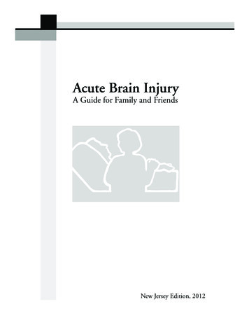
Acute Brain InjuryA Guide for Family and FriendsNew Jersey Edition, 2012
1999 CREDITS FOR IOWA EDITIONREVISED BY:IN COLLABORATION WITHMichele Wagner, R.N., M.S.N., C.N.R.N.Department of Nursing A.P.N.Lori Lindberg, M.S.W.Department of Social ServicesKaren Stenger, R.N., M.A., C.C.R.N.Department of Nursing A.P.N.Barbara Smith, M.S.Center for Disabilities and DevelopmentSue Lewis, B.S.Center for Disabilities and DevelopmentAnna Marie Guengerich, M.L.S.Center for Disabilities and Development1995 CREDITS FOR IOWA EDITIONWRITTEN BY:IN COLLABORATION WITH:Michele Wagner, R.N., B.S.N.Department of Nursing A.P.N.Laurie Ackerman, R.N., M.A.Neuroscience Department of NursingKaren Stenger, R.N., M.A.Department of Nursing A.P.N.Sue Witte, M.S.W.Department of Social ServicesLisa Butikofer, B.S., O.T.Department of Occupational TherapyEllen Swanson, R.D.L.D., M.S.Dietary ServiceLori Moore, M.S.W.Department of Social ServicesRosie Wilhelm, M.S.W.Department of Social ServicesPeggy Saehler, B.S., P.T.Department of Physical TherapyMary Johnson, R.N., M.S.N.Department of Nursing A.P.N. NeuroscienceMary Lohse Shepherd, B.S., P.T.Department of Physical TherapyDepartment of Speech and AudiologyJulie Stierwalt, Ph.D., A.B.D. C.C.C.-S.L.P.Department of NeurologyNeuroscience and Critical Care Nursing StaffILLUSTRATIONS AND DESIGN BY James Abel and Loretta PoppThe first edition of this book was made possible through donations from the Volunteer Program, theDepartment of Nursing, Multispecialty Services and the Department of Physical Therapy at University ofIowa Health Care.This New Jersey Edition was made possible through the New Jersey Traumatic Brain Injury Fund, administered by the New Jersey Department of Human Services, Division of Disability Services, and the Centerfor Disabilities and Development, Iowa’s nationally designated University Center for Excellence on Disabilities, a component of University of Iowa Health Care.2012 NEW JERSEY EDITION REVISED BY:Brain Injury Alliance of New Jersey825 Georges Road, 2nd FloorNorth Brunswick, NJ 08902Phone: (732) 745-0200 E-mail: info@bianj.orgCopyright 2008 The University of IowaWebsite: www.bianj.org
Acute Brain InjuryA Guide for Family and FriendsIntroductionThis booklet provides basic information about braininjury and its treatment. Please read it at your ownpace. As you learn more about the brain, you willhave many questions. Members of the health careteam will do their best to answer your questions.Definite answers may not be known because the longterm effects of brain injury can be difficult to predict.i
ii
Table of ContentsHow Does the Brain Work? . . 1What Types of Brain Injuries May Occur?. 3What Happens When the Brain Is Injured?. 6What is Intracranial Pressure?. 7What is Cerebral Perfusion Pressure?. 8How will the Patient Respond to a Brain Injury?. 8How are Brain Injuries Evaluated?. 9How are Brain Injuries Treated?. 10What Equipment Will You See When You Visit?. 12What Other Treatments May Be Used?. 14Who Will Help After Brain Injury?. 16How Will You React?. 18What are Suggestions for Coping?. 19What are the Signs of Improvement?. 20How Can You Help with Recovery?. 22What Happens After Acute Care in New Jersey. 25Resources. 27AppendixComa Scales. 33Index of Terms. 35iii
iv
Acute Brain InjuryA Guide for Family and FriendsHow Does the Brain Work?The brain controls the actions of the body and allows us to think, learn, andremember. The brain has three main sections: the cerebral hemispheres,cerebellum, and brain stem. There are left and right cerebral hemispheres.The cerebral hemispheres are divided into sections called lobes. Each section of the brain has special jobs to do and sections of the brain also work together. The left cerebral hemisphere controls the right side of the body and isusually responsible for speech. The right cerebral hemisphere controls the leftside of the body and is usually responsible for creative thinking.1
Protection and Oxygen for the BrainThe brain controls many important functions. It needs good protection andoxygen. The brain is protected and receives oxygen in the following ways:Skull: A hard bone that surrounds the brain tissueDura: A tough covering around the brain tissue and thespinal cordCerebrospinal Fluid (CSF) or Spinal Fluid: Fluid thatflows through the ventricles and around the brain andspinal cord. The ventricles are spaces inside the brain.The cerebrospinal fluid acts like a “shock absorber”for the brain.Blood: Provides oxygen and food for the brain2
What Types of Brain Injuries May Occur?Even though the brain is well protected, it may be injured. Every injury isdifferent. Most injuries are a result of bruising, bleeding, twisting, or tearingof brain tissue. Damage to the brain may occur at the time of injury. It mayalso develop after the injury due to swelling or further bleeding. Patients mayhave more than one type of brain injury.Skull Fracture: a break in the bone that surrounds the brain. Thesefractures often heal on their own. Surgery may be needed if there has beendamage to the brain tissue below the fracture.Contusion/Concussion: a mild injury or bruise to the brain which maycause a short loss of consciousness. It may cause headaches, nausea,vomiting, dizziness, and problems with memory and concentration. Thisinjury will not need surgery.Coup/Contre-Coup: A French word that describes contusions thatoccur at two sites in the brain. When the head is hit, the impact causes thebrain to bump the opposite side of the skull. Damage occurs at the site ofimpact and on the opposite side of the brain.3
Epidural Hematoma: a blood clot that forms between the skull and thetop lining of the brain (dura). This blood clot can cause fast changes in thepressure inside the brain. Emergency surgery may be needed. The size of theclot will determine if surgery is needed.Subdural Hematoma: a blood clot that forms between the dura andthe brain tissue. If this bleeding occurs quickly it is called an acute subduralhematoma. If it occurs slowly over several weeks, it is called a chronic subdural hematoma. The clot may cause increased pressure and may need to beremoved surgically.4
Intracerebral Hemorrhage: A blood clot deep in the middle of thebrain that is hard to remove. Pressure from this clot may cause damage to thebrain. Surgery may be needed to relieve the pressure.Diffuse Axonal Injury (DAI): Damage to the pathways (axons) thatconnect the different areas of the brain. This occurs when there is twistingand turning of the brain tissue at the time of injury. The brain messages areslowed or lost. Treatment is aimed at managing swelling in the brain becausetorn axons can not be repaired.Anoxic Brain Injury: An injury that results from a lack of oxygen to thebrain. This is most often from a lack of blood flow due to injury or bleeding.5
What Happens When the Brain Is Injured?Damage to the brainmay occur immediately,as a result of the injury,or it may develop asa result of swelling orbleeding that followsthe injury. The skull isusually filled like this:Normal BrainChanges can occur as a result ofbrain injury. The brain tissue mayswell causing it to take up moreroom in the skull. This is callededema. When this occurs, theswollen brain tissue will push theother contents of the skull to theside.Brain with EdemaThere may be bruising called contusionsor a collection of blood called ahematoma or clot. This may also pushthe other contents to one side.Brain with a Hematoma6
The flow of CSF may also become blocked. This will cause the open spaces(ventricles) to become enlarged. This is called hydrocephalus.Brain with HydrocephalusAny of these changes can cause increased intracranial pressure.What is Intracranial Pressure?To understand intracranial pressure, think of the skull as a rigid box. Afterbrain injury, the skull may become overfilled with swollen brain tissue, blood,or CSF. The skull will not stretch likeskin to deal with these changes. Theskull may become too full and increasethe pressure on the brain tissue. This iscalled increased intracranial pressure.7
What is Cerebral Perfusion Pressure?Blood flow to the brain is called cerebral perfusion pressure. Blood pressureand intracranial pressure affect the cerebral perfusion pressure. If the bloodpressure is low and/or the intracranial pressure is high, the blood flow to thebrain may be limited. This causes decreased cerebral perfusion pressure.Normal blood flowLimited blood flowHow will the Patient Respond to a Brain Injury?The patient’s responses may vary depending on the type of injury or pressurechanges in the brain. Possible responses include: agitation, confusion, decreased responses and coma.Coma: A state of unresponsiveness when patients do not speak orfollow commands, and are unaware of their surroundings. The lengthof time a patient remains in a coma varies.Patients may also have problems with speech, vision, or muscleweakness in their arms or legs.8
How Are Brain Injuries Evaluated?Patients with brain injury require frequent assessments and diagnostictests. These include: Neurological Exam: A series of questions and simple commandsto see if the patient can open their eyes, move, speak, and understandwhat is going on around them. For example: What is your name?Where are you? What day is it? Wiggle your toes. Hold up two fingers. A standard way to describe patient responses may be used. Mosthospitals use the Glasgow Coma Scale or Rancho Levels of Cognitive Functioning. You can read about these scales and what the scoresmean on p. 31. X-ray: A picture that looks at bones to see if they are broken (fractured). It can also be used to take a picture of the chest to look at thelungs. This test may be done at the bedside or in the X-ray departmentand takes between 5-30 minutes to complete. CT Scan (CAT Scan): An X-ray that takes pictures of the brainor other parts of the body. The scan is painless but the patient must lievery still. The test takes 15-30 minutes to complete. MRI (Magnetic Resonance Imaging) Scan: A large magnet and radio waves are used, instead of X-rays, to take pictures of thebody’s tissues. It is painless but noisy. The machine is shaped like along tube. The patient must lie still on a flat table in the middle of themachine. The test takes about 60 minutes to complete. Angiogram: A test to look at the blood vessels in the brain. Using acatheter, or small flexible tube, dye is put into an artery (usually in thegroin) that supplies blood to the brain. This test can tell if the bloodvessels have been damaged or are spasming. The test takes 1-3 hours.9
ICP Monitor: A small tube placed into or just on top of the brainthrough a small hole in the skull. This will measure the pressure insidethe brain (intracranial pressure). EEG (Electroencephalograph): A test to measure electrical activity in the brain. Special patches called electrodes are applied to thehead to measure the activity. The test is painless and can be done atthe bedside or in the EEG department. The length of the test varies.How are Brain Injuries Treated?The goals of brain injury treatment are to: Stop any bleedingPrevent an increase in pressure within the skullControl the amount of pressure, when it does increaseMaintain adequate blood flow to the brainRemove any large blood clotsTreatments will vary with the type of injury. The doctor will decide whichones are used. Some common treatments are:Positioning: Usually the head of the bed will be elevated slightly and theneck kept straight. This position may decrease the intracranial pressure byallowing blood and CSF to drain from the brain. Please do not change theposition of the bed without asking the nurse.Fluid Restriction: It may be necessary to limit the fluids that a patientreceives. The brain is like a sponge. It swells with extra fluid. Limitingfluids can help control the swelling. Please do not give fluids without askingthe nurse.Medications: There are several types of medications used to treat brain injury. Some of these include:10
Diuretics are used to decrease the amount of water in the patient’sbody. This makes less water available to the brain for swelling. Anticonvulsants are used to prevent seizures. Seizures occur as aresult of extra electrical activity in the brain. There are several typesof seizures. The most common type causes the patient to have jerking movements of the arms and legs followed by sleep. Other typesmay cause slight tremors of the face, or staring spells. Please notifythe nurse or doctor if you see any of these signs. Some patients have aseizure at the time of injury while others may develop seizures after theinjury. Barbiturates are given if the patient’s intracranial pressure is very highand hard to control. This medicine puts the patient into a deep “sleep”called a barbiturate coma. This may help prevent more swelling anddamage.Ventricular Drain (Ventriculostomy): A small tube is placed in theventricle. It measures and controls pressure inside the skull. It can be usedto drain some CSF (cerebrospinal fluid) from the brain.Ventilator: A machine used to support the patient in their own breathing,or give the patient breaths. When the ventilator gives extra breaths, the bloodvessels in the brain become smaller. This may help control the intracranialpressure.Surgery: There are three types of surgery used with brain injury: Craniotomy - The skull is opened to relieve the causes of increasedpressure inside the skull. Causes may be fractured bones, blood clots,or swollen brain tissue. Burr holes - A small opening is made into the skull to remove bloodclots. Bone flap removal - A piece of bone is removed from the skull to relieve pressure caused by swollen brain tissue.11
What Equipment Will You See When You Visit?Depending on the type of brain injury, different kinds of equipment will beused. Some common equipment is shown in the picture below. Ask a member of the health care team if you have any questions about equipment.Urinary “foley” catheter12
Monitor: A machine that shows heart rate, breathing, blood pressure, intracranial pressure, and cerebral perfusion pressure.Head Dressing: A bandage around the head used to keep the wound orincision clean and dry.ICP Monitor: A small tube placed into or just on top of the brainthrough a small hole in the skull. This will measure the amount of pressureinside the brain (intracranial pressure).Nasogastric Tube (NG): A tube placed through the nose into the stomach that can be used to suction the stomach or provide liquid formula directly into the stomach.Endotracheal Tube: A tube inserted through the patient’s nose or mouthinto the trachea (windpipe) to help with breathing and suctioning.EKG Lead Wires: Wires connected to the chest with small patches thatmeasure the heart rate and rhythm.Intravenous Catheter (IV) and Intravenous Fluid: A flexible catheterwhich allows fluid, nutrients, and medicine to be given directly into a vein.Ventilator: A machine used in the Intensive Care Unit to support the patient in their own breathing or give the patient breaths.Anti-Embolism Stockings (Frequently call TEDS): Long whitestockings used to help prevent the pooling of blood in the legs.Sequential Compression Stockings (Frequently called Kendalls):Plastic leg wraps that help prevent blood clots by inflating and deflatingaround the legs.Urinary “Foley” Catheter: A tube inserted into the bladder to drainand allow for accurate measurement of urine.13
What Other Treatments May Be Used?Antibiotics: Antibiotics are used to prevent and treat infections that occur. It is not unusual for people with brain injuries to get infections. Theymay get pneumonia, bladder infections, blood infections, or infections in thebrain or cerebrospinal fluid called meningitis.Chest PT and Suctioning: To prevent or treat pneumonia, staff mayuse a vibrating machine or may clap on the patient’s chest. This loosens thephlegm in the lungs. Then the patient will be asked to cough. If the patientis not able to cough up the phlegm they must be suctioned. When a patientis suctioned a catheter is placed in the back of the throat or into the lungs.Tracheostomy (Trach): If thepatient has a lot of lung secretions or ison a ventilator for a long time they mayneed a trach. A trach is a tube placedin the trachea (windpipe). It will makeit easier for the patient to cough upphlegm. It also allows the nurse tosuction the lungs.Initially the patient will be unable totalk while the trach is in place. As thepatient improves, a talking trach may beused. A trach is usually not permanent.Suctioning of the Stomach:Sometimes after brain injury, the stomach will stop working for a short time. This is called an ileus. Even thoughthe stomach may not be working itcontinues to make acid. The acid may damage the stomach lining and causestomach ulcers if they are not removed. A nasogastric tube (NG) will beplaced through the nose into the stomach. This tube will be used to helpremove stomach secretions. Medications may also be given to help preventstomach ulcers.14
Nutrition: Meeting nutrition and fluid needs is important after braininjury. Patients may be less active, yet have very high nutritional needs. Atfirst, nutrition may be supplied by an IV. When the stomach starts working,an evaluation of chewing and swallowing safety will be completed. if thepatient is too sleepy to eat, or unable to swallow, a small nasogastric feedingtube may be used for nutrition. The tube is placed through the nose into thestomach. Liquid formula will be given through the feeding tube. Feedingsmay be given continuously or several times a day. The dietician will assistwith food and fluid selection. Milkshakes and liquid formulas may also beused to provide extra calories and high protein nutrition. A feeding tubemay be used if the patient continues to be too sleepy to eat or unable toswallow. A gastrostomy tube is a feeding tube that goes in the stomach.A jejunostomy tube is a feeding tube that goes in the intestine.Bowel and Bladder Care: Patients may not have control of their bowelor bladder. Catheters or diapers will be used until bowel and bladder controlreturns.Skin Care: Some things that help prevent bedsores include turning thepatient, padding equipment, keeping skin clean and dry, using specialmattresses, and making sure the patient gets enough calories.Range of Motion (ROM) and Splints: Patients with brain injurymay not be able to move their joints as much as needed. This can cause tightmuscles and joints called contractures. Range of motion (ROM) exercisesand special splints for hands and feet help prevent contractures.Pain Control: Comfort measures and medication will be used for paincontrol. However, medications may be limited to types that do not causedrowsiness.15
Who Will Help After Brain Injury?Members of the health care team will work together with the patient, family,and friends during the hospital stay. Care will be centered on the individualneeds of the patient. Family and friends are important members of the team.Team y Speech PatholologistsDieticians& Other StaffSocialWorkersPatient: The patient is the most important member of the team. Care willbe planned based on how the patient responds to treatment.Family and Friends: You provide emotional support to the patient.Family and friends also provide the health care team with important factsabout the patient’s past history and can help watch for changes. Other teammembers will show you what you can do to help with the recovery process.Doctors: Neurosurgery doctors are specialists who help determine the typeof brain injury and its treatment. They may perform surgery on the brain.They will work with other doctors if the patient is in intensive care or has injuries to other parts of the body.16
Nurses: Nurses check patient’s vitals (temperature, blood pressure, heartand breathing rate) and watch for changes in strength and thinking. Theyhelp with daily cares such as eating and bathing. Nurses also coordinate careamong the members of the health care team.Social Workers: Social workers provide emotional support to help thepatient and family adjust to being in the hospital. They coordinate dischargeplanning, referral to community resources, and answer questions about insurance or disability.Physical Therapists (PT): Physical therapists evaluate and treat weaknesses in the patient’s strength, flexibility, balance, rolling, sitting, standingand walking. Treatment may include exercises or instruction in use of equipment such as walkers, canes, or wheelchairs.Occupational Therapists (OT): Occupational therapists evaluate thepatient’s ability to perform dressing, bathing, homemaking and activities thatrequire memory and organization. They provide treatment or equipmentneeded for safe independent living.Speech Therapists: Speech therapists test and treat speech, language,thinking and swallowing problems.Neuropsychologists: Neuropsychologists test thinking, memory, judgment, emotions, behavior and personality. This information can be used tohelp guide treatment. It will also help determine the amount of supervisionthat the patient needs when they leave the hospital.Dieticians: Dieticians assess nutritional needs. They work with the patientand other team members to help the patient meet their nutritional goals.Other staff members may work with the patient and family. These include: Respiratory therapists Activity Therapists Clergy Child Life Therapists Patient Representatives Vocational Counselors Music Therapists Recreation therapists17
How Will You React?When a friend or family member is hospitalized with a brain injury, it isnormal for you to have many different emotions. These emotions will comeand go at different times.Panic and FearPanic and fear are common reactions after a family member has a braininjury. Fears are intense because you are worried the patient may not survive.Until the patient becomes medically stable, your physical and emotional signsof panic may continue. These symptoms include rapid breathing, inability tosleep, decreased appetite and upset stomach. Some people may cryuncontrollably.Shock and DenialYou may feel that what is happening is not real. You may notice things goingon around you, but have trouble remembering information and conversationsor meetings with others. You may also have a hard time understanding theseriousness of the injury that has occurred.AngerMany people feel angry that they or their loved ones are in this situation.This may be justified. You may be angry with the patient for puttingthemselves in a situation where they could be hurt. You may also be angrywith family members, friends, or others involved in the accident. You maybe upset with the health care team for not doing or saying what you think isright. These are all normal reactions.GuiltGuilt is also a normal reaction. You may feel you could have done somethingto prevent the accident from happening, even when this may be far fromtrue. You may also think about past events and personal experiences with thepatient that you wish could have been different or better. If you are feelingangry with the patient, you may also feel guilty about your anger. Weencourage you to talk about your feelings with someone close to you or aprofessional staff member.18
IsolationDuring this time you may feel distant from others. In this strange situation,you may have a hard time relating to others. You may think that others willnot understand. You may isolate yourself because you think others are scaredor disapprove of your feelings. However, a crisis such as a brain injury is atime when it is helpful to accept comfort, support and assistance from others.HopeAs the patient begins to stabilize, your anxiety about survival will be combined with hope of recovery. Medical complications and slow recoverymay increase anxiety. However, hope may be brought about by the smallestchanges.Any of these emotions are normal reactions to a very stressful situation. Youmay find it helpful to discuss your feelings with friends, family, or the healthcare team. It may also be useful to write about your feelings and experiencesin a daily journal.What are Suggestions for Coping?People cope with stressful situations in different ways. What works for oneperson may not be helpful to another. We hope some of these suggestionswill help you get through this difficult time. Write important information down in a journal or notebook. You can use this to keep track of questions you want to askmembers of the health care team. It may also be useful to share withthe patient. Establish a “phone tree.” Name one person for family andfriends to call for information on the patient’s condition. Rotate family visits. If you need or want to leave the hospital, youmay want to have someone stay with the patient so you can feel reassured the patient is not alone. When someone offers to help, accept the offer. Try to bespecific about how this person can help.19
Express your feelings. Discuss your positive and negative feelingswith family members, friends, and staff. Be kind to yourself. Take time for a walk or have a meal witha friend. Also, try to leave the hospital for a meal or restful night ofsleep. It is very important to take care of yourself. By taking care ofyour own needs, you will be more prepared to make good decisions andsupport your loved one.What are the Signs of Improvement?“How long will it take for my loved one to get better?” “What will he or shebe like?” Your health care team may have a hard time answering these questions. Age, extent of damage, length of time since injury, and past mentaland physical health of the patient are factors used when predicting the extentof recovery.Most patients follow a general pattern of recovery after a severe brain injury.This pattern is divided into stages. It is important to know each patient isdifferent and may not follow the stages exactly. Patients vary in the amountof time spent at each stage and their recovery may stop at any stage.Recovery may be grouped into the following four stages:Stage 1: Unresponsiveness: During this stage the patient does notrespond consistently or appropriately. You may hear this stage referred toas a coma. You may notice different movements in the patient. These arereferred to as reflexive or generalized responses. Decerebrate: A reflex that causes straightening of the arms and legs. Decorticate: A reflex that causes bending of the arms andstraightening of the legs. Generalized responses: Random movement of the arms and legsfor no specific reason.20
Stage 2: Early Responses: During this stage the patient starts to respond to things that are happening to them. The responses will be moreappropriate, but may be inconsistent or slow. The patient will start tohave localized responses and follow simple commands. Some examplesof early responses to watch for are: Localized response: These are appropriate movements by the patient in response to sound, touch, or sight. Turning toward a sound,pulling away from something uncomfortable, or following movement with the eyes are examples. Following simple commands: Opening and closing eyes, stickingthe tongue out, or gripping and releasing hands when asked are examples.Stage 3. Agitated and Confused: At this stage the patient is responding more consistently. The patient will be confused about wherehe or she is and what has happened. The patient will have difficulty withmemory and behavior. The patient’s confusion may lead to yelling, swearing, biting, or striking out. Do not be alarmed if soft wrist and ankle tiesare used to protect the patient and prevent tubes from being pulled out.It is very important to remember this stage is a step toward recovery andthis behavior is not intentional.Stage 4. Higher Level Responses: The patient completes routinetasks without difficulty, but still needs help with problem solving, makingjudgments and decisions. The patient may not understand his or her limitations and safety is a big concern. Unusual or high stress situations makeactivities more difficult. The patient may seem more like the person youknew before. However, there may be personality changes.Unfortunately, there is no way to predict how long a person will remain inone stage or what the final outcome will be. The team will work during thehospital stay to achieve the best possible outcome.21
How Can You Help with Recovery?The family and friends of a personwith a brain injury are importantmembers of the team. Your knowledge about the patient’s emotional andphysical needs is valuable, and so isyour participation in helping take careof these needs.The following are suggestions for things you can do that correspond with thestages of recovery.Stage 1. Unr
and turning of the brain tissue at the time of injury. The brain messages are slowed or lost. Treatment is aimed at managing swelling in the brain because torn axons can not be repaired. Anoxic Brain Injury: An injury that results from a lack of oxygen to the brain. This is most often from a lack of blood flow due to injury or bleeding. 5