Transcription
Gedar Totuk et al. BMC Ophthalmology (2018) HNICAL ADVANCEOpen AccessA novel surgical technique for preventionof Ahmed glaucoma valve tube exposure:long scleral flap augmented with Tenonadvancement and duplicationO. M. Gedar Totuk1* , K. Kabadayi2, A. Colakoglu2, N. Ekizoglu2 and U. Aykan3AbstractBackground: To describe a new technique and present its long-term outcome for prevention of Ahmed glaucomavalve (AGV) tube exposure in patients with refractory glaucoma.Methods: Twenty-seven eyes of 24 patients (mean age, 50 years; age range, 16–78 years; 8 females, 16 males) withrefractory glaucoma who had the AGV implant were retrospectively reviewed. For AGV implantation, a long scleralflap combined with Tenon advancement and duplication was used. In this technique, a long scleral flap is createdto completely cover the extraocular part of valve’s tube, and the flap surface is covered with duplicated Tenon’stissue. The average follow-up after AGV implantation was 21.7 months (range, 12–36 months).Results: The mean intraocular pressure before the operation, which was 44.1 mmHg (range, 26–62 mmHg), decreased to14.2 mmHg (range, 8–20 mmHg) at the last follow-up visit, showing 67% reduction with AGV implantation. The meannumber of antiglaucomatous medications was 4.1 before the AGV implantation and decreased to 0.9 after the operation,showing 88% reduction. In 14 eyes (51.9%), there was no change in the best corrected visual acuity (BCVA), and in 11eyes (40.7%), the BCVA increased by 2 lines on the Snellen chart postoperatively. No patient developed postoperativehypotony, flat anterior chamber, diplopia, strabismus, erosion or exposure of the tube, or tube/plate migration.Conclusions: The long scleral flap augmented with Tenon advancement and duplication is an effective and safe surgicaltechnique for the implantation of AGV and preventing tube exposure in cases of refractory glaucoma.Keywords: Refractory glaucoma, Glaucoma valve tube exposure, Glaucoma valve implantation, Long scleral flap, Tenonduplication, Ahmed glaucoma valveBackgroundThe management of refractory glaucoma is often challenging. The common approach is to reduce intraocular pressure by using glaucoma drainage devices. In comparisonto glaucoma surgeries, the Ahmed glaucoma valve (AGV)implantation has been found to be more effective and safemethod for reducing intraocular pressure in patients withrefractory glaucoma [1–4]. Implantation of AGV can beused as a primary treatment option in refractory glaucomaor after failed conventional filtration surgeries [1].* Correspondence: melikegedar@gmail.com1Department of Ophthalmology, Bahcesehir University Faculty of Medicine,Yenisahra Mah. Batman Sok. No: 66-68 Sahrayicedit, Kadikoy, Istanbul, TurkeyFull list of author information is available at the end of the articleEarly and late hypotony, shallow anterior chamber,corneal-lenticular touch, choroidal detachment, hypotonymaculopathy, anterior chamber hyphema, suprachoroidalhemorrhage, peripheral iris synechiae, capsule fibrosisaround plate, erosion and exposure of the tube or plate,extrusion of the implant, endophthalmitis, and cataractare some of the complications associated with AGV [2, 5].AGV tube erosion almost always occurs at the proximallimbal conjunctiva if left uncovered, subsequently leadingto exposure of the tube and increased risk of endophthalmitis [5, 6]. Even when the tube is covered with graftmaterials like pericardial patch grafts, donor fascia latagraft, cadaveric dura mater, and preserved human sclera,the risk of exposure still exists. Grafts also carry the risk of The Author(s). 2018 Open Access This article is distributed under the terms of the Creative Commons Attribution 4.0International License (http://creativecommons.org/licenses/by/4.0/), which permits unrestricted use, distribution, andreproduction in any medium, provided you give appropriate credit to the original author(s) and the source, provide a link tothe Creative Commons license, and indicate if changes were made. The Creative Commons Public Domain Dedication o/1.0/) applies to the data made available in this article, unless otherwise stated.
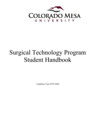
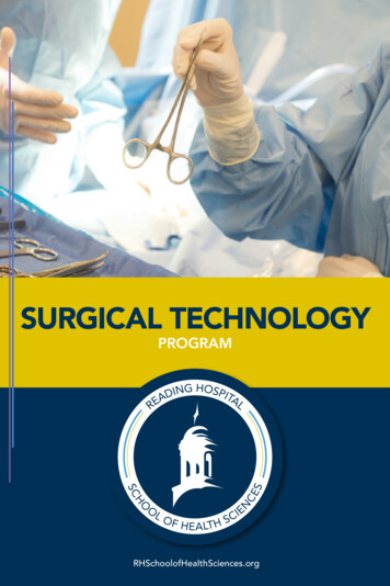
Gedar Totuk et al. BMC Ophthalmology (2018) 18:226virus transmission, corneal dellen formation, scleral melting, and high financial burden [7, 8]. Thus, there is no perfect surgical technique to prevent AGV tube exposure [9].We propose a new technique that completely buries thetube within the sclera using a covering of a long scleralflap with Tenon advancement and duplication to preventAGV tube exposure in patients with refractory glaucoma.In this study, we aimed to present this technique and itslong-term outcomes with regard to prevention of AGVtube exposure in patients with refractory glaucoma.MethodsStudy patientsTwenty-seven eyes of 24 patients (mean age, 50 years; agerange, 16–78 years; 8 females, 16 males) with refractoryglaucoma who underwent AGV implantation in our clinicbetween March 2014 and January 2018 were retrospectivelyreviewed. This study describes a novel surgical technique,however, this technique has been applied in our clinicwithin the routine clinical practice since 2014. Therefore,this study was performed as a retrospective review ofpatient records. For AVG implantation, a long scleral flapcombined with Tenon advancement and duplicationtechnique was used in all patients. All patients underwent acomprehensive assessment and ophthalmologic examination, including measurement of best corrected visualacuity (BCVA) on the Snellen chart and intraocularpressure (IOP) with applanation tonometry. Of the 27 eyes,5 had neovascular glaucoma with proliferative diabeticretinopathy, 5 had primary open angle glaucoma, 2 hadcongenital glaucoma, 15 had secondary glaucoma. Nineeyes (33.3%) had phakia, 1 (3.7%) had aphakia, and 17(63%) had pseudophakia. The demographics, diagnosis, andophthalmological findings of each eye before AGV implantation are summarized in Table 1.This study was approved by the Institutional EthicsCommittee of Bahcesehir University (Dec/6th/2017;2017–19/04) and conducted in accordance with the latestversion of the Declaration of Helsinki. According to theRegulation on Clinical Studies of Drugs and BiologicalProducts in Turkey (no: 29474), which was updated on13.09.2015, retrospective studies are not subject to therequirement of informed consent of patients. The Institutional Ethics Committee of Bahcesehir University, whichoperates in accordance with this regulation, waived therequirement of informed consent for this study (Dec/6th/2017; 2017–19/04).Surgical techniqueA fornix-based conjunctival flap was prepared with oneperpendicular relaxing incision in the superotemporalquadrant for the right eye or superonasal quadrant for theleft eye because the surgeon is right handed. The AGVmodel FP7 (New World Medical Inc., Rancho Cucamonga,Page 2 of 8CA, USA) was used for all surgeries. The implant wasexamined for integrity and primed by injecting 1 cc ofbalanced salt solution through the drainage tube. TheAGV implant was inserted under the conjunctiva andTenon’s capsule and sutured to the sclera at a distance ofabout 10 mm from the limbus with a 6/0 Vicryl suture.Two approximately 10-mm-long parallel scleral incisionswere made starting from the limbus ending at the plate, ofhalf thickness (around 250-μm thick), throughout the tubetract. Then, a 10-mm-long scleral flap was prepared with abevel up crescent knife (Fig. 1a). The drainage tube wastrimmed to the appropriate length permitting a 2–3-mminsertion in the anterior chamber (AC) with the beveled-upedge. The AC was then entered 2 mm posteriorly to thelimbus, under the scleral flap with a 23-G needle. The needle tract was parallel to the plane of the iris. The bevel-uptrimmed tube was inserted into the AC through the needletract without making contact with the iris or corneal endothelium. Then, the scleral flap was sutured with 10/0monofilament nylon sutures (Fig. 1b). Tenon advancementand duplication by blunt dissection into two layers wereapplied with the long scleral flap that was used to cover thetube (Fig. 1c). Finally, the conjunctiva was anchored to thelimbus with 8/0 interrupted Vicryl sutures (Fig. 1d). Asubconjunctival injection of corticosteroids and antibioticswas administered at the end of the procedure. Topicalcorticosteroids, nonsteroidal anti-inflammatory drugs,cycloplegics, and antibiotic regimen was started for allpatients postoperatively.AGV implantation with a long scleral flap covering withTenon advancement and duplication surgical techniquewas combined with intracameral anti-VEGF injections in8 patients with proliferative diabetic retinopathy (Table 1).A single surgeon (U.A.) performed all the operations.Statistical analysisThe study data were summarized for each eye using descriptive statistics such as mean, range, standard deviation, frequency, and percentage.ResultsThe mean follow-up period after AGV implantation was21.7 months (range, 12–36 months) (Table 1). In 2 eyes(7.4%), the BCVA decreased postoperatively by 2 lines onthe Snellen chart owing to suprachoroidal hemorrhageand uveitic reactivation. In 14 eyes (51.9%), there were nochanges in the BCVA, and in 11 eyes (40.7%), the BCVAincreased by 2 lines postoperatively (Table 2).The mean IOP, which was 44.1 mmHg (range 26–62) before the operation, decreased to 14.2 (range 8–20) mmHgat the last follow-up visit, showing 67% reduction withAGV implantation (Fig. 2). The mean postoperative IOPduring the follow-up period was 22.6 mmHg (range 10–35)(Table 2). Eleven (40.7%) cases with IOP over 18 mmHg
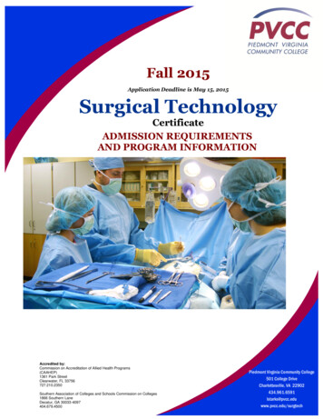
Gedar Totuk et al. BMC Ophthalmology (2018) 18:226Page 3 of 8Table 1 Patient demographics and clinical data before AGV implantation surgeryPatientAgeSexSideDiagnosisLensAdditional treatment1 50FRNVG PDRPhakicAC Lucentis2 50FLNVG PDRPhakicAC Lucentis3 50MRNVG PDRPseudophakicAC Lucentis411–20MLSEC GL (PPV silicon) PVRPseudophakicAC Avastin541–50MRSEC GL (PPV silicon) PDRPseudophakic6 50FRNVG PDRPseudophakicAC Avastin7 50FLNVG PDRPseudophakic/SubluxatedAC Avastin8 50MRPAOG/failed SLTPseudophakic/Degen myopia–9 50MRPAOGPhakic–10 50MLSEC GL (PPV silicon) PDRPseudophakic–1111–20FLSEC GL (CONG CAT/SEC IOLimplant/opaque cornea)Pseudophakic–1221–30MRSEC GL (PPV silicon)Phakic–1331–40FLCONG GLPhakic/Bullous keratopathy–1441–50MRSEC GL (PPV silicon)Pseudophakic–15 50FRSEC GL (Keratoplasty)Pseudophakic–1621–30MLSEC GL (PPV Silicon)Pseudophakic–1741–50FLTraumatic GL/Failed hydrusPhakic/Bullous keratopathy–18 50FRUveitic GL (HSV)Phakic–19 50MLSEC GL (PPV Silicon) PDRPhakicAC Avastin20 50MLSEC GL (anterior chamber IOL bullous keratopathy)Pseudophakic–2141–50MLSEC GL (PPV silicon) PDRPseudophakicAC Avastin22 50MLSEC GL (PPV silicon)Pseudophakic–2331–40MLCONG 2541–50MLPOAG/RPPseudophakic–26 50MRPOAG/failed TRABPhakic–2721–30FLTraumatic aphakic GLAphakic–AC anterior chamber, CAT cataract, CONG congenital, F female, GL glaucoma, HSV herpes simplex virus, IOL intraocular lens, L left, M male, NVG neovascularglaucoma, PDR proliferative diabetic retinopathy, POAG primary open angle glaucoma, PPV pars plana vitrectomy, PVR proliferative vitreoretinopathy, R right, RPretinitis pigmentosa, SEC secondary, SLT selective laser trabeculoplasty, TRAB trabeculectomyrequired additional treatment: 1 required diode cyclophotocoagulation and 10 were on 1–4 glaucoma medications(Table 1, Table 2).The mean number of anti-glaucomatous medications, which was 4.1 before the AGV implantation,decreased to 0.9 after the operation, showing 88%reduction (Table 2, Fig. 3].As a complication of AGV implantation, postoperativehyphema developed in 2 patients (7.4%), requiring AC lavage. In 3 eyes, the tube was occluded by the iris needed fortube repositioning, uveitic inflammatory debris, and siliconoil. In 2 eyes with neovascular glaucoma and proliferativediabetic retinopathy, intravitreal hemorrhage developedpostoperatively and additional intravitreal anti-VEGF andpars plana vitrectomy were required. In 1 eye, choroidal detachment resolved spontaneously. In 1 eye, suprachoroidalhemorrhage and choroidal detachment necessitated parsplana vitrectomy and silicone oil injection. Tenon’s cystsdeveloped in 2 eyes that resisted drainage and requiredcystectomy. In 2 eyes, secondary cataract formationrequired phacoemulsification (Table 1).No patient developed postoperative hypotony, flat anterior chamber, diplopia, strabismus, erosion or exposureof the tube, or tube/plate migration.DiscussionIn the present study, we described a novel technique ofcreating a long scleral flap augmented with Tenon
Gedar Totuk et al. BMC Ophthalmology (2018) 18:226Page 4 of 8Fig. 1 The long scleral flap combined with tenon advancement and duplication technique. First, a 10 mm long scleral flap is prepared with bevelup crescent knife between the two scleral incisions (a). The AC is entered with a 23G needle, tube was inserted into the AC, then the scleral flapwas sutured with with 10/0 monofilament nylon sutures (b). Tenon advancement and duplication technique over the long scleral flap thatcovered the tube is applied (c). Finally, the conjunctiva is anchored to the limbus with 8/0 interrupted vicryl sutures (d)advancement and duplication to prevent exposure of theAGV tube in patients with refractory glaucoma. Ourexperience with 27 eyes indicated that this technique iseffective in decreasing the IOP and requirement for antiglaucomatous medications without adversely affectingBCVA and not causing erosion or exposure of the tubeor tube/plate migration. Therefore, we suggest that thelong scleral flap augmented with Tenon advancementand duplication is an effective and safe surgicaltechnique for the implantation of AGV and preventionof tube exposure in cases with refractory glaucoma.The AGV was developed in 1993 and has become themost commonly used glaucoma drainage device with highsuccess rates of 60–90% in 1 year and 40–50% in up to4 years [1, 10–14]. It has been particularly used for themanagement of glaucoma refractory to standard filteringsurgery. Despite its well-documented efficacy in the treatment of refractory glaucoma, various early and late postoperative complications have been reported with AGVimplantation, one of which is exposure of the tubethrough conjunctival erosion. Since the exposed tube giveseasy access to microorganisms into the eye, it may lead toserious complications such as ocular inflammation, hypotony, poor vision, phthisis, and endophthalmitis [2–6].Tube exposure is usually the result of continuous microtrauma to the conjunctiva, which induces inflammatoryand/or immunologically mediated tissue damage. Youngerage, previous inflammation, diabetes, and inferiorly placedimplants are well-known risk factors for tube exposureassociated with glaucoma drainage devices [7, 15, 16].AGV, Baerveldt, and Molteno implants do not show significant differences in terms of tube exposure rates [17].Although various surgical techniques have beendescribed to prevent tube exposure, such as placementof patch graft (e.g., fascia lata, pericardium, donor sclera,or lyophilized dura mater patch grafts), long scleral tunnel, and/or doubling and advancement of Tenon’s tissue[8, 18–22], there is no consensus on the ideal technique.Our technique is AGV implantation using a long scleralflap augmented with Tenon advancement and duplication. The rationale behind this technique is to form twonatural barriers, which are the sclera and Tenon’stissues, which keep the silicon tube away from theconjunctiva throughout the whole tube tract, providing
Gedar Totuk et al. BMC Ophthalmology (2018) 18:226Page 5 of 8Table 2 Patient clinical data after AGV implantation surgeryPatient Follow-up Preop(Months) BCVAPostopBCVAPreopIOPPostopPostophighest IOP IOPIOP %Number of Number ofComplicationsReduction preop drugs postop drugsSEC operation1361MCF0.35034160.6840Tube occlusion by IV Lucentis,iris, SEC CATPHACO, tuberepositioning2361MCF0.13426180.4743HyphemaIV Lucentis,PHACO, 530None0512p HM5417100.8140None06271MCF0.35222170.6753IVHIV Lucentis,PPV727HM0.055418100.8150IVH, hyphemaAC Lavage,IV Lucentis,PPV820p p 4030190.5340Tenon’s cystCyst e CPC1223HM0.053828100.7430SEC CATSilicon extraction,PPV1324NLPP-3228170.4732Tenon’s cystCyst MCF1MCF6235200.6841HSV uveiticactivation and0tube 055728200.6550Tube occlusionwith silicon 27HMHM4020180.5520None0AC anterior chamber, BCVA best corrected visual acuity, CAT cataract, CPC cyclophotocoagulation, HM hand motions, HSV herpes simplex virus, IOP intraocularpressure, IV intravitreal, IVH intravireal hemorrhage, MCF meters counting fingers, NLP no light perception, P perception, PHACO phacoemulsification, Postoppostoperative, PPV pars plana vitrectomy, Preop preoperative, SEC secondaryprotection from mechanical trauma. The other advantage of this method is that it prevents inflammation orimmunological reaction against foreign materials such asgrafts. Besides, the placement of the tube under the longscleral flap helps prevent mechanical insult of the tubebending at the proximal 3-mm corneoscleral limbus,
Gedar Totuk et al. BMC Ophthalmology (2018) 18:226Page 6 of 8Fig. 2 The mean preoperative and postoperative intraocular pressure (IOP) of 27 study patients. Error bars indicated standard deviationsproviding a smooth tract for the tube. Fixing the tubeunder the sclera also prevents microdamage due tocontinuous movement of the tube.In the patch graft technique, although tube exposure issignificantly reduced, there is still a 5–14% risk of tubeexposure regardless of the graft and glaucoma drainagedevice type used [15, 16]. However, with our technique, theexposure rate was 0% during an average of 21.7 months offollow-up. Furthermore, considering the disadvantages ofvarious patch grafts like the risk of melting, mechanicaltrauma, immune atrophy and late exposure, infection, andrejection, along with the high cost of fibrin glue fixation [7,15, 23, 24], our technique can be considered superior topatch graft technique. In our technique, as we did not useany foreign material, there was no risk of inducing animmunologic reaction. As the scleral flap was sutured overthe tube, there was no continuous micromovement of theconjunctiva over the tube and no risk of mechanical traumathat leads to erosion. Our sutures in the scleral flap wereunder the duplicated Tenon’s tissue, and did not carry therisk of infection owing to their long distance from the ocular surface. The use of autologous material has the advantages of absence of immunologic reactions and low cost.In a meta-analysis by Stewart et al. [17], previouslypublished studies describing conjunctival erosion in patients with a glaucoma device (16 AGVs, 12 Baerveldt,Fig. 3 The mean number of preoperative and postoperative medications used for 27 eyes. Error bars indicated standard deviations
Gedar Totuk et al. BMC Ophthalmology (2018) 18:226and 17 Molteno implants) showed that tube exposure isnot a very late complication of the glaucoma drainagedevice surgery. A total of 3105 patients and 3255 eyes withan average follow-up of 26.1 3.3 months were included inthe analysis with the incidence of tube exposure 2.0% 2.6% (n 64), and an average exposure rate per month of0.09% 0.14%. Although the correlation between studylength and incidence of exposure was not significant, thereappeared to be a little increase in exposure incidence forstudies up to 2-year follow-up [17]. Based on thismeta-analysis, we conclude that our mean follow-upduration is sufficient enough to evaluate the effect of thistechnique on the development of tube exposure.Similar to the findings of previous studies [7, 15, 16], ourstudy population had no risk factors for tube exposure suchas younger age, inflammation prior to tube exposure, anddiabetes. Since we had no case with an inferiorly placedAGV implant, we could not determine the tube exposurerate of this novel technique in patients with inferiorlyplaced implants. We can only report that there was nodifference between the superotemporal quadrant and thesuperonasal quadrant placement in our study.Another previous technique of preventing tube exposureinvolves the use of a long scleral tunnel for the implantationof anterior tube parts of the glaucoma drainage device [18,19]. Long scleral tunnel technique was found to be superiorto the patch graft method in preventing tube exposure afterAGV implantation [25]. However, the long scleral tunneltechnique has the limitation of a long tunnel which complicates advancing the lancet through the curved contour ofthe sclera. Moreover, the entrance to the anterior chamberwith a lancet is more traumatic and carries higher risk ofleakage around the tube than entrance with a 23-gaugeneedle. In our novel technique, we created a half-thicknesssmooth flap in the sclera, so direct visualization of thelength of the scleral bed provided a more delicate tract andalleviated the risks posed by the blinded curved tunneltechnique. We entered the anterior chamber using a23-gauge needle safely with direct visualization.Some previous studies reported successful results withthe short scleral tunnel technique even in cases of congenital glaucoma [26]. However, we did not prefer the shortscleral tunnel or flap because we think that covering the entire length of the tube is the nontraumatic route and burying the tube completely in the sclera prevents the bendingof the proximal tube along the curvature of the globe.Brouzas [27] presented the double scleral tunnel in tandem technique, offering two short tunnels in order to overcome the length limitations of one tunnel and completelyburying the tube in the sclera, but tube exposure throughconjunctival erosion still occurred in 7.1% of the cases inthis study.Tamcelik [28] described a technique that combined shortscleral tunnel with Tenon advancement and duplicationPage 7 of 8and reported no tube exposure even at follow-up of 8 years.Our technique has the practical advantages of Tamcelik’stechnique and provides even lower complication rates. Inour technique, we made a watertight suture on the flapalong the path of the tube, so there was a lower risk ofleakage and subsequent hypotony. None of our patientsexperienced postoperative hypotony or shallow anteriorchamber. A large proportion of our patients experienced asignificant improvement in their visual acuity. In the vastmajority of our patients, visual acuity was low due toadvanced glaucomatous optic neuropathy. The relativeincrease in postoperative BCVA was the result of theresolution of the preoperative corneal edema along with adecrease in intraocular pressure.The possible limitation of our technique is the size ofthe filtration lake formed around the plate due to thepulling up and shortening of the Tenon’s capsule. Thinning of the Tenon layer by aging may cause failure ofthe surgery making the technique inapplicable. Thisshould be clarified with further long-term and largestudies. Our technique may not be ideal for all cases asscleral tissue may be very thin in patients who havepreviously undergone multiple surgeries.ConclusionsIn conclusion, the proposed long scleral flap augmentedwith Tenon advancement and duplication is an effectiveand safe surgical method for the implantation of AGVand preventing tube exposure in patients with refractoryglaucoma. It is also an easy technique with low cost.Although the rate of complications associated with thistechnique is low, we suggest that this new surgicaltechnique be further explored by surgeons in order todetermine its long-term safety and efficacy.AbbreviationsAC: Anterior chamber; AGV: Ahmed glaucoma valve; BCVA: Best correctedvisual acuity; IOP: Intraocular pressureFundingThe design of the study and collection, analysis, and interpretation of dataand in writing the manuscript was not funded.Availability of data and materialsData sharing is not applicable to this article as no datasets were generatedor analyzed during the current study.Authors’ contributionsOMGT, KK, AC, NE, UA have substantial contributions to the conception anddesign of the work; the acquisition, analysis, and interpretation of data forthe work; drafting the manuscript, revising it critically for importantintellectual content. OMGT, KK, AC, NE, UA have agreement to beaccountable for all aspects of the work in ensuring that questions related tothe accuracy or integrity of any part of the work are appropriatelyinvestigated and resolved. All authors read and approved the final version ofthe manuscript to be published.Ethics approval and consent to participateThis study was approved by the Institutional Ethics Committee of BahcesehirUniversity (Dec/6th/2017; 2017–19/04) and conducted in accordance with
Gedar Totuk et al. BMC Ophthalmology (2018) 18:226the latest version of the Declaration of Helsinki. According to the Regulationon Clinical Studies of Drugs and Biological Products in Turkey (no: 29474),which was updated on 13.09.2015, retrospective studies are not subject tothe requirement of informed consent of patients. The Institutional EthicsCommittee of Bahcesehir University, which operates in accordance with thisregulation, waived the requirement of informed consent for this study (Dec/6th/2017; 2017–19/04).Consent for publicationNot applicable.Competing interestsThe authors declare that they have no competing interests.Publisher’s NoteSpringer Nature remains neutral with regard to jurisdictional claims inpublished maps and institutional affiliations.Author details1Department of Ophthalmology, Bahcesehir University Faculty of Medicine,Yenisahra Mah. Batman Sok. No: 66-68 Sahrayicedit, Kadikoy, Istanbul, Turkey.2Bahcesehir University Faculty of Medicine, Istanbul, Turkey. 3FEBO, DunyagozHospital, Istanbul, Turkey.Received: 18 April 2018 Accepted: 28 August 2018References1. Souza C, Tran DH, Loman J, et al. Long-term outcomes of Ahmed glaucomavalve implantation in refractory glaucomas. Am J Ophthalmol. 2007;144:893–900.2. Riva I, Roberti G, Oddone F, et al. Ahmed glaucoma valve implant: surgicaltechnique and complications. Clin Ophthalmol. 2017;11:357–67.3. Bikbov MM, Khusnitdinov II. The results of the use of Ahmed valve inrefractory glaucoma surgery. J Curr Glaucoma Pract. 2015;9:86–91.4. Riva I, Roberti G, Katsanos A, et al. A review of the Ahmed glaucoma valveimplant and comparison with other surgical operations. Adv Ther. 2017;34:834–47.5. Minckler DS, Francis BA, Hodapp EA, et al. Aqueous shunts in glaucoma: areport by the American Academy of ophthalmology. Ophthalmology. 2008;115:1089–98.6. Al-Torbak AA, Al-Shahwan S, Al-Jadaan I, et al. Endophthalmitis associatedwith the Ahmed glaucoma valve implant. Br J Ophthalmol. 2005;89:454–8.7. Chaku M, Netland PA, Ishida K, et al. Risk factors for tube exposure as a latecomplication of glaucoma drainage implant surgery. Clin Ophthalmol. 2016;10:547–53.8. Quaranta L, Riva I, Floriani IC. Outcomes of using a sutureless bovinepericardial patch graft for Ahmed glaucoma valve implantation. Eur JOphthalmol. 2013;23:738–42.9. Giovingo M. Complications of glaucoma drainage device surgery: a review.Semin Ophthalmol. 2014;29:397–402.10. Christakis PG, Kalenak JW, Zurakowski D, et al. The Ahmed versus Baerveldtstudy: one-year treatment outcomes. Ophthalmology. 2011;118:2180–9.11. Huang MC, Netland PA, Coleman AL, et al. Intermediate-term clinicalexperience with the Ahmed Glaucoma valve implant. Am J Ophthalmol.1999;127:27–33.12. Papadaki TG, Zacharopoulos IP, Pasquale LR, et al. Long-term results ofAhmed glaucoma valve implantation for uveitic glaucoma. Am JOphthalmol. 2007;144:62–9.13. Topouzis F, Coleman AL, Choplin N, et al. Follow-up of the original cohort withthe Ahmed glaucoma valve implant. Am J Ophthalmol. 1999;128:198–204.14. Wilson MR, Mendis U, Smith SD, et al. Ahmed glaucoma valve implant vstrabeculectomy in the surgical treatment of glaucoma: a randomizedclinical trial. Am J Ophthalmol. 2000;130:267–73.15. Heuer DK, Budenz D, Coleman A. Aqueous shunt tube erosion. J Glaucoma.2001;10:493–6.16. Smith MF, Doyle JW, Ticrney JW Jr. A comparison of glaucoma drainageimplant tube coverage. J Glaucoma. 2002;11:143–7.17. Stewart WC, Kristoffersen CJ, Demos CM, et al. Incidence of conjunctivalexposure following drainage device implantation in patients with glaucoma.Eur J Ophthalmol. 2010;20:124–30.Page 8 of 818. Ozdamar A, Aras C, Ustundag C, et al. Scleral tunnel for the implantation ofglaucoma seton devices. Ophthalmic Surg Lasers. 2001;32:432–5.19. Ollila M, Falck A, Airaksinen PJ. Placing the Molteno implant in a long scleraltunnel to prevent postoperative tube exposure. Acta Ophthalmol Scand.2005;83:302–5.20. Gutiérrez-Díaz E, Montero-Rodríguez M, Mencía-Gutiérrez E, et al. Long-termpersistence of fascia lata patch graft in glaucoma drainage device surgery.Eur J Ophthalmol. 2005;15:412–4.21. Tanji TM, Lundy DC, Minckler DS, et al. Fascia lata patch graft in glaucomatube surgery. Ophthalmology. 1996;103:1309–12.22. Lankaranian D, Reis R, Henderer JD, et al. Comparison of single thicknessand double thickness processed pericardium patch graft in glaucomadrainage device surgery: a single surgeon comparison of outcome. JGlaucoma. 2008;17:48–51.23. Ainsworth G, Rotchford A, Dua HS, et al. A novel
Conclusions: The long scleral flap augmented with Tenon advanceme nt and duplication is an effective and safe surgical technique for the implantation of AGV and preventing tube exposure in cases of refractory glaucoma. Keywords: Refractory glaucoma, Glaucoma valve tube exposure, Glaucoma valve implantation, Long scleral flap, Tenon