Transcription
AL JOURRINNAPELALPERATNINATUAL JORClinical GuidelinesPerinatal Journal 2022;30(2):87–102 2022 Perinatal Medicine FoundationFirst trimester examination of fetal anatomy:clinical practice guideline by the WorldAssociation of Perinatal Medicine (WAPM) and thePerinatal Medicine Foundation (PMF)Nicola Volpe1 İD , Cihat fien2 İD , fiifa Turan3 İD , Waldo Sepulveda4 İD , Asma Khalil5 İD ,Daniel Rolnik6 İD , Valentina De Robertis7 İD , Paolo Volpe7 İD , Mar M. Gil8 İD , Petya Chaveeva9 İD ,Themistoklis Dagklis10 İD , Ritsuko K. Pooh11 İD , Przemyslaw Kosinski12 İD , Jader Cruz13 İD ,Erasmo Huertas14 İD , Francesco D’Antonio15 İD , Jesus Rodriquez Calvo16 İD , Ana Daneva Markova17 İD1Obstetrics& Gynecology Unit, Department of Medicine and Surgery, University of Parma, Azienda Ospedaliero-Universitaria Ospedale Maggiore diParma, Parma, Italy2Perinatal Medicine Foundation and Department of Obstetrics & Gynecology, Memorial Bahçelievler Hospital, Istanbul, Turkey3Department of Obstetrics, Gynecology & Reproductive Sciences, School of Medicine, University of Maryland, Baltimore, MD, USA4FETALMED Maternal-Fetal Diagnostic Center, Fetal Imaging Unit, Santiago, Chile5Fetal Medicine Unit, St. George University Hospital NHS Foundation Trust, London, UK6Department of Obstetrics & Gynaecology, School of Clinical Sciences, Monash University, Melbourne, Victoria, Australia7Fetal Medicine Unit, Di Venere and Sarcone Hospitals, ASL BA, Bari, Italy8Hospital Universitario de Torrejón, Madrid, Spain; School of Health Sciences, Universidad Francisco de Vitoria, Madrid, Spain9Fetal Medicine Unit, Shterev Hospital, Sofia, Bulgaria10Third Department of Obstetrics & Gynaecology, Faculty of Health Sciences, School of Medicine, Aristotle University of Thessaloniki, Thessaloniki, Greece11Fetal Diagnostic Center, CRIFM Clinical Research Institute of Fetal Medicine, Osaka, Japan12Department of Obstetrics, Perinatology & Gynecology, Medical University of Warsaw, Warsaw, Poland13Fetal Medicine Unit, Centro Hospitalar de Lisboa Central, Lisboa, Portugal14Instituto Nacional Materno Perinatal, Lima, Peru15Department of Obstetrics & Gynecology, Center for Fetal Care & High-Risk Pregnancy, University of Chieti, Chieti, Italy16Fetal Medicine Unit, University Hospital 12 de Octubre, Madrid, Spain17Department of Obstetrics & Gynecology, Medical Faculty, Skopje University, Skopje, North MacedoniaAbstractThis recommendation document follows the mission of the World Association of Perinatal Medicine (WAPM) in collaboration with thePerinatal Medicine Foundation (PMF). We aim to bring together groups and individuals throughout the world for precise standardizationto implement the ultrasound evaluation of the fetus in the first trimester of pregnancy and improve the early detection of anomalies and theclinical management of the pregnancy. The aim is to present a document that includes statements and recommendations on the standardevaluation of the fetal anatomy in the first trimester, based on quality evidence in the peer-reviewed literature as well as the experience ofperinatal experts around the world.Keywords: Fetal anatomy, first trimester, ultrasound, guideline.Correspondence: Cihat fien, MD. Department of Obstetrics & Gynecology, Memorial Bahçelievler Hospital, Adnan Kahveci Bulvar›, No 227, 34180Bak›rköy, Istanbul, Turkey. e-mail: csen@perinatalmedicine.org / Received: March 5, 2022; Accepted: March 23, 2022How to cite this article: Volpe N, fien C, Turan fi, Sepulveda W, Khalil A, Rolnik D, De Robertis V, Volpe P, Gil MM, Chaveeva P, Dagklis T, Pooh RK,Kosinski P, Cruz J, Huertas E, D’Antonio F, Calvo JR, Markova AD. First trimester examination of fetal anatomy: clinical practice guideline by the WorldAssociation of Perinatal Medicine (WAPM) and the Perinatal Medicine Foundation (PMF). Perinat J 2022;30(2):87–102. doi:10.2399/prn.22.0302001ORCID ID: N. Volpe 0000-0003-4209-5602; C. fien 0000-0002-2822-6840; fi. Turan 0000-0003-0919-6487; W. Sepulveda 0000-0002-3856-574X;A. Khalil 0000-0003-2802-7670; D. Rolnik 0000-0002-2263-3592; V. De Robertis 0000-0003-0196-0671; P. Volpe 0000-0002-1492-8868;M. M. Gil 0000-0002-9993-5249; P. Chaveeva 0000-0002-8041-2181; T. Dagklis 0000-0002-2863-5839; R. K. Pooh 0000-0002-1527-4595;P. Kosinski 0000-0003-3812-7560; J. Cruz 0000-0001-6002-9022; E. Huertas 0000-0002-9851-8419; F. D’Antonio 0000-0002-5178-3354;J. R. Calvo 0000-0002-3543-3063; A. D. Markova 0000-0002-7011-9205
Volpe N et al.IntroductionFrom aneuploidies screening to first trimesterfetal anatomyThe window of 11 0–13 6 weeks of gestation providesa great opportunity to evaluate the accurate dating andthe risk of fetal aneuploidy. Although it gives us anexcellent opportunity to look for basic anatomicallandmarks at early ages, the major focus of the 11 0 to13 6 weeks scan has been on aneuploidy screening.[1]First trimester combined screening has been proposedand established in different countries as an accurateand reproducible method to select a population offetuses at high risk for chromosomal abnormalities.[2,3]Such screening is based on the combination of severalparameters, including the nuchal translucency (NT)obtained by a targeted ultrasound scan performed at11 0 to 13 6 weeks.[1,3] The NT measurement reproducibility relies on a strict methodology and a welldefined certification and auditing system. The numberof certified operators has been increasing in the lastfew years, witnessing a significant diffusion of the firsttrimester scan worldwide. The consequent improvement of operators’ skills in first-trimester ultrasound,the increased knowledge of early fetal anatomy, theassociation between increased NT and fetal structuralabnormalities,[4–8] and the improving ultrasound technology allowing higher image resolution (software andhardware implementations, availability of the transvaginal probes), have led to increased detection of fetalstructural anomalies already in the first trimester.[5,9–17]A recent systematic review[18] has shown an estimateddetection rate of fetal structural abnormalities in thefirst trimester, ranging between 32% and 61%, according to the type of anomalies and population characteristics. In particular, the detection rate seems higherwhen focusing on major anomalies than all types ofanomalies (about 46% vs. 32% detection rate), andeven higher when scanning a high-risk rather than anunselected population of pregnant women (61% detection rate of structural anomalies). Such figures seem tobe widely variable in the literature according to different factors, such as operator skills (experience, training,knowledge of fetal embryology or use of indirect ultrasound markers of anomalies),[5,15,16,18–31] gestational age atthe time of examination,[28] route of ultrasound (transvaginal [TV] or transabdominal [TA]),[21,25,28,31] or time88Perinatal Journalallocated for the scan.[16,22,24,27,28,31] However, most of thestudies show exceedingly higher detection rates forspecific fetal organ system anomalies, such as majorbrain structural defects (acrania, alobar holoprosencephaly, cephalocele), major anterior wall defects(exomphalos, gastroschisis), and pathological bladderdilation (megacystis), leading to the definition of suchanomalies as “always detectable” already in the firsttrimester.[5,16] The increasing detection rates of fetalstructural anomalies in the first trimester, with few ofthem considered almost always detectable, togetherwith the establishment in different settings of a firsttrimester routine ultrasound evaluation due to the diffusion of the aneuploidy screening, seems to justify theimplementation of a fetal anatomical ultrasound surveyat such gestational age.[32]Implementing the first trimester ultrasoundevaluation of fetal anatomyThe systematic review from Karim et al. has shown asignificant improvement of detection rates for fetalstructural abnormalities when scanning high-riskfetuses and using an anatomical protocol with standardsonographic views.[18]Several ultrasound findings have been described aspotential markers of fetal structural abnormalities. Forexample, an increased fetal NT is associated with fetalaneuploidies or genetic syndromes and structuralabnormalities, reported in about 10% of cases withNT 99th percentile.[4] Moreover, increased NT andfetal tricuspid regurgitation and ductus venosus flowabnormalities have been associated with fetal majorstructural cardiac defects.[8,33] Recently, the ultrasoundappearance of cranial posterior fossa (CPF) structureshas been described as three anechoic spaces just abovethe occipital bone, in the same midsagittal viewobtained to measure the fetal NT. An abnormalarrangement of such spaces (visualization of only twospaces rather than three or abnormal ratio between thewidth of the anterior space and the two posterior ones)is predictive of open spina bifida, or cystic abnormalities of posterior fossa.[5,34–44] Therefore, such findingscould be considered ultrasound markers of fetal structural abnormalities, allowing the selection of a highrisk population of fetuses deserving a thorough ultrasound evaluation.
First trimester examination of fetal anatomy: clinical practice guideline by the WAPM and PMFAdopting an anatomical protocol with standardsonographic views also seems associated with higherdetection rates for fetal structural anomalies,[18] even ifdifferent protocols have been described in the literature.[5,15,16,45–48] In 2013, a comprehensive first-trimesteranatomic protocol has been proposed[48] combining thedata from four different studies,[49–52] suggesting a list offetal structures to be evaluated in the first trimester,and briefly describing their normal appearance, withonly a few details about the methodology to obtain anadequate ultrasound evaluation. Moreover, the sameguideline remarked that the second-trimester scanremains the standard of care for fetal anatomical evaluation. However, as mentioned, the detection rate offetal structural anomalies in the first trimester has beenincreasing in the last few years, together with theadvances of ultrasound technology and image resolution, with few fetal defects almost always detectablebefore 14 weeks. Therefore, the time has probablycome to offer a standardized evaluation of the fetalanatomy in the first trimester, rather than limiting theassessment to ultrasound markers, as establishing normal fetal anatomy should be one of the aims of pregnancy care.The detection of a fetal structural anomaly or anabnormality on the ultrasound views provided as standard anatomical protocol should prompt the referralfor a detailed evaluation of the fetal anatomy. Thediagnostic anatomical survey should include additionalviews and a more detailed assessment of the fetal structures, performed by perinatal expert, in optimal conditions (adequate ultrasound machine, time allocated tothe examination, route of examination). A referral center would represent the ideal setting for a thoroughevaluation for the diagnosis and management of fetalstructural abnormalities, including further genetic testing or imaging, appropriate multidisciplinary counseling and possible treatment.The concept of anatomical evaluation fordiagnosis in referral centersOne of the first published first trimester anatomic protocols[48] included structures suggested for routine ultrasound evaluation and some optional ones (face features,four chambers of the heart, bladder, kidneys, hands andfeet, and three-vessel cord), therefore creating at leasttwo levels of the anatomic survey: a basic, including theevaluation of suggested structures and a more detailedone, including also optional structures. However, theprerequisites of the two approaches have not beendescribed.The concept of different levels of anatomic evaluation includes different aspects: Advanced vs. basic training (what you are trained todo), involving the type of training, the corresponding certification of trainees, and the expertise byaccumulating experience. Routine vs. expanded anatomic protocol (what youare expected to do) is based not only on the difficulty and time required to obtain specific views of theanatomic structures but also on the possibility ofdetecting the corresponding abnormalities in thefirst trimester.Different protocols comprising a more or lessextensive assessment of fetal anatomy have been proposed over the last years.[5,15,16,45–48] As mentioned, thechoice of the structures and views to include is not necessarily related to the level of the sonographer’s expertise but could also be based on cost-effectiveness studiesor other considerations. To be more precise, an examiner may be capable of performing a detailed anatomicevaluation. Still, the protocol may not require a thorough examination for reasons such as limited time forthe ultrasound evaluation or as a part of a nationalstrategy based on cost-effectiveness studies or studieson maternal anxiety. In general, the aim of a routineultrasound anatomical evaluation should be to establishnormal fetal anatomy, whereas a referral center isexpected to provide diagnostic definition of fetal structural anomalies, such as further testing (if required)and management.According to the American Institute of Ultrasound inMedicine (AIUM), the specialized diagnostic examinationis an extension of the standard sonographic fetal assessment described in the AIUM-ACRACOG-SMFM-SRUPractice Parameter for the Performance of StandardDiagnostic Obstetric Ultrasound Examinations and theAmerican College of Obstetricians and Gynecologistpractice bulletin ‘Ultrasound in Pregnancy’.[53] Thedetailed obstetric ultrasound examination in the late firsttrimester is an indication-driven examination for womenVolume 30 Issue 2 August 202289
Volpe N et al.at increased risk of fetal abnormalities that are potentially detectable at such gestational age. In particular, a targeted early echocardiography could be performed forfetuses at high risk for congenital heart defects (maternal history, ultrasound markers of cardiac anomaly, suspected defect at the routine scan).[54–56] Similarly, indirect ultrasound signs of central nervous system anomalies or suspected defects at the routine anatomic surveycould be the indication for a targeted early neurosonogram.[42,57,58] Performance and interpretation of diagnostic examinations require adequate training, knowledge,imaging skills, and the ability to communicate the findings to the patient and referring physician effectivelyand appropriately. Thus, the performance of a detailed,advanced first-trimester ultrasound examination shouldbe rare outside referral practices with special expertisein identifying and diagnosing fetal anomalies in the firsttrimester. In addition, genetic counseling and diagnostic testing services should be available for patients diagnosed with fetal abnormalities in early weeks of gestation.Advantages and limitations of an early anatomyevaluationThe early detection of fetal anomalies yields significant advantages for the perspective of parents and theclinical management of the pregnancy. In particular,when further investigations are required, an earlydetection of the defect allows longer times for geneticanalysis, more detailed imaging, earlier detection ofassociated anomalies, or fetal treatment planning. Inaddition, if the parents opt for termination of pregnancy, an early procedure is usually safer, less traumatic,and allows more privacy to the patient. Moreover, incase of high-risk pregnancy due to structural anomalyof a previous fetus or child, when an early anatomicsurvey is possible, the absence of fetal abnormalities inthe first trimester is reassuring, reducing maternal anxiety.However, certain limitations of an early ultrasoundsurvey of the fetal anatomy need to be acknowledged.These include the small size of anatomical structuresdue to early gestational age[26–31] and the normal appearance of structures affected by some defects, showingabnormal anatomy only later in pregnancy (evolutiveor late-onset defects).[5,16,23–28,30] When dealing with suchsmall structures, increased maternal body mass index,90Perinatal Journaluterine fibroids or a shadowing abdominal scar have aneven greater impact on the quality of the images thanwould be expected in the second trimester. In case ofmaternal obesity, or in patients with previous abdominal surgeries (e.g. abdominoplasty, cesarean section,etc.) the abdominal wall tissue could significantly limitultrasound transmission, with poor visualization of thefetus, often forcing the operator to switch to the transvaginal route, with better image resolution, but limitedprobe maneuverability.A possible concern of the early detection of majorabnormalities could be the occurrence of a false positive diagnosis. An abnormal finding is usually associated to an increased maternal anxiety, often needingadditional ultrasound assessment, genetic testing, dedicated counseling, with additional clinical, economicaland psychological burden. A false positive findingcould therefore lead to unnecessary clinical efforts,invasive testing, or even termination of pregnancy, inthe worst-case scenario. The true occurrence of falsepositive cases is not well known due to the scarcity ofspecific data in the literature. However, low rates offalse positive diagnosis are reported by a big trialinvolving more than 39,000 pregnancies,[25] with general incidence of false positives 0.5%, but much lower inthe first trimester than in the second trimester ultrasound. Such numbers could vary according to the definition of structural abnormality, in particular whenconsidering isolated evolving anomalies with spontaneous resolution in prenatal life (e.g. mild megacystis,small bowel-only exomphalos, etc.).ScopeThe scope of the first-trimester anatomic survey isexpanding with advancing technology and expertise.The evaluation of fetal anatomy, including fetal heartand central nervous system, has evolved drastically inthe past decade[18,42,54] This is a continuous process thatneeds updating of the protocols as new data emerge(Table 1).The scope of this document is to propose a newmodel of standardized approach to the evaluation offetal anatomy in the first trimester of pregnancy,reached by a consensus of experts, in routine obstetriccare in low-risk pregnancies at 11–13 weeks of gestationto improve the prenatal detection of severe anomalies.
First trimester examination of fetal anatomy: clinical practice guideline by the WAPM and PMFTable 1. Summary of the structures recommended or suggested as part of the routine evaluation of the fetal anatomy in the first trimester, including the key features to check and the main anomalies potentially associated in case of abnormal features.OrganStructureHead andbrainSkullR/SSuggestedplane(s)RAxKey featuresPossible anomaliesOval uninterrupted shape,uniformly hyperechoicAcrania, cephaloceleHoloprosencephalyMidline falxRAxUninterruptedLateral ventricles/ CPRAxSymmetric, filled by CPVentriculomegalyCranial posterior fossaRSagThree similar anechoicspacesChiari malformation,cystic anomaliesNeckNuchal translucencyRSagThickness 95th centileMarker for anomaliesSpineVertebraeRSag, CoUninterrupted vertebral lineOpen spina bifida, kyphoscoliosisDorsal skinRSagUninterrupted skinMyelomeningoceleProfileRSagNo flat, no abnormal protrusions,regular chinMicrognathia, flat faceOrbitsSAx, CoAnechoic symmetric orbitsAn/microphthalmia, hypotelorismAnterior palateSCo, Ax, SagUninterrupted boneCleftThoraxLung fieldsRAxHomogeneous structure, shapecontinuity with abdomenPleural effusion, diaphragmatichernia,lung agenesis, CHAOS,severe skeletal dysplasiasHeartHeart activityRAxRegular, 150–170 bpmBradycardia, arrhythmiasFaceGIT andabdominalwallUrinary tractand genitaliaLimbsCardiac situsRAxApex pointing left, left-sided stomachIsomerismSize and positionRAxOccupies 1/3 of the chest, lies on themidline (2/3 of heart on its left)Diaphragmatic hernia, hypoplasticRt/Lt heart, ectopia cordis4 chambersRAxFour balanced chambers(consider Doppler)Hypoplastic Rt/Lt heart, valvularstenosis/atresia, AV septal defect3 vessels/archesSAxV-sign, balanced arches,Doppler suggestedCono-truncal anomalies,valvular stenosis/atresiaStomachRAxRound-shaped, anechoic,left-sidedDiaphragmatic hernia, esophagealor duodenal atresiaCord insertionRAx, SagNo bowel protrusionExomphalos, gastroschisis,body stalk anomalyBladderRAx, SagRound-shaped, anechoic,diameter 7 mmBladder exstrophy, bilateral renalagenesis, megacystis, LUTO,cloacal anomalyUmbilical arteriesRAxTwo arteries on bladder sides(Doppler)Single umbilical arteryKidneysSAx, CoTwo symmetric kidneys, homogeneousstructure, upper abdomenRenal agenesis, pelvic/horseshoekidney, cystic/hyperechoic kidneys,hydronephrosisGenital tubercleSSagFlat shape for female, upwardsposition for males–Active movementsR–Flexion/extensionNeuromuscular anomalies, FADSThree segmentsRAx, SagBones present, regularproportionsLimb reduction defects, skeletaldysplasiasHands/feetRAx, SagPresentLimb reduction defectsAx: axial; AV: atrio-ventricular; bpm: beats per minute; CHAOS: congenital high airways obstruction syndrome; Co: coronal; CP: choroid plexus; FADS: fetal akinesiadeformation sequence; GIT: gastro-intestinal tract; Lt: left; LUTO: lower urinary tract obstruction; R: recommended; Rt: right; S: suggested; Sag: sagittal.Volume 30 Issue 2 August 202291
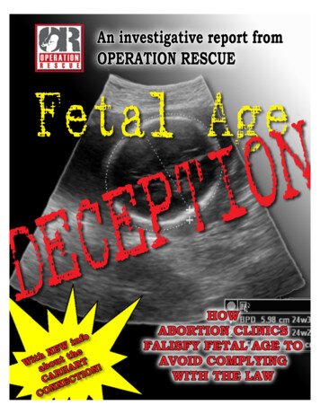
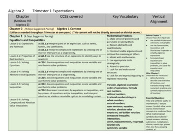
Volpe N et al.Technical issuesPreferred time of evaluation The first-trimester screening for chromosomal abnormalities has been developed for fetuses with the crown-rumplength (CRL) between 45 mm and 84 mm (11 0 to 13 6weeks).[1] Fetal growth and development at this stage ofpregnancy are rapid and differences in fetal anatomy during these three weeks are significant. As described, manyfetal structural abnormalities are already detectable during the ultrasound examination at 11 0 to 13 6 weeksof gestation. Therefore, the optimal timing for this scan,for both the technically appropriate measurement ofnuchal translucency and maximum detection of anomalies, was suggested to be 13 weeks of pregnancy, alsobased on the need for the second scan and number ofunsuccessful scans due to non-viable pregnancy.[59–61]However, in the subsequent years, technical advances ofultrasound machines and the implementation of perinatal specialist’s expertise improved the visualization ofanatomical structures showing similar visualization ratesat 12 and 13 weeks of gestation.[62] Moreover, the closerto the end of the first trimester the scan is performed, thelower is the probability of finding the fetus in a supineneutral position, optimal for NT measurement. Therefore,some data[63] suggest an optimum time for nuchal translucency measurement and anatomic evaluation at 12–13weeks.Ultrasound transducers (TA and TV, frequency) High-frequency ultrasound transducers increase the spatialresolution but decrease the penetration of the beam. Theselection of the optimal transducer and their frequencydepends on gestational age, maternal body habitus, theposition of the fetus, and the scanning approach used.Transabdominal transducers with 3–5 MHz are mainlyused; however, while they “penetrate” deeper, their resolution is lower than high frequency probes such as 4–8MHz and those of the transvaginal probe, which are oftencloser to the fetus and operate at higher frequencies,increasing images resolution. The examination is usually performed with grayscale 2Dultrasound. It may be essential to mention that harmonicand speckle-reduction filters may enhance image quality,mainly in patients with increased body mass index orabdominal scars. The use of transvaginal probes should always be considered if the fetus is in suboptimal position, or in case oflow transabdominal images quality. In such cases a transvaginal approach could be offered and performed if thepatient agrees.13 6 weeks were listed and group members were askedto answer the following questions: Should the following anatomical structures bealways evaluated, possibly, or never at the time ofthe first-trimester anatomy scan? Do you suggest one or more planes?Agreement among members was evaluated for eachanatomical structure and scanning plane.The evaluation of structures and planes that shouldalways be evaluated with an agreement among members exceeding 75% are referred to in this document as“recommended” as part of the first-trimester standard ultrasound examination of the fetal anatomy.The evaluation of structures and planes that should bepossibly evaluated with an agreement among membersexceeding 75% are referred to in this document as “suggested” as part of the first-trimester standard ultrasoundexamination of the fetal anatomy.The evaluation of structures and planes that shouldnever be evaluated with an agreement among members exceeding 75% are considered in this document asnot being part of the first-trimester standard ultrasound examination of the fetal anatomy.The same method was applied for the quantitativeassessment. The main fetal anatomical structuresreported in the literature as measurable were listed.Group members were asked if such anatomical structures should always be measured, possibly or never,and on which planes. According to the level of agreement among members, each measurement is referredto as recommended, suggested, or excluded, followingthe same criteria for the qualitative anatomical assessment.The members were asked to vote again after collegial discussion until consensus was obtained if noagreement was reached.First Trimester Examination of the FetalAnatomy in Routine PracticeHead and brainMethodsWith the scope of reaching a consensus among experts,a survey was conducted among group members.The main fetal structures that could be included inan anatomical ultrasound survey between 11 0 and92Perinatal JournalUnder normal conditions, the fetal skull appears as anoval-shaped hyperechoic bony structure. The two hemispheres, similar in size, are separated by a straight, uninterrupted midline echo (interhemispheric fissure) on theaxial planes. The choroid plexuses should fill the two lat-
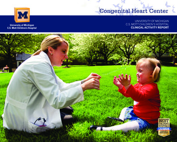
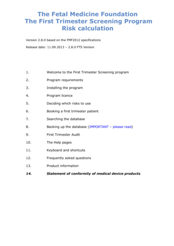
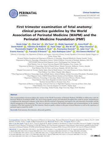
First trimester examination of fetal anatomy: clinical practice guideline by the WAPM and PMFeral ventricles on the sides of the midline (butterfly signon axial view)[64] occupying roughly half or more of theventricle length/area[65,66] (Fig. 1). On the midsagittalview, the anechoic round-shaped diencephalon is visiblein the middle of the fetal brain, and the cranial posterior fossa (CPF) structures are just posterior to it, including the brainstem (BS), the 4th ventricle (4V), and thecisterna magna (CM), appearing as three anechoicspaces, roughly similar in size (Fig. 2). The biparietaldiameter (BPD) could be measured on the axial view inselected cases, mainly for dating purposes.Recommendations Skull and head shape, midline echo, and brain hemispheres,including lateral ventricles and choroid plexuses, should alwaysbe evaluated at the routine first-trimester examination. Thesestructures should be preferably assessed on axial planes. The measurements of the biparietal diameter and headcircumference are not recommended on a routine basisbut could be helpful. The cranial posterior fossa should be evaluated routinelyon the midsagittal plane, showing three distinguishedanechoic spaces similar in size. The measurement of theratio between the width of the brainstem and the spacebehind it (BS/BSOB)[36] is not recommended on a routinebasis but could be helpful when the three spaces seemabnormal. Doppler studies should not be included in the standardevaluation of the fetal brain in the first trimester.Fig. 1. Axial view of the fetal head and brain. The hyperechoic ovalshaped skull is visible. The fetal hemispheres are separated bythe interhemispheric fissure (arrows). Lateral ventricles (*) containing choroid plexuses (C) are also visible.Technical issues The evaluation of the fetal structures requires adequate magnification: the fetal anatomical area, including the targetstructure, should occupy about 75% of the ultrasound image. The axial view of the fetal brain should be obtained withthe ultrasound beam perpendicular to the interhemispheric fissure, appearing as the midline echo, to evaluate its integrity adequately. In addition, the brain hemispheres should be equal in size, witnessing a proper axialrather than oblique approach, and the plane required forthe routine evaluation of the fetal anatomy should be justabove the thalami and midbrain to adequately visualizethe choroid plexuses and ventricles from frontal to occipital horns. An anterior approach could obtain the midsagittal viewof the fetal head (Fig. 2), with the ultrasound beamencountering the fetal face before reaching the intracranial structures. To be correctly midsagittal, the fetal profile should be visible, including forehead, nose (bone,overlying skin, and tip), rectangular-shaped palate, diencephalon, and anechoic structures in posterior fossa (BS,4V and CM). In addition, on a proper midsagittal plane,the bony process above the palate (zygomatic process ofthe maxilla) should not be visible. The nuchal translucency should be measured on this plane when the ultrasound beam is perpendicular to its lines.NeckUnder normal conditions, a thin subcutaneous collectionof fluid should be visible at the level of the fetal neck NT.No lateral cysts, septa or abnormally thick NT should bevisualized.Fig. 2. Midsagittal view of the fetal head and brain. It is possible tovisualize the diencephalon (D) and the cranial posterior fossastructures, including the brainstem (BS), the 4th ventricle (4V),and the cisterna magna (*) appearing as three anechoic spaces, roughly similar in size. The nuchal translucency (NT) is alsovisible as a fluid space behind the fetal neck. The fetal profileis well visible on this view, including the forehead (F), the nose(N), lips (L) and chin (C).Volume 30 Issue 2 August 202293
Volpe N et al.RecommendationRecommendations The NT sh
13Fetal Medicine Unit, Centro Hospitalar de Lisboa Central, Lisboa, Portugal 14Instituto Nacional Materno Perinatal, Lima, Peru 15Department of Obstetrics & Gynecology, Center for Fetal Care & High-Risk Pregnancy, University of Chieti, Chieti, Italy 16Fetal Medicine Unit, University Hospital 12 de Octubre, Madrid, Spain