Transcription
The Journal of ImmunologyNaive and Memory T Cells Induce Different Types ofGraft-versus-Host Disease1Suparna Dutt,* Diane Tseng,* Joerg Ermann,* Tracy I. George,† Yin Ping Liu,*Corrine R. Davis,‡ C. Garrison Fathman,* and Samuel Strober2*The goal of this study was to compare the ability of donor naive and alloantigen-primed effector memory T cells to inducegraft-vs-host disease after bone marrow transplantation in MHC-mismatched irradiated host mice. Purified CD4ⴙ naive(CD62LhighCD44low) T cells and CD4ⴙ effector memory (CD62LlowCD44high) T cells obtained from unprimed donors and donorsprimed to host alloantigens, respectively, were injected into host mice, and the rapidity, severity, and pattern of tissue injury ofgraft-vs-host disease was assessed. Unexpectedly, the naive T cells induced a more acute and severe colitis than the primed memorycells. Whereas the naive T cells expressing CD62L and CCR7 lymph node homing receptors vigorously expanded in mesentericlymph nodes and colon by day 6 after transplantation, the primed memory T cells without these receptors had 20- to 100-fold loweraccumulation at this early time point. These differences were reflected in the significantly more rapid decline in survival and weightloss induced by naive T cells. The primed memory T cells had a greater capacity to induce chronic colitis and liver injury andsecrete IL-2 and IFN- in response to alloantigenic stimulation compared with memory T cells from unprimed donors. Nevertheless, the expected increase in potency as compared with naive T cells was not observed due to differences in the pattern andkinetics of tissue injury. The Journal of Immunology, 2007, 179: 6547– 6554.Graft-vs-host disease (GVHD)3 is a major complication ofallogeneic hemopoietic cell transplantation that iscaused by mature donor T cells contained in the graftfrom untreated normal donors (1). The contribution of differentsubsets of donor T cells to the pathogenesis of GVHD has beenstudied extensively. Donor CD4 T cells induce injurious immuneresponses to alloantigens presented by MHC class II molecules onhost APCs, and donor CD8 T cells induces injurious responses toalloantigens presented by MHC class I molecules (2, 3). DonorCD4 and CD8 T cell responses may predominate in mousemodels of GVHD depending on the donor and host strain combinations involving major and/or minor histocompatibility Ag differences (2). Subsets of naturally occurring regulatory T cells contained in the graft, such as NK T cells and CD4 CD25 T cells,suppress GVHD induced by CD4 and CD8 T cells (4, 5).The role of naive and effector memory donor T cell subsets inthe induction of GVHD has been studied recently using untreatednormal donor mice (6 –9). These subsets can be separated according to the expression of the cell-trafficking molecule CD62L (L-*Department of Medicine, †Department of Pathology, and ‡Department of Comparative Medicine, Stanford University School of Medicine, Stanford University, Stanford, CA 94305Received for publication June 21, 2007. Accepted for publication September10, 2007.The costs of publication of this article were defrayed in part by the payment of pagecharges. This article must therefore be hereby marked advertisement in accordancewith 18 U.S.C. Section 1734 solely to indicate this fact.1This work was supported by grants from the National Institutes of Health (HL58250, HL-57743, and CA-49604) and a Stanford Vice Provost for UndergraduateEducation Faculty Grant.2Address correspondence and reprint requests to Dr. Samuel Strober, Stanford University School of Medicine, Center for Clinical Sciences Research Building, Room2215-C, 300 Pasteur Drive, Stanford, CA 94305-5166. E-mail address:sstrober@stanford.edu3Abbreviations used in this paper: GVHD, graft-vs-host disease; TCD, T cell depleted; BM, bone marrow; LP, lamina propria.Copyright 2007 by The American Association of Immunologists, Inc. 0022-1767/07/ 2.00www.jimmunol.orgselectin), and the T cell activation molecule, CD44 (10). Naive Tcells express the CD62Lhigh CD44low phenotype, whereas the effector memory T cells express the CD62LlowCD44high phenotype(10, 11). The differences in the patterns of receptor expression aredue to the down-regulation of CD62L and the up-regulation ofCD44 on the naive T cell surface after antigenic exposure (10, 11).In normal untreated mice, it is thought that CD4 and CD8 Tcells with the effector memory phenotype are derived from naiveT cells that have been exposed to environmental Ags (12, 13) or byhomeostatic proliferation of naive CD4 T cells (14). It is not clearwhether environmental Ags in the mouse cross-react with mouseMHC and/or minor histocompatibility Ags.Although highly purified naive T cells from untreated normaldonors induced acute lethal GVHD, purified effector memory Tcells from the same donors failed to induce GVHD in irradiatedMHC-mismatched or matched murine hosts used for investigation(6 –9). The failure of the latter subset to induce GVHD may reflectimportant differences in the biology of naive and effector memoryT cells, or may instead reflect the lack of cross-reactivity betweenenvironmental Ags and host histocompatibility Ags. In the absence ofcross-reactivity, effector memory T cells from untreated normal donors would be directed to “third-party Ags” and would fail to specifically recognize host alloantigens unless the donors had been primedto the alloantigens. In a previous study using an MHC-matchedmodel, effector memory donor CD4 T cells isolated from mice undergoing chronic GVHD induced potent GVHD in secondary recipients (15). However, the potency of primed effector memory cells anunprimed effector memory cells was not compared, nor was the distribution and kinetics of tissue injury induced by primed CD4 memory T cells compared with purified donor CD4 naive T cells. NaiveCD4 T cells failed to induce lethal GVHD in the dose rangetested in the MHC-matched strain combination chosen for thelatter study (15). Thus, the CD4 T memory cells were considerably more potent than naive CD4 cells (15).To make a direct comparison of naive and memory T cells in asetting in which naive T cells induce vigorous GVHD, we studied
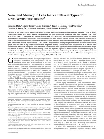
6548MEMORY T CELLS INDUCE GVHDthe ability of purified naive and effector memory CD4 T cellsfrom untreated C57BL/6 (H-2b) donors and from C57BL/6 donorsprimed to BALB/c (H-2d) alloantigens for their capacity to inducelethal GVHD in MHC-mismatched irradiated BALB/c hosts. Wehave previously shown that acute lethal GVHD induced by CD4 T cells in this strain combination is associated with acute colitisthat is dependent upon early migration of the CD4 T cells to themesenteric lymph nodes facilitated by the cell trafficking molecules CD62L and 4 7 integrin (16). Subsequently, the donorCD4 T cells migrate to the colon, and induce severe injury tothe colonic crypts, associated with diarrhea and death.In the current study, we found that CD4 effector memory Tcells from unprimed donors failed to induce lethal GVHD, but thatthe same cell subset from host alloantigen-primed donors induceduniformly lethal GVHD. Surprisingly, the naive T cells induced amore severe and rapid type of GVHD with acute colitis than theprimed memory T cells.indicated in the text and figures. Mice were kept on antibiotic water (25 g/ml neomycin/0.3 U/ml polymyxin B; Sigma-Aldrich) for the first 28days. Survival and the signs of GVHD, hair loss, hunched back, swollenfaces, and diarrhea were monitored daily and body weight was measuredweekly.Materials and MethodsCell preparation and acute GVHD induction were performed as describedabove. For day 6 analysis, lymphocytes from tissues of individual micewere used for flow cytometric analysis. Single-cell suspensions from mesenteric lymph nodes, peripheral lymph nodes (bilateral axillary and inguinal nodes were pooled), and spleen were filtered through fine nitex membranes to remove aggregates. Mononuclear cells from the liver wereisolated according to the method by Lan et al. (18). Lamina propria (LP)lymphocytes were purified as described (19).AnimalsWild-type C57BL/6 (H-2b) male mice, 5– 8 wk old, and male BALB/c(H-2d) mice, 5–10 wk old, were purchased from the breeding facility of theDepartment of Comparative Medicine (Stanford University, Stanford, CA).All mice were housed in a specific pathogen-free facility. Care of allexperimental animals was in accordance with institutional and NationalInstitutes of Health guidelines.Abs and FACSThe following reagents were used for flow cytometric analysis: unconjugated anti-CD16/32 (2.4G2), anti-CD4 FITC (RM4-5), anti-TCR- allophycocyanin (H57-597), anti-CD62L allophycocyanin, anti-CD62L biotin(Mel-14), anti-CD44 PE (IM7), anti-LPAM-1 PE ( 4 7 integrin complex)(DATK32), anti-H-2Kb FITC (AF6-88.5), and anti-CD4 Cy7 allophycocyanin (GK1.5) mAbs. The latter were purchased from BD Pharmingen.Anti-CCR7 biotin (4B12) was purchased from eBioscience. All stainingswere performed in PBS/1% calf serum in the presence of purified antiCD16/32 at saturation to block unspecific staining via FcRII/III. Propidiumiodide (Sigma-Aldrich) was added before analysis to exclude dead cells.All sorts and analyses were done on a modified dual-laser FACSVantage(BD Biosciences) or on the six-color dual-laser FACS LSR in the SharedFACS Facility, Center for Molecular and Genetic Medicine (Stanford University), using FlowJo software (Tree Star) for data analysis.Histopathology and pathologic scoring for GVHD severityHistopathological specimens from the liver and large intestines of hostswere obtained at 7 and 72 days after transplantation and fixed in formalinbefore embedding into paraffin blocks. Tissue sections of 4- to 5- m thickness were stained with H&E. Microscopic images were obtained using anEclipse E1000M microscope (Nikon) with SPOT RT digital camera andacquisition software (Diagnostic Instruments) with a final magnification of 300 for all images. Image processing was performed with Photoshop CS(Adobe) with standard adjustments of brightness, contrast, and color balance to the entire image. Histologic assessment of liver and colonic GVHDwas performed in a blinded fashion using the histopathological scoringsystem described by Kaplan et al. (17). The mean GVHD score representsthe mean SD among five animals per group.Cell distribution studiesCytokine assayCytokine production after 60 h of allogeneic stimulation was assessed insupernatants of cultures of sorted naive and effector memory CD4 T (1 105 cells) from primed and unprimed C57BL/6 donors as responders andirradiated (3000 cGy) Thy1.2-depleted BALB/c splenocytes as allogeneicstimulator cells (5 105 cells). Five different cytokines were analyzed ina multiplex assay system using fluorescently labeled microsphere beads(Beadlyte Mouse Multi Cytokine Detection system; Upstate Biotechnology) and cytokine levels were quantitated using the Luminex 100 system.Statistical analysisKaplan-Meier survival curves were made using Prism (GraphPad Software). Statistical differences in animal survival were analyzed by log-ranktest. Differences in donor type T cell recovery in tissues of hosts and cytokine production were analyzed using the two-tailed Student t test. For alltests, p ⱕ 0.05 was considered significant.Cell preparationsResultsSingle-cell suspensions were prepared from spleens, washed twice, andfiltered through a fine nitex membrane. The samples were then enriched forCD4 cells with anti-CD4 magnetic microbeads using the MidiMACSsystem (Miltenyi Biotec). After staining with anti-CD4 FITC, anti-CD62Lallophycocyanin, and anti-CD44 PE, cells were sorted into CD4 CD62LhighCD44low and CD4 CD62LlowCD44high populations on a modified dual-laser FACSVantage (BD Biosciences). For preparation of T celldepleted bone marrow (TCD BM), BM cells were obtained from the femurand tibia, and single-cell suspensions were filtered through nitex membranes. Suspensions were stained with anti-Thy1.2 biotin mAb (5a-8;Caltag Laboratories) and streptavidin-magnetic beads (Miltenyi Biotec),and passed over two consecutive MACS LS-separation columns (MiltenyiBiotec). TCD BM contained 0.01% T cells, as determined by stainingwith anti-TCR- allophycocyanin. Thy1.2-depleted splenocytes were usedas allogeneic stimulator cells.Expression of chemokine receptors and activation molecules onnaive and effector memory CD4 T cellsImmunizationFive-week-old male C57BL/6 mice were immunized by i.p. injection of50 106 splenocytes from age matched male BALB/c mice followed bya dose of 10 106 splenocytes 1 wk later. Four weeks after immunization,splenocytes from the primed C57BL/6 mice were used for further studies.GVHD modelAcute GVHD was induced as described previously (5). In brief, BALB/chosts were lethally irradiated (800 cGy) from a 200 Kv x-ray source andinjected with donor cells via tail vein within 24 h. All mice received 2 106 TCD BM cells for reconstitution with or without CD4 T cells asNaive and effector memory CD4 T cells can be separated according to the expression of CD62L and CD44 surface markers, because the former express the CD62LhighCD44low pattern and thelatter express the CD62LlowCD44high pattern (10). Fig. 1A showsthe flow cytometric analysis of CD62L vs CD44 on gated CD4 Tcells from the spleen of untreated C57BL/6 mice (top panel). Approximately 70% were CD62LhighCD44low and 18% wereCD62LlowCD44high (left upper and right lower quadrants). Thesetwo populations of cells were sorted and reanalyzed for purity. Thesorted cells were 99% pure naive or effector memory type according to the patterns shown in the two lower panels of Fig. 1A.We analyzed the gated naive and effector memory cells fromuntreated (unprimed) and alloimmunized (primed) C57BL/6 micefor the expression of CCR7, 4 7, CD25, CD69, and CXCR3 surface markers as judged by flow cytometry in Fig. 1B. Alloimmunized mice were given 50 106 BALB/c spleen cells i.p. followedby a booster injection of 10 106 spleen cells 1 wk later. Thespleen cells from alloimmunized C57BL/6 mice were harvested 4wk later.Both the unprimed and primed mice showed a narrow peak ofintense staining of the chemokine receptor CCR7 among gated
The Journal of Immunology6549FIGURE 1. Unprimed and primed CD62LhighCD44low and CD62LlowCD44high CD4 T cells express comparable levels of different surface markers. A,Splenocytes from C57BL/6 donors were sorted into CD62LhighCD44low (naive) and CD62LlowCD44high (effector memory) CD4 T cell populations. B,C57BL/6 splenocytes from primed and unprimed animals were stained with anti-CD4, anti-CD44, and anti-CD62L mAbs. Gated CD62LhighCD44low andCD62LlowCD44high CD4 T were analyzed for CCR7, 4 7, CD25, CD69, and CXCR3 expression. Histogram plots of the respective markers are overlaidfor CD62LhighCD44low CD4 T cells (bold line) and CD62LlowCD44high CD4 T (broken line).naive CD4 T cells as compared with the broader peak of lessintense staining of effector memory cells (Fig. 1B). In contrast, theeffector memory T cells from primed and unprimed mice showedboth a dull and bright peak of staining for the activation markerCD69, and the naive cells showed only a dull peak (Fig. 1B). Asimilar bimodal pattern was observed in effector memory T cells,and a single dull or negative peak in naive T cells for the 4 7integrin and CXCR3 chemokine receptors (Fig. 1B). In addition,there was a slight increase in the bright staining of CD25 amongthe effector memory (11.5%) as compared with naive (4%) T cells.In general, the staining patterns were similar using both primedand unprimed mice. The results indicate that CCR7 is down-regulated and 4 7, CD69, and CXCR3 are up-regulated on effectormemory as compared with naive T cells.Ability of naive and effector memory CD4 T cells fromprimed and unprimed donors to induce GVHDPrevious studies have shown that purified naive T cells fromunprimed donors induce lethal GVHD in irradiated allogeneichosts and that purified effector memory cells do not (6 –9). In thecurrent study, we compared the ability of purified effector memoryCD4 T cells from both unprimed C57BL/6 donors and C57BL/6donors primed to host alloantigens for their capacity to inducelethal GVHD in MHC- and minor Ag-mismatched BALB/c hosts.The hosts were conditioned with a single dose of myeloablativetotal body radiation and injected i.v. with 2 106 TCD BM fromunprimed donors and equal numbers (0.125 106) of sorted naiveor effector memory CD4 T cells from primed and unprimed donors 24 h later. The latter cell dose had been previously determinedto be on a sensitive portion of the dose-response curve for inducinglethal GVHD (16). The sorted cells were collected by first gatingon CD4 cells then using the thresholds shown in Fig. 1A to separate CD62LhighCD44low (naive) and CD62LlowCD44high (effectormemory cells). As shown in Fig. 2, naive CD4 T cells fromunprimed donors induce severe GVHD, and all the hosts died byday 65 with clinical signs of GVHD including diarrhea, loss of fur,and weight loss. Hosts given TCD BM cells uniformly survived 100 days, and their body weights returned to pretransplant levelswithin one month.In contrast, effector memory CD4 T cells from unprimed donors induced less severe GVHD and 80% hosts survived for atleast 100 days (Fig. 2A). The difference in survival of hosts givenunprimed naive or effector memory cells was significantly different( p 0.0001). The attenuated GVHD induced by effector memory
6550MEMORY T CELLS INDUCE GVHDFIGURE 2. Naive CD4 T induce a more rapid and severe type of GVHD than primed CD4 effector memory T cells. Lethally irradiated BALB/c hostmice were given i.v. injections of 2 106 TCD BM cells from unprimed C57BL/6 donors with or without CD62LhighCD44low or CD62LlowCD44high CD4 T cells from primed or unprimed donors. The data were pooled from two independent experiments. There were 8 –10 hosts in each group. A, Survival ofirradiated hosts given TCD BM cells from unprimed donors with or without 0.125 106 sorted naive and effector memory CD4 T cells from primed andunprimed donors. B, Percentage of starting body weight of host mice given TCD BM with or without sorted naive and effector memory CD4 T cells asin A. Brackets show SEs of the mean. , Analysis was stopped for a given group when there were two hosts remaining positive.T cells was reflected in the weight loss of hosts which was significantly less severe ( p 0.0001) as compared with the group givennaive T cells (Fig. 2B).BALB/c hosts given TCD BM and sorted naive T cells fromprimed donors developed severe GVHD with marked weight loss,and all hosts died by day 40 (Fig. 2A). There were no significantdifferences ( p 0.742) in survival and weight loss, in the groupsgiven primed or unprimed sorted naive T cells. However, therewere highly significant differences in survival ( p 0.0004) andweight loss ( p 0.0002) in the groups given primed or unprimedsorted effector memory T cells (Fig. 2). Whereas 80% of the hostsgiven unprimed effector memory T cells survived at least 100 days,all hosts given primed effector memory T cells died by 100 days(Fig. 2A). In addition, the serial body weights of hosts givenunprimed effector memory cells were constant over 100 days andsimilar to pretransplant values, but body weights of hosts givenprimed effector memory T cells gradually fell over a period of atleast 80 days (Fig. 2B). The difference of body weights was significant ( p 0.0002). All recipients had complete donor chimerism ( 95%) as detected by staining with anti H-2Db mAb (datanot shown).The marked early weight loss and acute GVHD in hosts givenunprimed naive T cells was reflected in the microscopic changes inthe tissue sections of the colon 7 days after transplantation. Fig. 3Cshows that the colon from hosts given naive T cells from unprimeddonors had a marked dropout of crypts, loss of crypt goblet cells,and a marked mononuclear cell infiltrate between crypts. In contrast, the colon of hosts given unprimed effector memory T cellsshowed no crypt dropout, retention of goblet cells, and little or nomononuclear cell infiltrate (Fig. 3D). The colon of hosts givennaive T cells from primed donors showed a pattern of severe cryptinjury (Fig. 3A). Interestingly, the colon of hosts given effectormemory T cells from primed donors showed minimal injury at day7 with a mild infiltrate and occasional abscesses at the crypt bases(Fig. 3B). The normal appearance of colon sections from controlhosts given irradiation and no transplant (Fig. 3E), and from control hosts given irradiation and TCD BM alone (Fig. 3F) are shownfor comparison. Sections of the liver and skin of all hosts werenormal at day 7 (data not shown).To compare late changes in the histopathology of the liver andcolon in hosts given effector memory T cells from unprimed andprimed donors, tissues were harvested from groups of hosts at 72days after transplantation. Fig. 4A shows that liver from a representative host given effector memory cells from primed donors hada portal infiltrate consisting of lymphocytes, macrophages, andfewer neutrophils. This infiltrate is centered primarily on bile ductswith some clusters adjacent to portal veins. Fig. 4B shows a representative example of a subset of portal tracts that is invested bysmall numbers of mixed inflammatory cells centered primarily onthe bile ducts that was observed in hosts given unprimed effectormemory T cells. The liver of hosts given TCD BM cells showedvery mild infiltrates of lymphocytes, plasma cells, and neutrophilscentered on small bile ductules (Fig. 4C). In Fig. 4D, the colon ofa representative host given primed effector memory cells, showeda hyperplastic mucosa with dilated crypts that contain necrotic celldebris. Few mucin containing goblet cells remained. The LP wasmoderately expanded with neutrophils, lymphocytes, and macrophages. This inflammation extends into the submucosa. In Fig. 4E,the LP of a representative host given unprimed effector memory Tcells is mildly expanded by lymphocytes, plasma cells, and neutrophils. Goblet cells are retained.Fig. 4F shows the colon of host given only TCD BM cells.The colon had a mild diffuse infiltrate of lymphocytes, plasmacells, and neutrophils in the deep LP. Goblet cells were retainedin hosts given unprimed effector memory T cells or no T cells.The mean GVHD histopathologic scores of colon was significantly increased ( p 0.02) in the group given primed effectormemory T cells as compared with groups that receivedunprimed effector memory T cells or no T cells (Fig. 4G). Themean histopathologic score of the liver was higher in the groupgiven primed effector memory cells as compared with thegroups given no T cells or unprimed effector memory T cells(Fig. 4G). However, the difference was at the border of statistical significance ( p 0.056). Hosts with primed or unprimedeffector memory T cells had no microscopic abnormalities ofGVHD in the skin (data not shown).Accumulation of naive and effector memory CD4 T cells fromprimed and unprimed donors in host tissues at day 6Our previous studies have shown that the acute colitis associatedwith lethal GVHD in BALB/c hosts given C57BL/6 TCD BM andsorted CD4 T cells was dependent on the ability of the donorT cells to migrate to the mesenteric lymph nodes by day 2 aftertransplantation (16). The rapid nodal migration preceded the
The Journal of ImmunologyFIGURE 3. Histopathologic changes of acute severe colitis inducedwith CD4 T naive T cells but not CD4 T effector memory T cells.Representative tissue sections were obtained from hosts that received0.125 106 CD62LhighCD44low or CD62LlowCD44high CD4 T cells fromprimed or unprimed donors and 2 106 TCD BM cells from unprimeddonors. A, Colon of a host given naive CD4 T cells from primed donors.Severe acute graft vs host disease is present with numerous crypt abscesses(ⴱ) and erosion with exudate on surface of mucosa. Arrow highlights theaccompanying mononuclear cell infiltrate. B, Colon of a host given primedeffector memory CD4 T cells. A mild inflammatory infiltrate separatescrypts which are lined by goblet cells with abundant mucin. A focal abscess(ⴱ) is shown in one crypt. C, Colon of a host given unprimed naive CD4 T cells. Crypts are shortened and separated by a prominent inflammatorycell infiltrate composed of lymphocytes and plasma cells (arrows). ⴱ, Cryptabscess and there are decreased numbers of goblet cells lining crypts. D,Colon of a host given unprimed effector memory CD4 T cells. Gobletcells are retained in crypt walls with normal architecture and minimal inflammatory cell infiltrate. E, Colon of an irradiated control host showingnormal appearing colonic mucosa and scant inflammatory infiltrate. F, Colon of a control host given only TCD BM cells showing unremarkablecolon with back-to-back crypts containing numerous goblet cells. Inflammatory infiltrate is minimal. Tissue sections were stained with H&E. Eachpanel is representative of three hosts examined.accumulation of donor T cells in the colon on day 6. Targetedinactivation of genes encoding both CD62L and 4 7 trafficking molecules of donor T cells prevented nodal and colonicaccumulation (16). Sorted effector memory CD4 T cells havereduced expression of both CD62L and CCR7 and increasedexpression of 4 7 receptors as compared with sorted naiveCD4 T cells (Fig. 1).Because it is unclear how these changes affect migration tothe mesenteric lymph nodes and colon, we compared the accumulation of the sorted CD4 T cells from primed and unprimeddonors in the host tissues at day 6. Fig. 5 compares the accumulation of total CD4 T cells to that of the sorted naive andeffector memory CD4 T cells from untreated donors. Accumulation of the total CD4 T cells was not significantly different ( p 0.05– 0.3) from that of sorted naive CD4 T cellsfrom primed or unprimed donors in spleen, peripheral lymphnodes, mesenteric lymph nodes, colon, and liver (Fig. 5). Incontrast, the accumulation of sorted unprimed effector memoryCD4 T cells was 20-fold reduced in the spleen ( p 0.01)6551FIGURE 4. Histopathologic changes of tissue injury are increased 72days after transplantation of primed vs unprimed CD4 effector memory Tcells. Representative tissue sections were obtained from hosts that received0.125 106 CD62LlowCD44high CD4 T cells from primed or unprimeddonors and 2 106 TCD BM cells from unprimed donors. A, Liver of hostgiven primed effector memory CD4 T cells. There is a portal infiltrate(arrows). B, Liver of host given unprimed effector memory CD4 T cells.The arrows indicate mild portal infiltrates. C, Liver of host given TCD BM.Occasional small clusters of inflammatory cells surround small bileductules (arrow). D, Colon of host given primed effector memory CD4 Tcells. The mucosa is hyperplastic, and LP has heavy cell infiltrate (arrowheads). Numerous crypts are dilated and contain necrotic cell debris (ⴱ).Apoptotic cells are frequent (arrows). E, Colon of host given unprimedeffector memory cells. The LP is mildly infiltrated (arrowheads) The arrows indicate apoptotic cells; ⴱ, degenerate cell debris. Goblet cells areretained. F, Colon of host given TCD BM cells. The LP has mild infiltrate(arrowheads). Crypts contain some necrotic cell debris (ⴱ). Occasional apoptotic cells are found (arrows). Light microscopy, H&E stain. Each panelis representative of five hosts examined. G, Mean ( SD) of histopathologic GVHD scores of liver and colon from the three groups (n 5) pergroup.and 100-fold reduced in the peripheral and mesenteric lymphnodes, colon, and liver as compared with total CD4 T cells( p 0.01– 0.001) (Fig. 5). Interestingly, the accumulation ofprimed effector memory CD4 T cells was not significantlydifferent ( p 0.07– 0.78) from that of unprimed effector memory CD4 T cells except in the liver. In the liver, the accumulation of primed cells was 100-fold higher than that ofunprimed cells ( p 0.02).
6552FIGURE 5. Absolute number of donor T cells in the spleen, mesentericlymph nodes, peripheral lymph nodes, liver, and colon of irradiated hostsday 6 after the injection of 0.5 106 total CD4 T cells orCD62LhighCD44lowCD4 T cells or CD62LlowCD44highCD4 T cells.Bars show the means of the absolute number of donor T cells, and bracketsshow SEs of groups of mice given T cells from primed or unprimed donors.Means are from two separate experiments, with three mice in each experiment. #, The absolute number of donor T cells was 0.001 106.ⴱ, Statistically significant difference between primed and unprimed group(p ⱕ 0.05). All hosts received 2 106 wild-type (WT) TCD BM cells.Comparison of immune responses of sorted donor naive andeffector memory cells to host allostimulation in vitroThe potency of donor T cell induction of GVHD in vivo is dependent upon both the magnitude of the immune response to hostalloantigens, and the ability of the donor T cells to migrate to hosttissues to encounter alloantigens and migrate to subsequent sites oftissue injury. To examine the magnitude of the alloresponse ofdonor T cells to host stimulation in absence of the cell migration,we incubated the sorted responder CD4 T cell subsets fromprimed and unprimed donors with host stimulator cells in the MLR(16). In all instances, a constant number of sorted C57BL/6 donorcells (1 105) were incubated with a constant number of irradiated BALB/c host splenocytes (5 105), and the concentrations ofIL-2 and IFN- were measured in the supernatant 60 h later.Control cultures contained syngeneic donor stimulator cells.Fig. 6 shows that sorted naive CD4 T cells from unprimed andprimed donors secreted mean concentrations of IL-2 that wereFIGURE 6. Cytokine responses of unprimed andprimed C57BL/6 donor naive or effector memory CD4 T cells to host stimulator cells in the MLR. A, Mean SEM concentrations of IL-2; B, mean SEM concentrations of IFN- in the supernatants of triplicate MLRcul
fector memory T cells express the CD62LlowCD44high phenotype (10, 11). The differences in the patterns of receptor expression are due to the down-regulation of CD62L and the up-regulation of CD44 on the naive T cell surface after antigenic exposure (10, 11). In normal untreated mice, it is thought that CD4 and CD8 T