Transcription
The Sensitivity of Children to Electromagnetic FieldsLeeka Kheifets, Michael Repacholi, Rick Saunders and Emilie van DeventerPediatrics 2005;116;303-313DOI: 10.1542/peds.2004-2541This information is current as of May 17, 2006The online version of this article, along with updated information and services, islocated on the World Wide Web 2/e303PEDIATRICS is the official journal of the American Academy of Pediatrics. A monthlypublication, it has been published continuously since 1948. PEDIATRICS is owned, published,and trademarked by the American Academy of Pediatrics, 141 Northwest Point Boulevard, ElkGrove Village, Illinois, 60007. Copyright 2005 by the American Academy of Pediatrics. Allrights reserved. Print ISSN: 0031-4005. Online ISSN: 1098-4275.Downloaded from www.pediatrics.org at Sarasota Memorial Hospita on May 17, 2006
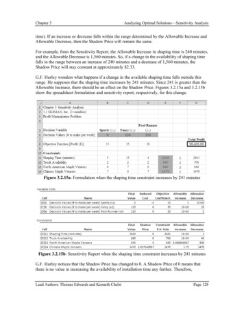
SPECIAL ARTICLEThe Sensitivity of Children to Electromagnetic FieldsLeeka Kheifets, PhD*; Michael Repacholi, PhD‡; Rick Saunders, PhD‡; and Emilie van Deventer, PhD‡ABSTRACT. In today’s world, technologic developments bring social and economic benefits to large sections of society; however, the health consequences ofthese developments can be difficult to predict and manage. With rapid advances in electromagnetic field (EMF)technologies and communications, children are increasingly exposed to EMFs at earlier and earlier ages. Consistent epidemiologic evidence of an association betweenchildhood leukemia and exposure to extremely low frequency (ELF) magnetic fields has led to their classification by the International Agency for Research on Canceras a “possible human carcinogen.” Concerns about thepotential vulnerability of children to radio frequency(RF) fields have been raised because of the potentiallygreater susceptibility of their developing nervous systems; in addition, their brain tissue is more conductive,RF penetration is greater relative to head size, and theywill have a longer lifetime of exposure than adults. Toevaluate information relevant to children’s sensitivity toboth ELF and RF EMFs and to identify research needs,the World Health Organization held an expert workshopin Istanbul, Turkey, in June 2004. This article is based ondiscussions from the workshop and provides background information on the development of the embryo,fetus, and child, with particular attention to the developing brain; an outline of childhood susceptibility to environmental toxicants and childhood diseases implicatedin EMF studies; and a review of childhood exposure toEMFs. It also includes an assessment of the potentialsusceptibility of children to EMFs and concludes with arecommendation for additional research and the development of precautionary policies in the face of scientificuncertainty. Pediatrics 2005;116:e303–e313. URL: www.pediatrics.org/cgi/doi/10.1542/peds.2004-2541; children,environmental risk, policies, sensitive periods, mobilephones, electromagnetic fields, power lines.ABBREVIATIONS. ELF, extremely low frequency; IARC, International Agency for Research on Cancer; RF, radio frequency; EMF,electromagnetic field; WHO, World Health Organization; CNS,central nervous system; ALL, acute lymphoblastic leukemia;AML, acute myeloblastic leukemia; SAR, specific absorption rate.From the *Department of Epidemiology, University of California School ofPublic Health, Los Angeles, California; and ‡Radiation and EnvironmentalHealth, World Health Organization, Geneva, Switzerland.Accepted for publication Feb 2, 2005.doi:10.1542/peds.2004-2541The opinions in this paper are the sole responsibility of the authors and donot represent the position of the World Health Organization.Conflict of interest: Dr Kheifets worked and consulted for Electric PowerResearch Institute and Utilities (Palo Alto, CA), and Dr Saunders workedfor the National Radiation Protection Board (Oxfordshire, United Kingdom).Address correspondence to Leeka Kheifets, PhD, Department of Epidemiology, UCLA School of Public Health, 73-284 CHS, 50 Charles E. Young DrS, Los Angeles, CA 90095-1772. E-mail: kheifets@ucla.eduPEDIATRICS (ISSN 0031 4005). Copyright 2005 by the American Academy of Pediatrics.Children in both industrialized and developingcountries are exposed to a large variety ofenvironmental agents including indoor andoutdoor air pollution, water and food contaminants,chemicals (eg, pesticides, lead, mercury), and physical agents such as ultraviolet radiation and excessivenoise. Changes in exposure to these agents are beinglinked to real or perceived increases in the incidenceof certain childhood diseases, such as asthma, leukemia, and brain cancer, and in some behavioral andlearning disabilities. Environmental exposures canbe particularly harmful to children because of theirspecial vulnerability during periods of developmentbefore and after birth.Exposure to electric and magnetic fields from 0 to300 GHz has been increasing greatly as countriesincrease their capacity to generate and distributeelectricity and take advantage of the many new technologies, such as telecommunications, to improvelifestyle and work efficiency (Fig 1). Evidence of anassociation between childhood leukemia and exposure to extremely low frequency (ELF) magneticfields has led to their classification by the International Agency for Research on Cancer (IARC) as a“possible human carcinogen”1 based on consistentepidemiologic data and lack of support by laboratorystudies in animals and cells. The reason why theresults of the childhood leukemia studies are consistent is still being investigated, but one possibility isthat children may be more sensitive to radiation insome or all parts of the electromagnetic spectrum.Concerns about the potential vulnerability of children to radio frequency (RF) fields from mobile telephony were first raised by an expert group in theUnited Kingdom2 on the grounds that children havea longer lifetime of exposure than adults, and from aphysiologic point of view, they have a developingnervous system, their brain tissue is more conductivethan that of adults because it has a higher watercontent and ion concentration, and they have greaterabsorption of RF energy in the tissues of the head atmobile telephone frequencies. This topic was discussed further at a European Cooperation in theField of Scientific and Technical Research (COST) 281workshop,3 in a report of the Health Council of theNetherlands,4 and in a recent report from the UnitedKingdom’s National Radiological Protection Board.5To evaluate the available information relevant tochildren’s sensitivity to electromagnetic fields(EMFs) and to identify research needs, the WorldHealth Organization (WHO) held an expert workshop in Istanbul, Turkey, in June 2004. This article isbased on discussions and recommendations from theworkshop and provides background information 1PEDIATRICS Vol. 116 No. 2 August 2005Downloaded from www.pediatrics.org at Sarasota Memorial Hospita on May 17, 2006e303
Fig 1. Electromagnetic spectrum. VDUs indicates video display units.the development of the embryo, fetus, and child,with particular attention to the developing brain; anoutline of childhood susceptibility to environmentaltoxicants, childhood diseases implicated in EMFstudies, and exposure to ELF and RF fields, with afocus on children. After a brief presentation of theEMF science most pertinent to effects on childrenand a review of several proposed mechanisms, thepotential sensitivity of children to EMFs is discussed.Finally, recommendations are outlined on the protection of children through the development of precautionary approaches in the face of scientific uncertainty.FROM EMBRYO TO ADOLESCENCEEmbryo, Fetal, and Childhood DevelopmentDevelopment proceeds from conception to adulthood through a number of different stages in whichthe developmental processes are markedly different,and their susceptibility to environmental teratogensvaries. The prenatal period of development is divided roughly into 3 periods: the preimplantationperiod, extending from fertilization to the settling ofthe embryo into the uterine wall; a period of organogenesis, characterized by the formation of themain body structures; and the fetal period, duringwhich growth of the structures already formed takesplace. Additional developmental changes take placeafter birth. Postnatal changes are characterized byslower growth and maturation of existing organ systems, notably the central nervous system (CNS), thehemopoietic and immune systems, the endocrineand reproductive systems, and the skeletal system.The completion of sexual development at the end ofthe second or the beginning of the third decade ofhuman life marks the completion of this period ofgrowth and maturation. Essentially, however, thenature of the toxicant and the timing and magnitudeof exposure determine the risk of any adverse effectsin terms of both severity and occurrence. Vulnerabile304ity can vary quite rapidly during the prenatal period,whereas slower changes occur postnatally.6During the first 2 weeks of embryonic development (known as the “all-or-none period”), the embryo is very sensitive to the lethal effects of toxicagents and much less sensitive to the induction ofmalformation. Many of the cells are still omnipotential stem cells, and if the embryo survives a toxicexposure it can recuperate without an increased riskof birth defects or growth retardation. During thenext 6 to 8 weeks of development, major organogenicevents occur and toxic agents with teratogenic potential can cause major malformations of the visceralorgans, the CNS, the face, and the limbs. From the8th to the 15th week, neuron proliferation, differentiation, and migration in the CNS are particularlyvulnerable.7 Genitourinary and other malformations,gonad cell depletion, and neurodevelopmental problems may occur if the thresholds for these effects areexceeded. During the late fetal period, effects ongrowth of the fetus and susceptible organs such asthe CNS diminish, but vulnerability to deleteriouseffects remains high compared with adults.Development continues after birth, but now thisprocess largely entails the maturation of existing organ systems, although growth is still occurring. Neurobiologists long believed that neurogenesis in thehuman ends during the first months of postnatal life,but recent rodent and primate studies demonstratethat there is lifelong neuron production in someparts of the CNS.8 However, with some particularexceptions, most adult neurons are already producedby birth. The number of connections (synapses) between neurons in the human brain peaks at 2 yearsand decreases by 40% to the adult number duringadolescence8 as experience is acquired and “redundant” connections lost. This reflects the balance between the formation of new synapses (synaptogenesis) and synapse elimination, a “pruning” back ofexcess synapses between neurons, which are keySENSITIVITY OF CHILDREN TO ELECTROMAGNETIC FIELDSDownloaded from www.pediatrics.org at Sarasota Memorial Hospita on May 17, 2006
processes in the development of the postnatal “hardwiring” of the brain. Another important neurologicevent that occurs postnatally is myelination, whichfacilitates the transmission of information within theCNS and occurs most rapidly from birth to 24months but may also continue into the second decade. Unfortunately, the susceptibility of these processes to environmental agents has not been studiedextensively and thus is not well understood. However, because developmental processes are vulnerable to disruption by agents that may not be toxic tomature systems, it is reasonable to expect that thelater stages of brain development present specialrisks.8Other threshold effects that can result from postnatal exposures include interference with fertilityand endocrine function, alterations in sexual maturation, and interference with the development of theimmune system. Endocrine disrupters, exogenoussubstances that mimic the action of hormones (particularly steroids), may alter the function of the developing endocrine system and have adverse effectson the reproductive organs, liver, kidney, adrenalglands, CNS, immune system, cardiovascular system, and bones.9Exposure to toxic agents with mutagenic and carcinogenic potential, such as ionizing radiation, cancer chemotherapeutic drugs, and some chemicals,poses theoretical, stochastic risks for the induction orprogression of cancer during embryonic and childhood development. However, although many agentshave been alleged to be responsible for cancer andgenetic disease, such effects will only result fromagents that have either mutagenic properties or theability to produce more subtle effects on carcinogenicprocesses, such as the stimulation of excessive cellproliferation or an influence on cell-to-cell communication, apoptosis, or DNA repair.males exposed in utero developed masculine behaviors, and males showed exaggerated male matingbehaviors. These observations suggest that thesechemicals masculinized by mimicking steroid hormones or by changing hormone levels.Of perhaps more specific interest are toxic exposures that affect the nervous system of the fetus,infant, and child. Because development of the nervous system is very specific in pattern and timing,exposure to various agents at critical periods of development can cause long-lasting or permanent injury. For instance, exposure to ethanol or methylmercury has been shown to affect neuronal proliferationin rodents and in other experimental models. Someagents such as ethanol, lead, methylmercury, andsome pesticides seem to affect synaptogenesis. Eachof the multiple processes of neural development hasbeen shown to be affected by specific toxic agents,often at low doses but at critical periods of development.The timing of exposure might be critical as well:for ionizing radiation, excess risk for leukemias andbrain and thyroid cancer is higher for exposures thatoccur in childhood; the risk of breast cancer washighest for Japanese women exposed to ionizing radiation from the atomic bomb during puberty, although the risk also increased in women who were 10 years old (an age at which girls have little or nobreast tissue) at the time of the explosion.11 Similarly,sunburns in childhood seem to be particularly potentin increasing the risk of skin cancer later in life.12Exposure in childhood may also increase the risk ofdisease later in life simply because the duration ofexposure can be much longer if it starts early. Thereis evidence, for instance, that the younger a person iswhen starting smoking, the higher the risk of lungcancer.13Children’s Susceptibility to Environmental ExposuresSome diseases are limited to the embryo, child, oradolescent; other diseases that occur in children andadults manifest themselves differently in children.Of particular relevance to EMF exposure are childhood leukemia and brain cancer. There is consistentevidence from epidemiologic studies of a risk ofchildhood leukemia associated with exposure to environmentally high levels of ELF magnetic fields.There is no explanation for this effect from laboratory studies. An increased risk of brain cancer hasbeen investigated in relation to ELF exposures andhas been raised particularly in the context of mobilephone use and the absorption of RF signals by thebrain, although there is no convincing evidence suggesting an increased risk. To put potential EMF effects in perspective and determine how EMFs mightbe involved in the development of these diseases, weprovide a brief overview of rates and risk factors forthem.Several aspects of exposure and susceptibility warrant a focus on children. In some exposure scenarios,children may receive higher doses than adults, resulting from higher intake and accumulation or differences in behavior. Greater susceptibility to sometoxicants and physical agents has been demonstratedin children. Because the period from embryonic lifeto adolescence is characterized by growth and development, deleterious effects can occur at lower levelsand be more severe or lead to effects that do notoccur in adults; on the other hand, children can bemore resilient because of better recuperative capacities.Toxic exposures in utero have produced effectsthat are quite surprising, given the period or level ofexposure. Cassidy et al10 reported that exposure tothe persistent organochlorine chlordane in utero atquite low levels causes significant long-term alterations in sexual behavior. These effects were evidentat levels of exposure very similar to those experienced in homes in the United States when chlordaneand heptachlor were universally applied as termiticides. Both of these chemicals produced markedchanges in sexually dimorphic functions in rats; fe-Childhood Diseases Relevant to EMF ExposureChildhood LeukemiaLeukemias are the most common cancer to affectchildren, accounting for 25% to 35% of all childhoodmalignancies. The biological heterogeneity of childhood leukemia is well documented; the major 541Downloaded from www.pediatrics.org at Sarasota Memorial Hospita on May 17, 2006e305
phologic types are acute lymphoblastic leukemia(ALL) and acute myeloblastic leukemia (AML).The rate of leukemia for children 15 years old hasbeen estimated to be 4 per 100 000 per year in thedeveloped world and 2.5 per 100 000 per year in thedeveloping world.14 In developed countries, the incidence of leukemia rises rapidly after birth, peakingat 3 years of age before declining and then risingsteadily again throughout life. Thus, unlike manycancers, it has a short latency and a peak incidenceearly in life15 that has resulted in many etiologichypotheses, most notably those involving exposureto infections.16Subtypes of AML and ALL are frequently characterized by genetic alterations, including changes inchromosome number (hyperdiploidy or hypodiploidy) and chromosomal translocations that may involve chimeric or fusion genes.17,18 These genes include MLL, TEL, and AML1, all of which can fusewith many other genes and, in the case of TEL andAML1, with each other. There is strong evidence thatthis rearrangement may originate in utero, supported by data obtained from studies of identicaltwins or children with concordant ALL. Screening ofnewborn blood samples suggests that 1% have theTEL-AML1 gene fusion, 100 times the proportion ofchildren that will develop ALL with a TEL-AML1gene fusion before the age of 15 years. This impliesthat the conversion of the preleukemic clone to overtdisease is low and that development of childhoodALL is a multistep process requiring at least 1 prenatal event in combination with additional prenataland/or postnatal events. Although the “first hit,” theinitiating in utero event, is believed to be common,the “second hit,” possibly occurring postnatally, israre and therefore acts as the rate-determining stepin development of the disease.As with most other cancers, the mechanism bywhich leukemia arises is likely to involve gene-environment interactions, the environmental exposuresbeing derived from both endogenous and exogenoussources. Accordingly, it is important to identify exposures that either cause DNA damage and inducechromosome breaks that are repaired inadequatelyor act as promoters and/or progressers, ultimatelyleading to the overt expression of the disease. Exposures acting before birth and early in life have longbeen thought to be important determinants of leukemia; it is unfortunate that the evidence regarding themajority of suggested exposures is limited and oftencontradictory. Ionizing radiation given at large dosesis one of the few known risk factors for leukemia.Brain CancerCNS tumors account for 20% of all malignanciesin children 15 years old19 but account for 2% ofcancers in adults. CNS cancers in children occur intissues of mesodermal or embryonic origin, but inadults they occur in epithelial tissues. Another difference between childhood and adult tumors is thatadult tumors tend to occur in the cerebral hemispheres, whereas the majority of pediatric tumors arebrainstem gliomas.The international incidence rates of childhoode306CNS tumors (0 –14 years) vary between developedand developing nations, with the higher rates observed in most Westernized countries reaching 3 per100 000 per year compared with 1 to 2 per 100 000 inother parts of the world.19 Over recent decades,steady rises in the incidence of childhood CNS tumors have been observed in several populations ofthe United Kingdom, the United States, Japan, andAustralia. The debate continues over whether theseincreases are “real” or an artifact of improved diagnostic practice and case finding by cancer registries.The causes of CNS cancers are largely unknown,although up to 5% may be explained by geneticpredisposition, associated with disorders such asneurofibromatosis type I.20,21 Having a parent or sibling with a CNS tumor also increases the risk. Theidentification of environmental risk factors for CNStumors has generally been inconsistent.20,21 Again,ionizing radiation given in therapeutic doses is oneof the few known risk factors for CNS tumors.CHILDRENⴕS EXPOSURE TO RF AND ELF FIELDSIn evaluating the potential role of environmentalexposures in the development of childhood diseases,it is important to consider not only the fact thatchildhood exposures can be different from exposuresduring adulthood but also the fact that they can behighly age dependent. Exposures of interest duringthe preconception and gestation periods include residential and parental exposures to ELF and RF fields,including mothers’ exposure from use of domesticappliances and mobile phones. Infants and toddlersare exposed mostly at home or at day care facilities.Among preteens, exposure sources expand to include mobile-phone use and sources at school, withan increased use of mobile phones in adolescence.Here we focus on 2 major exposure scenarios: residential ELF and RF exposures and exposure frommobile phones.Residential ExposureEveryone is exposed to ELF electric and magneticfields at home.22 High-voltage power lines are a major source of exposure for children who live nearthem; however, only 1% of children live in closeproximity to high-voltage lines. For most children,exposure to low-level fields from primary and secondary distribution wiring is continuous; short-duration and intermittent exposure to higher fields results from proximity to domestic appliances. ELFexposure also occurs at school, during transport, andeven during mobile-phone use. Typical averagemagnetic fields in homes seem to be 0.05 to 0.1 T.Generally, magnetic fields in homes vary from country to country; geometric-mean fields are 35 nT inthe United Kingdom and 70 nT in the United States.This difference results from the supply voltage usedin the United States (110 V) being approximately halfthat used in the United Kingdom (220 V), leading toapproximately twice the electric current and magnetic field exposure. The fraction of homes with average fields above certain thresholds likewise varies;for example, 1% to 2% of homes in the United Kingdom and 10% in the United States have fields of 0.2SENSITIVITY OF CHILDREN TO ELECTROMAGNETIC FIELDSDownloaded from www.pediatrics.org at Sarasota Memorial Hospita on May 17, 2006
T. Exposure to appliances has been estimated to be30% of total exposure. Maximum fields experiencedare typically in the tens of microtesla. There is evidence that younger children use appliances less (andspend less time outside the home), so their personalexposure is closer to and correlates better with thefields in the home.RF fields are produced by radio and televisionbroadcasts, mobile phones and base stations, andother communications infrastructure. Radio and television signals are broadcast to a large area fromcomparatively few sites. Mobile-phone base stationscover a smaller area and produce much lower emissions but are now much more common than radioand television stations (tens of thousands in manycountries). Because of the width and angle of the RFsignal beam and perturbation by the earth and building materials, there is little correlation between fieldstrength and distance to the source. Typical powerdensities outdoors would be 0.01 to 1 mW · m 2 butcould be orders of magnitude higher (ie, ⱖ100 mW ·m 2). Depending on where the measurements aretaken, base stations can be the largest individualsource of RF fields, but other sources such as radio ortelevision transmitters can result in comparable orgreater exposures. Indoor levels are often lower byorders of magnitude, because buildings screen fields.A European median indoor power density of 0.005mW · m 2 has been reported.Background environmental levels are the primarysource of RF exposure for very young children. Potential sources of residential RF exposure to childrenare wireless in-house communications (eg, wirelessmonitors used in children’s cribs, cordless phones,Wi-Fi) and mobile-phone use by someone in closeproximity to a child, creating passive exposure. Because children 5 years of age usually spend most oftheir time at home, residential exposure can be asufficient predictor of individual exposure.22,23 RFexposure may be estimated more easily for childrenthan for adults, because the variety of exposuresources is smaller. When they reach adulthood, today’s children will have a much higher cumulativeexposure to RF fields than today’s adults.At present, population exposure to RF fields hasbeen much less characterized than ELF fields, partlybecause of technical challenges (lack of adequatemeasuring equipment), the rapid evolution of mobile-phone technology (frequency, coding schemes),and new patterns of use (duration of calls, shortmessage services). However, the main reason ELFfields are better understood than RF fields is thatthey have been studied more.Mobile-Phone UseModern children will experience a longer period ofexposure to RF fields from mobile-phone use thanadults, because they started using mobile phones atan early age and are likely to continue using them.Data from a multinational case-control study of potential causes of adult brain cancer show that boththe prevalence of regular mobile-phone users anddaily use are highest in the younger age groups (eg,19% of younger subjects made calls for 30 minutesa day, compared with 10% of older subjects).24,25Moreover, several recent trends (such as increasedpopularity, reduced price, and advertising to children) have led to increased mobile-phone use amongchildren.26 A steep increase in mobile-phone ownership among children has been reported in severalpublic-opinion surveys.27 For example, in Australia 90% of 6- to 9-year-olds reported sometimes usingtheir parents’ mobile phones, and in Germany approximately one third of 9- to 10-year-olds reportedowning a mobile phone. Clearly, mobile phones arethe dominant source of RF exposure for teens andpreteens.HEALTH-RISK ASSESSMENTThe workshop addressed the potential sensitivityof children at all stages of development from conception through to sexual maturity. The nature of anyadverse health effect that ensues from exposure to anenvironmental toxicant depends not only on the timing and magnitude of the exposure but also on themechanisms by which the toxicant interacts with thedeveloping tissue or organ. As a consequence, it isnot possible to generalize about the possible healtheffects that might ensue from exposure to an agentposing unknown risks to health by drawing parallelswith other toxic agents unless they have very similarmechanisms of interaction. Instead, it is necessary toexamine the experimental and epidemiologic evidence by formulating and testing hypotheses on thebasis of an examination of the known and possibleinteraction mechanisms.Health Risks to Children From ELF FieldsExposure to ELF EMFs induces electric fields andcurrents within the body; guidance on exposure isbased on avoiding the risks to health that result fromthe interaction of the induced fields and currentswith electrically excitable nerve tissue, particular thatof the CNS (see, for example, refs 28 and 29). Presentguidance on occupational exposure is based on abasic restriction on induced current density in theCNS of 10 mA · m 2, which approximates an electricfield in CNS tissue of 100 mV · m 1. Guidance onpublic exposure incorporates an additional safetyfactor, reducing the basic restriction to 2 mA · m 2(20 mV · m 1). The basic restrictions are linked toexternal field strengths (reference levels) through dosimetric calculation, which is based on realistic anatomic human models and measurements of the dielectric properties of human tissue. For generalpublic exposure, the corresponding reference levelsfor power-frequency electric and magnetic fields areof the order of 5 kV/m and 100 T, respectively.Dosimetric calculations have not been conductedextensively for children and have not been undertaken for pregnant women and their unborn children. In general, adults exposed to ELF electric ormagnetic fields have higher internal electric-fieldstrengths and current densities than children becauseof size and shape differences. However, the distributions are different, and in children some tissues havehigher field strengths and current densities for thesame external field. Furthermore, children have 541Downloaded from www.pediatrics.org at Sarasota Memorial Hospita on May 17, 2006e307
nificantly higher internal field strengths and currentdensities from contact currents than do adults. Dosecomputations using anatomically correct models ofchildren30 reveal that modest, imperceptible currentinto the hand (10 A) produces 50 mV · m 1 averaged across the lower-arm marrow of a small childand approximately ⱖ130 mV · m 1 in 5% of thattissue. During pregnancy, the magnitude and distribution
and trademarked by the American Academy of Pediatrics, 141 Northwest Point Boulevard, Elk publication, it has been published continuously since 1948. PEDIATRICS is owned, published, PEDIATRICS is the official journal of the American Academy of Pediatrics. A monthly Downloaded from www.pediatrics.org at Sarasota Memorial Hospita on May 17, 2006


