Transcription
Nicu et al. BMC Oral Health (2018) EARCH ARTICLEOpen AccessCharacterization of oral polymorphonuclearneutrophils in periodontitis patients: acase-control studyElena A. Nicu1,2*† , Patrick Rijkschroeff1†, Eva Wartewig1, Kamran Nazmi3 and Bruno G. Loos1AbstractBackground: Maintaining oral health is a continuous and dynamic process that also involves the immune system.Polymorphonuclear neutrophils (PMNs) migrate from blood circulation and become apparent in the oral fluid.Controversies exist regarding the specific role of the oral PMNs (oPMNs) in the presence of chronic oral inflammation,such as periodontitis. In this study we characterized cell counts, activation status, apoptosis, and reactive oxygenspecies (ROS) generation by oPMNs and circulatory (cPMNs), and the salivary protease activity, in subjects withand without periodontitis.Methods: Venous blood and oral rinse samples were obtained from 19 patients with untreated periodontitis and 16control subjects for PMN isolation. Apoptosis and expression of cell activation markers CD11b, CD63, and CD66b wereanalyzed using flow cytometry. Constitutive ROS generation was detected using dihydrorhodamine123. Additionally,ROS production in response to stimulation was evaluated in samples incubated with 10 μM phorbol myristate acetate(PMA) or Fusobacterium nucleatum. Total protease activity was measured using substrate PEK-054.Results: Periodontitis patients presented with over 4 times higher oPMN counts compared to controls (p 0.007),which was a predictor for the total protease activity (r2 0.399, P 0.007). More oPMNs were apoptotic in periodontitispatients compared to the controls (P 0.004). All three activation markers were more expressed on the oPMNs comparedto the cPMNs (p 0.05), and a higher expression of CD11b on the oPMNs from periodontitis patients was observedcompared to the control subjects (P 0.024). Constitutive ROS production per oPMN was higher compared to the cPMN(P 0.001). Additional analysis showed that the oPMNs retained their ability to respond to stimulation, with no apparentdifferences between the periodontitis and control subjects.Conclusions: Higher numbers of oral PMNs, being more apoptotic and having increased levels of degranulation markerswere found in periodontitis compared to periodontal health. However, since the oPMNs in periodontitis were responsiveto ex vivo stimulation, we conclude that the oPMNs are active in the oral ecosystem. It is currently unknown whether theoPMN counts, which correlated with the detected protease levels, are detrimental in the long term for the oral mucosaintegrity.Trial registration: This study was retrospectively registered at the ISRCTN registry (trial ID ISRCTN15252886). Registrationdate August 11, 2017.Keywords: Periodontitis, Polymorphonuclear neutrophilic granulocytes, Reactive oxygen species, Apoptosis,Degranulation* Correspondence: e.nicu@acta.nl†Elena A. Nicu and Patrick Rijkschroeff contributed equally to this work.1Department of Periodontology, Academic Centre for Dentistry Amsterdam(ACTA), University of Amsterdam and VU University Amsterdam, GustavMahlerlaan 3004, 1081, LA, Amsterdam, The Netherlands2Opris Dent SRL, Sibiu, RomaniaFull list of author information is available at the end of the article The Author(s). 2018 Open Access This article is distributed under the terms of the Creative Commons Attribution 4.0International License (http://creativecommons.org/licenses/by/4.0/), which permits unrestricted use, distribution, andreproduction in any medium, provided you give appropriate credit to the original author(s) and the source, provide a link tothe Creative Commons license, and indicate if changes were made. The Creative Commons Public Domain Dedication o/1.0/) applies to the data made available in this article, unless otherwise stated.
Nicu et al. BMC Oral Health (2018) 18:149BackgroundOral health is often confused with the absence of oral disease. However, oral health is a dynamic state, maintainedas long as the active equilibrium between oral microbiota,salivary defense mechanisms, and host immune responsesexists [1, 2]. Despite the heavy colonization with oralmicroorganisms, overt infections rarely occur in theoral cavity.Polymorphonuclear neutrophils (PMNs) are primary determinants in the host response to microbes. They constitute the most abundant population of white blood cells,and are capable of recognizing, binding, internalizing andkilling microorganisms [3]. PMNs are recruited to inflammatory sites and are crucial for the clearance of infection.The majority of the existing studies focused on PMNsretrieved from venous blood, which may not necessarilyreflect the PMNs’ functional contribution on the mucousmembranes within the oral cavity. Importantly, thecurrent knowledge is mainly derived from studying PMNsin diseased states, and little is known about their contribution to oral health maintenance [4–10].Previously, our group characterized PMNs in the oralcavity (oPMNs) from a large group of systemically and orally healthy young individuals [11]. There, we establishedthat on average 1.0 106 oPMNs can be purified after 4rinses, and the PMNs were capable of reactive oxygen species (ROS) production in response to bacterial stimulationex vivo. We also studied the oPMN in edentulous subjectsand found that in the absence of teeth, the number ofoPMNs was significantly decreased [12]. Moreover, theoPMNs in edentulous individuals were basically non-functional, being either apoptotic or impaired and not capableof responding to bacterial stimulation ex vivo. The absence of functional oPMNs might compromise the resilience of the edentulous oral cavity.In a healthy mouth, low numbers of PMNs constantlymigrate through the oral epithelia and become apparentin the oral fluid. The sulci around the teeth form an important source for oPMNs; for a long time it has beenknown that with gingival inflammation the number ofoPMNs is increased [4, 7, 13]. Interestingly, Dutzan etal. characterized PMNs in biopsies of both gingiva andbuccal mucosa. They concluded that the PMN numberswere correlated with the levels of chemotactic factors inthe tissues and found higher PMN numbers in periodontitis patients compared to non-periodontitis at both ofthese locations [14].Periodontitis is a chronic inflammatory disease of thesupporting tissues of the teeth, initiated in susceptiblesubjects by an aberrant immune response to dentalplaque, and exacerbated by a dysbiotic biofilm. Althoughthe PMN’s primary role is protective against microbialinvasion, continuous recruitment and migration ofPMNs through the periodontal tissues might contributePage 2 of 9to the collateral damage and tissue breakdown in periodontitis [15]. Our knowledge about the PMNs that have“travelled” from the gingiva into the periodontal pocket,along the biofilm, into the oral cavity is limited and it isnot known whether the oPMN in periodontitis patientscan still contribute to oral health maintenance. Olderstudies only reported on oPMN numbers in periodontitis[13, 16], while more recent studies showed the possibilityto study the phenotype and functional capabilities ofoPMNs [6, 8, 17, 18]. The oPMNs may represent a distinctsubset of peripheral PMNs, which acquire specific traits inperiodontitis patients.To gain more knowledge on the role of oPMNs in periodontitis, this study aimed to characterize the oPMNsnumbers and function, in patients with untreated periodontitis and compare those to cells from subjects without periodontitis. Since all oPMNs are migrated cells fromthe peripheral blood circulation, we also included theanalysis of circulatory PMNs (cPMNs) to investigatepossible changes of PMN features that may occur whenPMNs journey from blood into the oral cavity.MethodsThe study was approved by the Medical Ethical Committee of the VU University Medical Center, The Netherlands(2012–210#B2012406, March 29th 2013) and conductedin accordance to the Declaration of Helsinki [19]. All subjects were informed about the purpose of the study, received written information and had given written consentprior to inclusion. This study was retrospectively registered at the ISRCTN registry (trial ID ISRCTN15252886).Registration date August 11, 2017.Study participantsParticipants were recruited from October 2014 untilMay 2016, at the Academic Centre for DentistryAmsterdam (ACTA). Patients referred to the Department of Periodontology with untreated periodontitiswere selected based on the presence of radiographicbone loss of 1/3 of the root length on minimum 2non-adjacent teeth, on peri-apical long cone radiographs.Control subjects were screened for their periodontalcondition using the Dutch Periodontal Screening Index(DPSI) [20]. Healthy controls were defined as subjectshaving a maximum probing depth of 4–5 mm in theabsence of gingival recession ( DPSI 3-). Additionally,the absence of alveolar bone loss was confirmed on bitewing radiographs not older than 12 months. Subjectswere excluded if they presented with a history of pathologic conditions that are known to systemically affectPMN numbers and function (such as hematological disorders, diabetes mellitus, antibiotics use within the last6 months, recent history of illness or fever, allergies, alcoholism and pregnancy). Furthermore, individuals with
Nicu et al. BMC Oral Health (2018) 18:149less than 20 teeth, removable partial dentures, nightguards, orthodontic banding, (peri) oral piercings or apparent oral lesions were also excluded.Bacterial cultureFusobacterium nucleatum (F.n.) strain DSM 20482 wasgrown anaerobically (80% nitrogen, 10% carbon dioxideand 10% hydrogen) in brain-heart infusion (BHI) brothsupplemented with 5 μg/ml hemin (Sigma-Aldrich Chemie B.V., Zwijndrecht, Netherlands) and 1 μg/ml menadione (Sigma). Bacteria were isolated from brothcultures by centrifugation, washed twice in sterilephosphate-buffered saline (PBS), prior to dilution withsterile PBS to give a final suspension of 4 108 cells/mlwhich was stored at 20 C.Isolation and purification of PMNsFor each participant, the oPMNs and cPMNs were isolated and subsequently analyzed on the same day without delay. The collection procedure was based onpreviously described protocol [11]. All participants wereinstructed neither to gargle nor to clear their throat during the sampling procedure. The oPMNs were obtainedby 4 serial rinses of the oral cavity with 20 ml of sterilesodium chloride solution (0.9% NaCl) for 30 s, with a4½ min intermission. The pooled sample was kept onice in a 50 ml centrifuge tube (Sigma) until the end ofthe collection procedure. One ml from the first rinse ofeach participant was pipetted into a 2 ml microtube andstored at 80 C for further analysis (see below Proteaseactivity). Venous blood samples were drawn from theantecubital fossa of all subjects into 9 ml sodium heparinblood collection tubes (BD Vacutainer , Breda, theNetherlands) and maintained at room temperature untilthe PMN isolation procedure. The isolation procedureof oPMNs and cPMNs was carried out as described before [11]. Cell counts were obtained using a Muse CellAnalyzer (Merck Millipore, Darmstadt, Germany) andverified with a Bürker-Türk counting chamber andTrypan Blue exclusion for the viability of the PMNs.Analysis of PMN apoptosisCell death was analyzed by means of flow cytometry,using the commercially available apoptosis detection kit(BD Pharmingen FITC Annexin V Apoptosis DetectionKit, BD Biosciences, San Diego, CA, USA). In brief,PMNs were incubated for 15 min in the dark at roomtemperature and fixated according to the manufacturer’sinstructions. Flow cytometric analysis was performedwithin 1 h on a BD Accuri C6 flow cytometer (BDBiosciences) and the Accuri CFlow Plus software wasused for data acquisition and analysis. The percentagesof propidium iodide positive (PI ) PMNs were calculated, indicating apoptotic or damaged cells.Page 3 of 9Analysis of membrane-bound markers of PMN activationPMN activation was analyzed for the expression of clusters of differentiation (CD) markers CD11b, CD63, andCD66b on the Accuri C6 flow cytometer (BD Biosciences). The low affinity immunoglobulin-Fcγ receptorIIIb (CD16b) was used as a PMN identification marker.PMNs were gated according to CD16 expression and thesideward scatter profile, and analyzed for the mAb ofinterest as described previously [12]. Briefly, PMNs wereincubated on ice for 30 min with phycoerythrin(PE)-conjugated monoclonal antibodies anti-CD11b andanti-CD63 (BD Pharmingen, Breda, Netherlands) orfluorescein isothiocyanate (FITC)-conjugated monoclonal antibody anti-CD66b (BD Pharmingen) according tothe manufacturer’s instructions. FITC and PE conjugatedmouse IgG1 were used as isotype control antibodies withthe same concentration as the specific antibodies. Afterincubation, PMNs were washed with PBS and resuspended in PBS containing 1% paraformaldehyde. CD16positive PMNs were gated and analyzed for the expressions of the mAb of interest (CD11b, CD63, or CD66b).Data are expressed as mean fluorescence intensity (MFI)after correction for the non-specific binding of the isotype controls.ROS analysisNon-stimulated samples were incubated for 30 min with2 mM dihydrorhodamine123 (DHR) in PBS, in a shaking(50 rpm) waterbath at 37 C. Stimulation was achieved byadding phorbol myristate acetate (PMA, Sigma) at a finalconcentration of 0.1 μg/ml or non-opsonized F.n. to thecell suspension in a ratio of PMN:F.n. of 1:20. Results wereexpressed as the MFI or the fold increase in MFI (the ratioof MFI of the PMA or F.n.-stimulated samples divided bythe MFI of samples without stimulation).Protease activityTo quantify the protease activity in the oral rinses,one ml aliquots from the first rinses of each participant were retrieved from 80 C storage and thawed onice. The protease activity was determined using black96-wells microplates (F Bottom, Greiner Bio- One GmbH,Frickenhausen, Germany). Each microwell was filled with70 ml of PBS, and 8 μM protease substrate PEK-054([FITC]-NleKKKKVLPIQLNAATDK-[KDbc]), a substratefor total protease activity [21]. As a positive control,trypsin from bovine pancreas was added in duplicate intwo-fold serial dilutions, and sterile PBS was used as anegative control. Samples from periodontitis patients andcontrols were defrosted and 30 μl was added to eachmicrowell. The increase in fluorescence was monitoredover 60 min using a fluorescence microplate reader (Fluostar Galaxy, BMG Laboratories, Offenburg, Germany) withan excitation wavelength of 485 nm and an emission
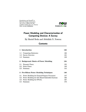
Nicu et al. BMC Oral Health (2018) 18:149Page 4 of 9wavelength of 530 nm. Relative fluorescence (RF) valueswere obtained for periodontitis patients and controls. Thetotal protease activity was defined in RF per Unit (RF U).Statistical analysisAll analyses were performed with the SPSS Statistics23.0 software (IBM, Chicago, Illinois, USA). Means, standarddeviations (SD), range and frequency distributions werecalculated. Normal distribution of data was tested with theKolmogoroff-Smirnov test and when needed, log transformation of data sets was performed before proceeding withparametric statistics. The characteristics of patient andcontrol groups were compared with the Student’s T-test orthe chi-square test, where appropriate. Within groups,comparisons between non-stimulated and stimulatedsamples (PMA or F. nucleatum) were performed using thePaired T-test. A further, linear regression analysis wasperformed to explore a possible relationship between theoPMN numbers and the measured PMN parameters. Inthese analyses, the log-transformed experimental data foroPMN counts were entered as dependent variables, andtotal protease activity was entered as the independent variable. A P-value 0.05 was considered statistically significant.ResultsStudy populationA total of 19 periodontitis patients and 16 control subjects were included in this study. A description of thecharacteristics of the participants is provided in Table 1.No significant differences were observed between thetwo groups in relation to age, sex distribution andTable 1 Characteristics of the study population stratified accordingto periodontal conditionControl(n 16)Periodontitis(n 19)p-valueAge44.1 11.647.9 12.40.354Sex (male)8 (50%)11(55%)0.765Ethnicity (Caucasian)12 (75%)7 (37%)0.024Smoking (current smoker)4 (25%)11 (58%)0.050Body mass index23.5 2.227.9 5.00.006# of teeth27.7 3.026.6 3.30.303# of teeth with 50%bone loss09.8 6.0–cPMNsa19.3 11.217.6 9.00.873oPMNsb1.8 3.28.1 11.50.007Counts ( 106)Results for demographic, dental characteristics and counts of oPMNs and cPMNs.Values represent means standard deviations, or numbers (%) of subjects. P-valuescalculated by χ2-test or Student’s T-test; (n/a not applicable). P-values 0.05 wereconsidered statistically significant and are shown in boldaTotal cPMNs obtained from a 6 ml tube of blood, after the isolation andpurification stepsbTotal oPMNs collected after 4 30 s rinsing with 4½ minutes intermission, afterthe isolation and purification stepsnumber of teeth. Periodontitis patients were more oftenof non-Caucasian descent, smoked and had a higherbody mass index (BMI) than the control group. Withinthe periodontitis group, an average of 9.8 teeth showed aradiographic bone loss of 50% of the root length.Isolation of PMNsThe yield of cPMN from venous blood was comparablein healthy controls and periodontitis patients (mean 19.3versus 17.6 106, P 0.873, Table 1). On average, 4.5times more oPMN were retrieved from periodontitispatients (mean 8.1 106 cells per sample) than from thecontrol group (mean 1.8 106 cells per sample, P 0.007, Table 1).Apoptosis assayThe apoptotic cPMNs (PI ) accounted for a mean of2.8% in the control group. The periodontitis patientsshowed a higher mean percentage of PI cPMNscompared to the control group (mean 12.3%, P 0.026,Fig. 1). Similar to the profile of the circulatory cells, thePI oPMNs percentages were higher in periodontitispatients (56.2%) than in controls (39.9%, P 0.004,Fig. 1). Additionally, more PI cells were found amongstthe oPMNs compared to the cPMNs in both patients andcontrols (P 0.001, Fig. 1).Degranulation assayThe expression of CD11b, CD63, and CD66b on thecPMNs was low and comparable in healthy controls andin periodontitis patients (p 0.05, Fig. 2a, b, and c,respectively). The oPMNs were more activated thancPMNs, as they expressed 3 to 36-fold higher levels ofall three activation markers than the cPMNs, both inhealthy controls and in periodontitis patients (all P 0.001, Fig. 2). CD11b was more expressed by oPMNs derived from periodontitis patients than by oPMNs derivedfrom controls (P 0.024, Fig. 2a). The expression ofCD63 and CD66b on the oPMNs was not statisticallydifferent between the periodontitis patients and controls(CD63 P 0.054, Fig. 2b; CD66b P 0.483, Fig. 2c).ROS assayNon-stimulated cPMNs demonstrated comparable levelsof ROS production between the control group and theperiodontitis group (Fig. 3a). Upon stimulation with either PMA or F. nucleatum, ROS production from thecPMNs increased 24–26 fold for the control subjects (bothP 0.001, Fig. 3b), whereas the periodontitis patientsshowed an increase of 33–34 fold compared to thenon-stimulated cPMNs (both P 0.001, Fig. 3b). The samepattern was observed for the oPMNs’ non-stimulated andstimulated ROS production levels. Comparable ROS levelswere produced by oPMNs originating from controls and
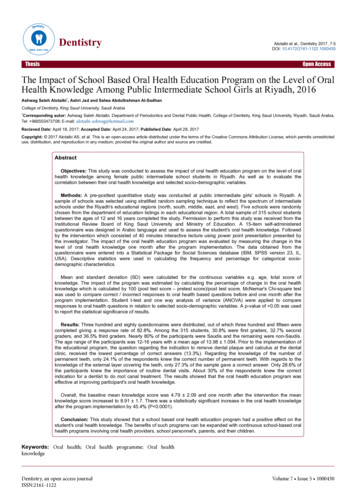
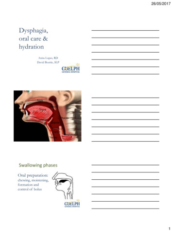
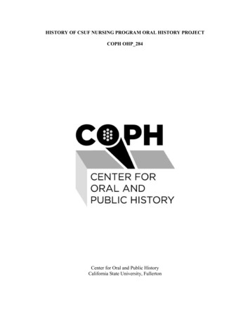
Nicu et al. BMC Oral Health (2018) 18:149Page 5 of 9Fig. 1 Apoptosis analysis of PMNs from controls (n 16) andperiodontitis subjects (n 19) quantified by flow cytometry. Thepercentages of propidium iodide positive (PI ) PMNs were calculated,representing apoptotic cells. The different cell populations areexpressed as a percentage of the total PMN cell population and aregiven as mean SD. *Comparisons periodontitis group versus controlgroup, Student’s T-test, *P 0.05. #Comparisons oPMN versus cPMNwithin subjects, Paired T-test, #P 0.001periodontitis patients (Fig. 3a and c). The oPMNs nonstimulated ROS levels were 3–5 higher compared to thenon-stimulated ROS levels produced by cPMNs (both P 0.001, Fig. 3a). Upon stimulation with PMA, only theoPMNs’ from the periodontitis group showed a significant increase of ROS production (2 fold increase,P 0.012, Fig. 3c), while ROS levels were not significantly induced by PMA in the control group. Uponbacterial stimulation with F. nucleatum, oPMNs fromboth the control group and the periodontitis groupproduced higher levels of ROS (control 2.4 fold increase,P 0.001; periodontitis 2.2 fold increase, P 0.038, Fig. 3c).Protease activityThe total protease activity was analyzed from the firstrinse samples and showed a 3.1 times higher activity inthe periodontitis group compared to the control group(P 0.001, Fig. 4a). Additionally, the number of oPMNswas a significant predictor for the total protease activity(r2 0.386, P 0.001). Further subanalysis revealed thatthis relation was only found amongst the periodontitispatients (r2 0.399, P 0.007, Fig. 4b), and not in thecontrol subjects (r2 0.063, P 0.349, Fig. 4b).DiscussionThe purpose of the current investigation was to studythe numbers and function of the oPMNs in subjects withperiodontitis. Normally, oPMNs contribute to oralhealth maintenance and form a part of the innate oraldefense mechanisms. However in periodontitis theseFig. 2 Mean fluorescence intensity (MFI) of cellular expression of (a)CD11b, (b) CD63 and (c) CD66b, on PMNs isolated from controls(n 16) and periodontitis subjects (n 19). PMNs were gatedaccording to CD16 expression and the sideward scatter profile.Expressions of surface markers of interest were corrected for thenon-specific binding of the isotype control antibodies. Data aremean SD. *Comparisons periodontitis group versus controlgroup, Student’s T-test, *P 0.024. #Comparisons oPMN versuscPMN within subjects, paired T-test, all #P 0.001
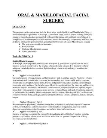
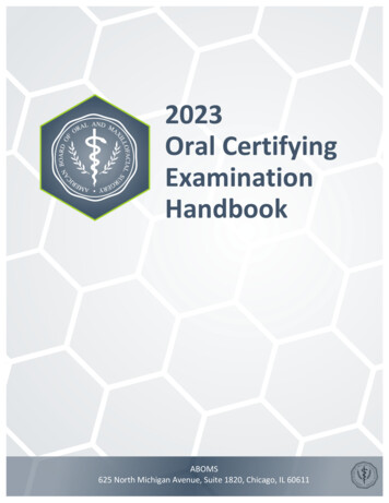
Nicu et al. BMC Oral Health (2018) 18:149Page 6 of 9Fig. 3 Mean fluorescence intensity (MFI) of reactive oxygen species(ROS) produced by PMNs isolated from controls (n 16) andperiodontitis subjects (n 19). a Non-stimulated ROS production wascalculated and expressed as mean SD. b Stimulated ROS productionproduced by cPMNs when incubated with phorbol myristate acetate( PMA) or F. nucleatum ( F.nuc). c Stimulated ROS productionproduced by oPMNs when incubated with phorbol myristate acetate( PMA) or F. nucleatum ( F.nuc). Ratios were labeled as fold increaseand were calculated for the stimulated ROS samples (MFI of thePMA-stimulated or F.n.-stimulated samples, divided by the MFI of thenon-stimulated ROS samples). #Comparisons oPMN versus cPMN withinsubjects, Paired T-test, #P 0.001. γComparisons to correspondingnon-stimulated samples (panel a), Student’s T-test, all γP 0.05cells may be left with limited or depleted functional capabilities. The results of the present study showed a significant higher number of oPMNs in patients withuntreated periodontitis compared to subjects withoutperiodontitis. Confirming our previous results [11], theoPMNs showed increased expression of degranulationmarkers and unstimulated ROS release compared to thecPMNs; this was observed in both the periodontitis andcontrol subjects. However, the periodontitis patientsshowed more apoptotic oPMNs and increased degranulation compared to the control group. Combined, thesefindings suggest that as the PMNs migrate through oraltissues and “travel” along the biofilms surrounding theteeth, these cells age, use their granule resources andlose ROS production capabilities; these traits are morepronounced in periodontitis.On average, we found 4.5 times more oPMNs in thepatient group. Our results confirm previous reports ofincreased PMN numbers in the presence of gingival inflammation and deepened pockets [4, 5, 7, 13, 18].Higher numbers of oPMN in periodontitis is most likelyexplained by an increased periodontal inflamed surfacearea (PISA), which has been reported to reach 5–20 cm2[22, 23]. In response to the inflamed oral surroundings,PMNs leave the blood circulation, enter the periodontalspace and mix in the oral fluid. Moreover, as the oralbiofilm acquires pathogenic traits, more chemokines andcytokines are released into the periodontal tissues,resulting in increased recruitment of PMNs [24, 25].We found more apoptotic oPMNs in periodontitis patients compared to controls. One explanation for thisfinding might be that there are more cPMNs beingprogrammed for apoptosis in periodontitis and that thesecells appear in patients’ rinse samples. We suggest thatmore apoptotic cPMN could be a systemic characteristicof the periodontitis patients, and therefore also found inthe oral cavity. More apoptotic oPMN in periodontitis isin contrast to the recent results of Lakschevitz andco-workers who suggested lower numbers of apoptoticoPMN in periodontitis compared to controls [8]. In ourprotocol, the participants repeated the oral rinsing 4
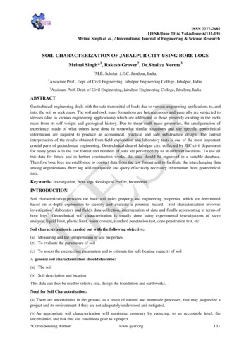
Nicu et al. BMC Oral Health (2018) 18:149Fig. 4 Total protease activity measured from oral rinses originatingfrom controls (n 16) and periodontitis subjects (n 16). a Totalprotease activity was measured as relative fluorescence per unit (RFU) and expressed as mean SD. *Comparisons periodontitis groupversus control group, Student’s T-test, *P 0.001. b Total proteaseactivity was only predicted by oPMN counts in periodontitis patients(dashed line, r2 0.3989, P 0.0065) and not in the control group(straight line, r2 0.0629, P 0.3486)times, while the subjects from Lakschevitz et al. rinsed 6times consecutively [8]. In pilot experiments precedingthe current study, we observed that the viability of oPMNsincreased by 17.7% between the first rinse and the fourthsuccessive rinse, suggesting that fresh, new PMNs are retrieved as the oral rinse is repeated (data not shown).Furthermore, Lakschevitz et al. based their conclusionsabout oPMN viability on a small sample subset (N 3, forthis sub-analysis) [8], whereas we analyzed nineteenperiodontitis patients and observed inter-individualdifferences.Upon PMN activation, the cytoplasmic granules fusewith the PMN cellular membrane and an upregulationof the granular markers (CD11b, CD63 and CD66b) becomes measurable. The increased expression of thesePage 7 of 9CD markers in our study confirms that the oPMNs haveundergone migration and degranulation relative to thecPMNs. Additionally, the upregulated expression ofCD11b and the tendency for increased CD63 in periodontitis patients’ oPMNs, suggest a higher release ofgranular content, possibly related to activation duringthe migration through the inflamed periodontal tissuesand along the subgingival biofilm. Important to note isthat periodontal inflammation can be present in variousinflammatory states, ranging from a pre-inflammatorystate within stressed tissues, to a full-fledged inflammatory state within damaged tissues. The results from thisstudy corroborate the recent findings by Fine et al., whoalso found degranulated oPMNs in periodontitis [18].Intrinsic increase of ROS production by cPMN hasbeen proposed for patients with chronic periodontitis asa susceptibility trait [26–29]. In our study, we could notconfirm this trait, since our results showed similar unstimulated and stimulated ROS levels originating fromperiodontitis patients and healthy controls. A plausibleexplanation for the discrepancy with the literature couldbe the application of different methods used to analyzeROS production. While most studies have used luminolenhanced chemi-luminescence for the measurement oftotal ROS generation (intra- and extracellular), our studyhas used flowcytometric analysis. In contrast to thechemi-luminescence method, which measures variousROS species in a multi-well assay, the flowcytometricanalysis used in this study detects only hydrogen peroxide at a single cell level. However, the flowcytometricmethod is preferred because it is applicable in situationswith low cell density, such as in our oral rinse samples.Another possible reason may lie in the socio-demographic differences between study populations. Matthews et al. demonstrated that when periodontitispatients were compared with age-, and sex-matchedcontrols, only the unstimulated cPMNs produced higherlevels of ROS, while the stimulated levels were comparable between the measured groups [29]. In the presentstudy, age and sex distribution were not differentbetween the periodontitis and control subjects, howeverthe ethnicity, BMI and smoking frequencies were different. Ethnic background can influence PMN numbersand ROS activity [30, 31]. Furthermore, it has beensuggested that PMNs in obese subjects may be in aprimed state compared to non-obese subjects, and canparticipate in the pathogenesis of obesity-related diseases,such as periodontitis [32]. Another study demonstratedthat cigarette smoke extract could lower the PMN ROSproduction capabilities in response to F. nucleatum specifically [33].Our group previously evaluated ROS levels of oPMNsand/or cPMNs in response to F. nucleatum [11, 12, 34].Several PMN receptors are involved in active ROS
Nicu et al. BMC Oral Health (2018) 18:149production, like Toll-like receptors and protein kinase C agonists, from which Toll-like receptors are the most efficientin ROS generation [35, 36]. While PMA stimulation actsvia protein kinase C, F. nucleatum is able to activate thePMNs’ Toll-like receptors (TLR-2, TLR-4, TLR-9) [30]. F.nucleatum stimulation may therefore result in a differentoPMN ROS response than PMA stimulation. The ROSlevels that were recently reported in chronic periodontitispatients [18] were acquired using an extensive flowcytometric gating strategy in order to identify PMN sub-popula
instructions. Flow cytometric analysis was performed within 1 h on a BD Accuri C6 flow cytometer (BD Biosciences) and the Accuri CFlow Plus software was used for data acquisition and analysis. The percentages of propidium iodide positive (PI ) PMNs were calcu-lated, indicating apoptotic or damaged cells.