Transcription
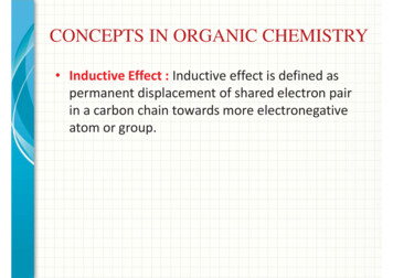
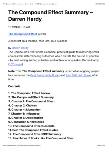
SCIENTIFIC ARCHIVES OF DENTAL SCIENCESVolume 2 Issue 12 December 2019Soraya DendougaResearch ArticleThe Effect of Preparation in Dentin on Porcelain Veneers’ SurvivalHigher Education at the College of Dental Medicine, Algeria*Corresponding Author: Soraya Dendouga, Higher Education at the College of Dental Medicine.Received: October 21, 2019; Published: November 28, 2019AbstractIntroduction: For this study we wanted to evaluate the possible influence of the depth of the preparation on the longevity ofporcelain veneers.Materials and Methods: Our study is a randomized clinical trial; on the comparison of 264 veneers stuck on 73 patients. 139veneers have a preparation in dentin. The patient’s recruitment was done according to well-defined inclusion criteria. Theinformation was reported on a clinical record.The study lasted 64 months with regular checks of 6 months, 12 months, 18 months, 24 months, 36 months, 48 months and 60months.Results: In this study, the maxillary teeth prepared according to different depth on dentin shown that ceramic veneers have stableesthetic qualities; they are biologically acceptable in so far as the recommendations of current preparations are respected.The Chi square test revealed that there is statistically no significant difference between the three methods.“Preparation without palatine return or window, preparation in slight return and preparation in slight return”.Conclusion: The depth preparation for ceramic veneers have no influence on porcelain Veneers’ survival.Keywords: Dentin; Porcelain Veneers’IntroductionTissue preservation is a prerequisite for any modern dentistrytreatment to better ensure the long-term resistance of restorations,maintain pulp vitality and have the possibility to re-intervene inthe future [1,2]: veneers meet these expectations.The veneers allow to have a biomimetic restoration whichFigure 1: Veneer compared to a contact lens [18].allows a homogeneous distribution of the stresses withoutalteration of the tooth. They are described as biomimetic becauseof their biology, which is essentially guaranteed by the minimumsize that does not alter the dentine nucleus and gives the tooth itschromatic tone and saturation [3].The depth of the preparation is a key element that has givenrise to reflection: for some authors, it is preferable to remain inthe enamel while for others one can reach the dentine without anyparticular concern.Figure 2: Illustration of the thinness of the ceramic,hence the name of a thin ceramic film [18].Citation: Soraya Dendouga. “The Effect of Preparation in Dentin on Porcelain Veneers’ Survival”. Scientific Archives Of Dental Sciences 2.12 (2019): 75-85.
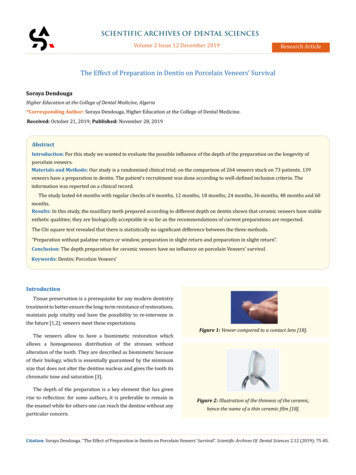
The Effect of Preparation in Dentin on Porcelain Veneers’ SurvivalSeveral studies have been carried out in the world withsometimes divergent results.Our study is a randomized clinical trial carried out in the dentalprosthesis department. This study covered 264 veneers in 73patients between 2015 and 2019.Several parameters were evaluated: the type of coronarypreparation, the frequency of the failures, the satisfaction of thepatients and the depth of the dental preparation which will bedeveloped in this article.Materials and MethodsThe subjects are recruited from among those who come to thedental prosthesis service at the Mustapha University HospitalCenter in Algiers.Diagnosis of abnormality; color; form; Minor structure ormalposition will be observed with the naked eye during the clinicalexamination.Selection criteriaCriteria for inclusionIncluded in the study is any previous tooth showing: A typical color anomaly: amelogenesis imperfecta due totetracyclines; fluorosis; stains due to age, dye of externalorigin (tea, coffee or tobacco) by infiltration of tissues;dyschromic teeth without loss of substance;An anomaly of form: microdontics; atypical form cases ofconoid incisors;Criteria for non-inclusion76We will not include in our study anyone: Judged unable to understand the essay, uncooperative orunstable. Presenting a contra-indication to the glued ceramic facetsnamely:Elderly teeth;Teeth abraded;Presence of parafunctions;Adverse occlusal relationships;Bad oral hygiene.Dental preparation method for ceramic veneersIt differs from conventional preparations in that the retention ofthe prosthetic element is ensured only by gluing.Materials needed for preparationThe protocols of the preparations use the concept of controlledtissue penetration through the use of diamond burs calibrated forthis purpose.Several authors have proposed several set of burs. Among theseauthors Bernard Touati, and Galip Gurel. A set of 3 strawberries isenough for a good preparation.Diamond ball mill which delimits the desired depression in thedentine; Fillet cutter which allows to eliminate the grooves and todraw the cervical limit: Red ring cutter: fine grain for finishing.An abnormality of structure or texture: dysplasia,dystrophy, erosion, attrition, mechanical or chemicalabrasion and coronary fracture;A minor malformation type rotation.Our study will concern both men and women, but in any case,we will include only those patients deemed reliable, cooperativeand likely to be followed regularly:Figure 3: Set of retained cutter for preparation General condition: healthy patient presenting no general Oral hygiene: must be satisfactory;Materials needed for preparationthe incisivo-canine block.coronary preparations for bonded ceramic veneers. or local pathology;Type of teeth: to reduce the variants, it will be limited tofor ceramic veneers [4].Our study is a randomized clinical trial comparing three types ofCitation: Soraya Dendouga. “The Effect of Preparation in Dentin on Porcelain Veneers’ Survival”. Scientific Archives Of Dental Sciences 2.12 (2019): 75-85.
The Effect of Preparation in Dentin on Porcelain Veneers’ SurvivalDuring this test, the allocation to the different treatment groupsis randomly made by lot, thus ensuring the comparability of thesegroupsThe same operator will proceed to the draw as follows: Thedistribution in all the different groups will be based on chance.77The clinical stage begins with the establishment by thepractitioner of a clinical file to list the information necessary forthe study. This sheet is completed by the taking of photographsbefore any dental preparation, to allow us to archive the case andto give the necessary information to the prosthetist.Before any prosthetic intervention, we start with preoperativesteps: such as motivation to hygiene; restoration of the oralcavity: descaling; pulpal vitality tests; drilling of the gingivodental grooves and repair of old fillings if necessary.Once this stage is complete, study models are made frompreliminary impressions made using an irreversible hydrocolloidFigure 4: Vestibular view.type alginate.The study of these models will give us the dental formula andinformation regarding static occlusion: overjet, overbite, molarclass, canine class, incisal covering etc. These templates will bearchived and constitute the patient's record.Then we go to the preparation of the teeth, for that we can usetwo methods:Figure 5: Proximal view [5].1.The technique of progressive reduction: use of grooves and2.Or the technique of masks.a silicone key;We opted for the technique of progressive reduction; thetechnique of masks being applied when several teeth are involvedin faceted restorations to align the teeth for example; while in ourstudy we have sometimes had to treat a single tooth.We begin with the realization of a high viscosity silicone keyfor this we must cover at least one tooth on each side of the toothconcerned by the preparation to have good stability. Then we willFigure 6: Palatal view [5].From left to right preparation in semi-jacket; preparation inslight return; preparation without palatine return.Technique of realization of the coronary preparationsAll clinical stages of the ceramic veneers performed in the study(dental preparation, bonding and control) were made by the samepractitioner.The ceramic veneers were made by a single prosthetist whoused the same technique and the same ceramic to avoid therealization bias.cut vertically this key to check its good adaptation.Once the wrench is made, we go to the size of the concernedtooth using an air turbine; under water jet whatever the case;using burs from a Komet France box marketed under the name"cabinet for ceramic veneers". This box contains three burs:1.A ball mill whose references are (801, 314, 018);3.A flame red ring cutter for finishing whose references are2.A milling cutter green ring whose references are (6856,314, 018);(868, 314, 012).The realization of the horizontal grooves is done by means ofthe ball mill which will determine the depth of the preparation.Citation: Soraya Dendouga. “The Effect of Preparation in Dentin on Porcelain Veneers’ Survival”. Scientific Archives Of Dental Sciences 2.12 (2019): 75-85.
The Effect of Preparation in Dentin on Porcelain Veneers’ SurvivalThis depth is considered sufficient when the mandrel of the burwill come into contact with the tooth surface. The cutter used hasa diameter of one millimeter and its penetration until contact withthe mandrel will correspond to a depth of 0.5 mm.Each tooth has three planes on the buccal surface: a cervicalplane, a median plane and an incisal plane. A groove is made on78Dentin hybridization is indicated when there is dentinalexposure or type I (without palatal return) is an enamel preparation;only type II and III are concerned, but to avoid any kind of bias inour study we preferred to apply this dentin treatment to all teethregardless of the importance of tooth preparation.To do so we will use a dentine bonding agent compatible witheach plane. In some cases where the teeth are small (distancethe adhesive that will be used later during bonding (Excite, IvoclarThen we place the green ring leave cutter and remove persistentThe final imprints were taken according to Wash techniquetaken between the free edge and the collar) only two horizontalgrooves will be made.enamel bridges between the different grooves. The bur used is aworking end mill, its use at the cervical level will lead to a draft ofthe preparation limit which initially will be at a distance from thegingival ring.Once the enamel bridges have been removed, the dentalfluorosis has disappeared, giving way to a dental surface ofordinary hue, and in this case we opt for a supragingival cervicallimit, so that the hue remains dark and then, in this case, it is betterVivadent, lot No. L23818) in accordance with the manufacturer'sinstructions.using a high viscosity silicone relined by a low viscosity silicone.Temporary restorations were made at the chair, modelingwith the fingers of the light-curing composite (Arabesk, Voco, lotno. 1236135). We took care to stay away from the gum to avoidbleeding that may compromise the session collage.Occlusion is checked during the various mandibular excursions.The patient will be released only after giving a number ofto make a juxta-gingival limit.instructions and emphasizing the importance of good oral hygiene.return).elaborated with the same low-melting ceramic (Finesse All-This kind of preparation concerns type I (without palatalWith regard to type II; the dental preparation described abovewill be continued by reducing the free edge with the bur. PalatineFor the realization in the laboratory, all the facets wereCeramic, Ceramco, U.S.A).It was treated according to the lamination technique using theconcavity will be reduced palatally by always using the sameconventional method of lost wax and following the manufacturer'sensuring a continuity between the buccal face and the palatal facecylinders, they were preheated in a conventional preheatingstrawberry and at the same time a fine leave will be drawn whichwill form the limit at this level. The preparation is completed byto have a sufficient thickness for the ceramic.For type III (semi-jacket) the preparation is the same as that oftype II but it will be pushed more in the palatal direction to endjust before the cingulum.For these last two types of preparation type II and type III wewill take care to objectify with articular paper the impact of theopposing tooth to prevent the tooth-ceramic junction from beingat this level.For all three types of dental preparation, the finishing will bedone in the same way and will consist in the elimination of all thesharp angles using a flame mill with fine granulometry red ring.Once the preparation is complete, the silicone key is positionedand the homothetic aspect will be checked.instructions. The models were cast with extra-hard plaster andare used in one piece. After placing the wax models in the coatingfurnace (type 5636, KaVo Dental GmbH, Biberach, Germany) at afinal temperature of 850 C. ceramic was pressed into the preheatedhollow mold (EP 500, Ivoclar Vivadent, Schaan, Liechtenstein) at910 - 920 C.Coating cylinders and ingots were placed in the center of thepress furnace and pressed at a temperature of 1050 C. The pressedfacets were separated from the casting rods with a disc. Facetadjustment has been verified on the master models.Two glazing procedures were performed in a porcelain bakingoven. The intrados of the facets is treated with 5% hydrofluoricacid; contained in the cabinet of the ceramic used; for 60 secondsand neutralized in a bath of bicarbonate then the facets are dried.We go to facet gluing for this we start by depositing theprovisional by means of a probe. If the composite persists on theCitation: Soraya Dendouga. “The Effect of Preparation in Dentin on Porcelain Veneers’ Survival”. Scientific Archives Of Dental Sciences 2.12 (2019): 75-85.
The Effect of Preparation in Dentin on Porcelain Veneers’ Survival79tooth we will do a surfacing with a strawberry being careful not to480 mW/cm (Optilux, Demetron, Danbury, CT, USA) at the incisalFor the choice of glue we have taken as reference the work ofThe complete polymerization is done section by section for 40touch the tooth. Afterwards we clean the dental surfaces and trythe facets.Hikita., et al. who evaluated the adhesion strength to enamel anddentin, different adhesives tested with different adhesives. Thisstudy shows that the best results are achieved by the bondingcomposites requiring the use of an adhesive system preceded bya total etching.A dual composite has therefore been used: a mixed adhesiveresin, which can offer, thanks to porosities, a potential forthe absorption of additional stresses to that of the onlyphotopolymerizable resins [6,7].Once the facets are deemed satisfactory, they are washed withwater to remove the "liquid strip" which is water soluble, airdried and then brushed with a silane agent (Monobond S, IvoclarVivadent) (the facet being already etched in the laboratory), itis allowed to act for 1 minute then it is dried. An adhesive layer(Excite DSC, Ivoclar Vivadent) is again added to the silanizedsurface without light curing, the thus prepared facet is protectedfrom light in a metal can.All veneers were glued with Variolink II (Variolink II, IvoclarVivadent, lot # M10683, lot # M34435, lot # P22457), a dual curingcomposite prepared according to the manufacturer's instructions.The preparations were isolated using cotton rolls combinedwith effective salivary aspiration. The surfaces of the teeth werecleaned with a rubber cup lined with a fluorine-free polishingpaste mounted on a contra-angle, then washed Dried and etchedfor 15 seconds with a 37% phosphoric acid gel (Total Etch, IvoclarVivadent) and then with a jet of water, they are washed for at least15 seconds.The surface thus treated is dried in air, avoiding desiccationof the dentin. The adhesive (Excite DSC, Ivoclar Vivadent) was2edge to stabilize the facet and to be able to easily remove excesscomposite.seconds, starting with the proximal areas. Once the polymerizationis complete, the edges of veneer are checked with a probe for anyexcess of composite particularly at the interdental spaces.Checking occlusion in static and dynamic state. Afterequilibration, all surface alterations are repolished.Before releasing the patient, care should be taken to mentionthe contact of the opposing tooth with the facet: the contact canbe made: On the palatal side; On the incisal edge; On the ceramic to 1/3 incisal;On the medium 1/3 ceramics;No contact.Patient appreciation will also be mentioned to assess the degreeof satisfaction that may be: none; medium or good.Control sessionsPatients are invited to follow-up sessions after: 6 months, 12months, 24 months, 36 months, 48 months and 60 months.During these appointments, facets were inspected visually andevaluated according to the criteria studied.Clinical casePatient aged 23 presented to our service following an aestheticdiscomfort: dental fluorosis type II of Dean. This patient, wellinformed, asked for a treatment by ceramic facets.In this stage of fluorosis prosthetic restoration is the rule andceramic veneers are the most conservative form.applied with microbrushes, rubbing gently for 20 - 30 seconds, andthen spread with an air jet for at least 10s to facilitate penetrationinto the dentine.Always thanks to the jet of air the residual solvents willevaporate and also allow the formation of an adhesive film whichwill leave the dental surface shining.The dual composite is mixed just before use. It is applied,polymerized for 10 seconds with light with an energy density ofFigure 7: Case presentation: Dean's type II fluorosis.Citation: Soraya Dendouga. “The Effect of Preparation in Dentin on Porcelain Veneers’ Survival”. Scientific Archives Of Dental Sciences 2.12 (2019): 75-85.
The Effect of Preparation in Dentin on Porcelain Veneers’ Survival80ResultsIn this study 264 veneers were placed in 73 patients representedby 52 women and 21 men. The average age of patients at the startof treatment was 27 years with a range of 13 to 49 years.The veneer’s number varies from "one" to "six" per patient. Allveneers were performed at the level of the upper anterior block.Figure 8: Penetration grooves.Once the treatment is finished, patients are invited to comeback if there is any problem with their facets or abutment teeth.Patients will be checked after 6 months; 12 months; 24 months;36 months; 48 months; 60 months. The information collectedduring these checks is reported on the clinical sheet.All the patients participated in the various controls, only onepatient, who benefited from two restorations on the central incisor,did not participate in the study and this from the second recall (12months), which is equivalent to an abandonment or loss of sight ofVestibular view1.40% of patients (0.76% of restorations).Descriptive studyNumber of veneers depending on the type of prepared toothTooth typeNumbersPercentageCentral incisor12045,45%Total264100,00%Lateral incisorPalatal viewFigure 9: Coronary preparation.Canine8632,58%5821,97%Table 1: Number of veneers by type of prepared tooth.120 central incisors (11 and 21) were treated with ceramicveneer which represents 45.45% of all veneers. 32.58% representsthe percentage of lateral treated veneers while the caninesrepresent only 21.97% of the whole.Distribution of facets according to the characteristics ofprepared teethCharacteristics of the teethNumbersHealthy, alive26Angular fracture, alive1Obturated, aliveObturated, mortifiedFluorosis, aliveFluorosis; mortifiedFigure 10: Final result after veneers 410,20%0,40%1,10%100,00%Table 2: Frequency of facets according to thecharacteristics of prepared teeth.Citation: Soraya Dendouga. “The Effect of Preparation in Dentin on Porcelain Veneers’ Survival”. Scientific Archives Of Dental Sciences 2.12 (2019): 75-85.
The Effect of Preparation in Dentin on Porcelain Veneers’ SurvivalA healthy tooth is any tooth with no loss of substance or textureanomaly. These teeth, although healthy, have been restored byfacets for aesthetic purposes: improving the hue or correcting aslight malposition.For angular fractures, it is a class IV of black not exceeding 2 mm81The veneers were stuck on 208 unclosed teeth whichcorresponded to 78.79% of all the teeth while 56 of the facets were21.21% glued on closed teeth.Distribution of veneers according to the limit of thepreparationof amplitude. The majority of teeth were included in our sample at68.30%. The remaining teeth are distributed as follows: 10.20%Limit of the preparationNumbersPercentageIn the enamel12547,30%live plugged, 10.20% mortified plugged; 1.10% mortified fluorosisand 0.40% have angular fracture and are alive.Pillar al264In the dentine139Total26421,21%The limit of the preparations in the dentin are majority for 125facets which is equivalent to 53% against a frequency of 47% of100,00%the preparations in the enamel.unsealed tooth character closed tooth.Distribution of the type of preparation according to the limitsof the preparationType of dental preparationSituation of the limit of preparationType IIn the enamelIn the dentin13Type IIIn the enamelIn the dentin4349Type IIIIn the enamel08“Slight return palatine”“Semi-jacket”100,00%Table 4: Frequency of veneers according to the limit of preparation.Table 3: Frequency of veneers according to the“Without return palatine”52,70%NumbersTotalTotalIn the dentinTotalPercentage748785,10%14,90 0%Table 5: Frequency of the type of preparation according to the limits of the preparation.Ceramic veneers are restorations whose retention is ensured byIn the present study, the Kaplan-Meier analysis was done in twogluing. This bonding can be at the level of the enamel, the dentinedifferent ways: the first analysis is patient-related, the second wasSince the structure of enamel and dentin is different, theOur study is a clinical trial that seeks to evaluate the effectivenessor at the level of both.question has been asked whether the behavior of ceramic facetscan be different for these two structures.Analytical studyAlthough the Kaplan-Meier statistics were initially designed todeal with individuals, the use of the tooth as a statistical unit in theperson's place is justified [13].done by taking the restoration as a statistical unit.of one type of preparation compared to two other types and this bycomparing the results obtained in each group.Given an observed difference between groups, there are twopossibilities either this difference is only due to chance, or thisobserved difference is real.We applied the test of "Chi2" to check the dependence or notbetween the variables and the different groups. This probability isCitation: Soraya Dendouga. “The Effect of Preparation in Dentin on Porcelain Veneers’ Survival”. Scientific Archives Of Dental Sciences 2.12 (2019): 75-85.
The Effect of Preparation in Dentin on Porcelain Veneers’ Survivalcalled "p". The value of p is the threshold from which the observeddifference is considered to be statistically significant. It means thatit is real and has a small chance of being due to chance.When p is 0.05 the difference is significant.When p is 0.05 the difference is not significant.Analysis of facets according to the limit of the preparationSurvival rate of facets according to the limit of the preparation.82Occurrence of secondary caries according to the limit of thepreparationLimit of the preparationSecondarycariesIn enamelIn able 6: Frequency of caries according to the limit of preparation.The incidence of caries was recorded evenly between dentinaland enamel preparations (50%, 50%).The Chi-square test gives a p 0.91 (p 0.05) and thus theincidence of caries is not related to the depth of dental preparation.The change of color of the facet according to the limit of thepreparationFigure 11: Survival curve of ceramic facets accordingto the depth of the preparation (enamel/dentin).In our study, which included 264 facets, 125 facets presented anenamel preparation of which 6 showed an event while 139 veneershad a preparation at the dentin level, 8 of which showed an event.The analysis of these results gives: Survival rate of ceramic facets with an intra-amolarpreparation of 95.20% (95% CI, [91.50%, 99.00%]) and anaverage survival time of 58.57 months (IC at 95%, [56.65months, 60.50 months]).A survival rate of ceramic facets with a dentinal preparationof 92.70% (95% CI, [87.60%, 98.10%]) and an averageLimit of preparationColor change ofthe veneerIn enamelIn otal247100%17100%264100%Table 7: Frequency of changing the color of the faceaccording to the situation of the preparation.The hue of a veneer is the result of three elements: the shadeof the stump, the color of the gluing composite and the hue of theceramic used.So, if we talk about changing the color of the facet, we generallysurvival time of 60.90 months (95% CI), [58.80 months,exclude the ceramic that does not change and we look for the sideAn overall survival rate of ceramic facets according to theThe Chi2 test gives a p 0.01 (p 0.05) and therefore the depth63.00 months]).preparation depth of 93% and an average survival time of61.1 months (95% CI, [59.59 months, 62.60 months]).The Log rank test gives a value p 0.79 (p 0.05) andtherefore the difference is not significant between the twoof the stump or collage composite.of the preparation affects the change in color of the veneer: whenthe depth is in the dentin there is probability of change of color ofthe veneer.preparation depths as to the occurrence of the events.Citation: Soraya Dendouga. “The Effect of Preparation in Dentin on Porcelain Veneers’ Survival”. Scientific Archives Of Dental Sciences 2.12 (2019): 75-85.
The Effect of Preparation in Dentin on Porcelain Veneers’ SurvivalDiscussionParticularity of the enamelEnamel is the hardest of dental tissues; its elasticity andpermeability are practically nil; its density is 3 while it is 2.30for dentin and 2.01 for cementum. The enamel consists of 2.30%water, 96% mineral elements and 1.70% of organic elements.Enamel is produced by ameloblasts, which are responsible forits structure. The enamel is made up of mineral elements calledenamel prisms separated by the interprismatic substance which ismineral and organic.This structure is more visible if the enamel is etched withphosphoric acid.83At a higher magnification, we see the variability of thedisposition of the hydroxy-apatite crystals.The mode of adhesion to the enamelUntreated enamel has a very low surface energy and is notsuitable for bonding. It is covered with a bio film of polysaccharidescoming from saliva. The enamel milling eliminates the aprismaticsuperficial enamel that is not conducive to bonding [16] as wellas this bio film. Enamel etching with 37% phosphoric acid willincrease the surface energy of the enamel and create a micro-retentive surface to a depth of 10 to 20μ which can be optimallypenetrated by the resins. Hydrophobic di-acrylics.Particularity of dentinDentin has a very heterogeneous chemical composition: to theinorganic part of hydroxyapatite (70%) is added a rather largeorganic part, consisting of collagen (18%) and water (12%).The hydroxy-apatite, arranged in an irregular manner, containsthe organic matrix composed mainly of collagen fibers.The dentine is traversed by canaliculi or tubuli, which containthe extensions of the odontoblasts and the pulpal fluid which filtersthe pulp up to the amelo-dentinal junction.Figure 12: Surface of a tooth observed in SEM [19].We visualize very well the twisted appearance of the prismsof the enamel. Some prism groups appear in longitudinal section,others in cross-section.Figure 13: Enamel prisms in cross-section [19].The tubuli increase in number and in diameter as we approachthe pulp: at 1mm distance from the pulp, we can count about40000/mmon the occlusal side and 80000/mm2 on the cervicalside. Their diameter varies from 1.8 μm at the occlusal level to 3.5μm at the cervical level.This microstructure varies continuously due to physiologicaland pathological changes.Figure 14: Transverse disposition of dentinal tubilis [19].Citation: Soraya Dendouga. “The Effect of Preparation in Dentin on Porcelain Veneers’ Survival”. Scientific Archives Of Dental Sciences 2.12 (2019): 75-85.
The Effect of Preparation in Dentin on Porcelain Veneers’ Survival 84A survival rate of ceramic facets with a dentinal preparationof 92.70% (95% CI, [87.60%, 98.10%]) and an averagesurvival time of 60.90 months (95% CI), [58.80 months, 63.00months]).An overall survival rate of ceramic facets according to thepreparation depth of 93% and an average survival time of61.1 months (95% CI, [59.59 months, 62.60 months]).The Chi2 test gives a value p 0.795 (p 0.05) and therefore theFigure 15: Electron microscopic image of dentinal tubuliconstituting dentin [19].The mode of adhesion to dentinDentin bonding is more complex and difficult than enamelbonding. Therapeutic approaches have changed the treatmentphilosophy: The dentin must be etched, which does not hurt the pulp; View the presence of water in the dentine; hydrophilic resinsmust be used so that they can penetrate into the smeareddentinal surface despite its wet state [5,9].During the mechanical preparation of a tooth (milling); a thickdentine sludge of 1 to 5 μm is formed consisting of soft elementswhich seal the dentinal tubules.Etching dissolves the mineral components of this smear mudand the hydroxyapatite of the underlying dentin a few micronsdeep [10].The application of a hydrophilic primer on the thus opendentinal tubili will facilitate the diffusion of the adhesive into thecollagen network. This infiltration into collagen will result in theformation of a mixed layer called a "hybrid layer" or a resin-dentineinter-diffusion zone [11].Incidence of the limits of t
for ceramic veneers [4]. Materials needed for preparation Our study is a randomized clinical trial comparing three types of coronary preparations for bonded ceramic veneers. Citation: Soraya Dendouga. "The Effect of Preparation in Dentin on Porcelain Veneers' Survival". Scientific Archives Of Dental Sciences 2.12 (2019): 75-85.