Transcription
Title: Methods to assess helicase and translocation activities of human nuclear RNAexosome and RNA adaptor complexesRunning head: Assays for nuclear RNA exosome and adaptor complexesAuthors:M. Rhyan Puno1Christopher D. Lima1,2*Affiliations1Structural Biology Program, Sloan Kettering Institute, Memorial Sloan Kettering Cancer Center,1275 York Avenue, New York, NY 10065, USA2Howard Hughes Medical Institute, 1275 York Avenue, New York, NY 10065* Correspondence: Email: limac@mskcc.org; Ph: (212) 639-8205; FAX: (212) 717-30471
AbstractThe nuclear RNA exosome collaborates with the MTR4 helicase and RNA adaptor complexes toprocess, surveil, and degrade RNA. Here we outline methods to characterize RNA translocationand strand displacement by exosome-associated helicases and adaptor complexes usingfluorescence-based strand displacement assays. The design and preparation of substratessuitable for analysis of helicase and decay activities of reconstituted MTR4–exosomecomplexes are described. To aid structural and biophysical studies, we present strategies forengineering substrates that can stall helicases during translocation, providing a means tocapture snapshots of interactions and molecular steps involved in substrate translocation anddelivery to the exosome.Keywords: RNA exosome, helicase, translocase, RNA-protein complex, DExH-box, SF22
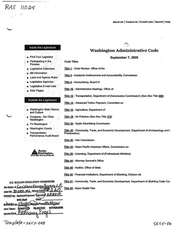
1. IntroductionThe RNA exosome is a conserved, eukaryotic 3′-to-5′ ribonuclease protein complex that isresponsible for several aspects of nuclear and cytoplasmic RNA metabolism, ranging fromgeneral RNA turnover to processing and quality control of various coding and noncodingtranscripts (Houseley, LaCava, & Tollervey, 2006; Puno, Weick, Das, & Lima, 2019). Thehuman nuclear RNA exosome is composed of 9 core subunits with three S1/KH-domainproteins (EXOSC1–3) atop a hexameric ring of PH-domain proteins (EXOSC4–9) (Fig. 1). Theexosome core contains a central channel that guides single-stranded RNA to two RNAdegrading enzymes: DIS3, a processive 3′-to-5′ RNAse II-like exoribonuclease and EXOSC10,a distributive 3′-to-5′ RNase D-like exoribonuclease that associate with the exosome core (Gerlach et al., 2018; Liu, Greimann, & Lima, 2006; Makino, Halbach, & Conti, 2013; Wasmuth,Januszyk, & Lima, 2014; Weick et al., 2018; Zinder & Lima, 2017).Several functions of the nuclear RNA exosome require the helicase MTR4 (also known asSKIV2L2) (Weick & Lima, 2021). MTR4 is a multi-domain protein with a core structurecomposed of two RecA-like ATPase modules (RecA1 and RecA2), a winged helix (WH) fold anda C-terminal helical bundle (HB) domain (Gerlach et al., 2018; Jackson et al., 2010; Weick et al.,2018; Weir, Bonneau, Hentschel, & Conti, 2010). Inserted within WH is an arch-shapedprotrusion consisting of an elongated stalk and a globular Kyprides-Ouzounis-Woese (KOW)domain that interacts with structured RNA and/or protein adaptors for exosome recruitment (Jackson et al., 2010; Taylor et al., 2014; Thoms et al., 2015). Nuclear cofactors MPP6 and/orC1D/EXOSC10 tether MTR4 to the exosome (Fig. 1) (Gerlach et al., 2018; Wasmuth, Zinder,Zattas, Das, & Lima, 2017; Weick et al., 2018).3
MTR4 is a central component of several nuclear RNA adaptor complexes that identify, captureand prime exosome substrates for decay. In human, these include the Nuclear ExosomeTargeting (NEXT) complex, the TRF4-2/PAPD5-ZCCHC7-MTR4 polyadenylation (TRAMP)complex, and the Poly(A) Exosome Targeting (PAXT) connection or Polysome Protectorcomplex (PPC) (Fig. 1) (Lubas et al., 2011; Meola et al., 2016; Ogami et al., 2017). NEXT,composed of an RNA binding protein RBM7, a zinc-knuckle protein ZCCHC8, and the MTR4helicase, functions in turnover of unwanted transcripts produced by pervasive transcription andsurveillance of aberrant RNA species that emerge from faulty transcription. PAXT/PPC core is abinary complex between MTR4 and ZFC3H1 and is loosely associated other cofactors thatinclude poly(A) binding protein PABPN1. PAXT/PPC primarily targets processed polyadenylatedtranscripts. In contrast to its S. cerevisiae counterpart, less is known about endogenous targetsof human TRAMP.The ability of MTR4 and RNA adaptor complexes to disrupt RNA structures and displaceproteins deposited on RNA is key to their function in promoting exosome-mediated decay (Punoet al., 2019; Puno & Lima, 2018; Weick & Lima, 2021). In this chapter, we present methodsused in our laboratory to characterize helicase activities of exosome-associated helicases andRNA adaptor complexes to determine mechanisms and kinetic models underpinning substrateselection, translocation, unwinding, and targeting to the exosome. These assays may also proveuseful in studies aimed at assessing helicases and RNA adaptor complexes for their ability todisplace proteins bound to RNA. We also provide a protocol for analyzing helicase-dependentRNA decay by MTR4–exosome complexes. Finally, we describe strategies in designingsubstrates for trapping helicases during translocation to generate samples for biophysical andstructural analysis.2. RNA helicase assays with MTR4 and RNA adaptor complexes4
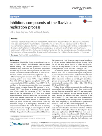
Assays that detect strand displacement are widely used to query translocation and/or helicaseactivity in vitro. These assays measure the ability of a helicase to displace and release anoligonucleotide strand (reporter strand) from a duplex region of a substrate. Unwinding polarityand requirements for a single-stranded translocation strand can be assessed by designingduplex substrates with single-stranded regions extending from either 5′ or 3′ ends.Several approaches are employed to monitor strand displacement using radiolabeled orfluorescently modified oligonucleotides. In a gel shift assay, the displaced reporter strand iscaptured by a complementary DNA oligonucleotide to prevent reannealing to the translocationstrand while capturing the product for detection using native polyacrylamide gel electrophoresis(Fig. 2A and B). This is a common and accessible approach, since it utilizes materials andequipment available in most molecular biology laboratories, but it is limited by the number ofsamples and individual time points that can be conveniently taken manually. Throughput andtemporal resolution are enhanced by using Fluorescence Resonance Energy Transfer (FRET)based assays that enable detection of strand displacement in real time (Özeş, Feoktistova,Avanzino, Baldwin, & Fraser, 2014). Kinetic data can be obtained in minutes instead of hours asis often required for shift assays where assays are followed by gel electrophoresis andscanning. FRET substrates typically consist of a fluorescent reporter strand annealed to atranslocation strand conjugated with a quencher dye (Fig. 2A). Upon strand displacement, thefluorophore is now distant from the quencher resulting in an increase in total reporterfluorescence.A variant of the FRET-based method, the molecular Beacon Helicase Assay (MBHA), employsa displacement strand that forms a stem loop after being released from the translocation strand.The displacement strand (molecular beacon) is modified with the fluorophore and quencher5
moieties at opposite ends (Belon & Frick, 2008; Özeş et al., 2014). Strand displacement resultsin a decrease in total fluorescence that can be detected in real time (Fig. 2A and C). Themolecular beacon cannot reanneal to the translocation strand, so this approach effectivelyeliminates the need for a capture strand in the reaction. It also bypasses the synthesis of twomodified oligonucleotides, reducing cost and expanding the size range of synthetic translocationstrands that can be used.The assays described in this section are performed under multiple turnover conditions wherebythe helicase sequentially binds multiple substrates. For single turnover conditions, a helicasetrap oligonucleotide is included in the reactions to prevent rebinding of dissociated helicase tothe substrate (Pang, Jankowsky, Planet, & Pyle, 2002). Enzyme titrations are performed withconcentrations generally ranging from 10-fold above and below the estimated RNA bindingconstant and can be used to calculate maximal rate and apparent substrate affinity (halfmaximal rate concentration).2.1 Equipment C1000 Touch Thermal Cycler (Bio-Rad) Nanodrop 2000 (Thermo Fisher Scientific) SpectraMax M5 microplate reader (Molecular Devices) ThermoMixer (Eppendorf) Typhoon FLA 9500 scanner (GE Healthcare) XCell SureLock Mini-Cell tank (Thermo Fisher Scientific)2.2 Materials6
Annealing buffer (10 mM Tris-HCl pH 7.0, 100 mM potassium acetate) 10X Assay buffer (100 mM Tris pH 7.0, 50% v/v glycerol, 15 mM MgCl 2) ATP·MgCl2 solution (200 mM ATP, 200 mM MgCl2, pH adjusted to 7.0 using KOH andsupplemented with in 1 mM Tris-HCl pH 7.0) (See Subsection 2.5 for details) Corning 3693 half-area 96-well white plate DNA capture strand (see Table 1, 8 µM Capture1 in annealing buffer) Lyophilized RNA (see Table 1; synthetic oligos obtained from IDT or Dharmacon) 2X Helicase solution (purified helicase in 20 mM Tris pH 7.0, 100 mM NaCl, 0.1 mM TCEP) Helicase diluent (20 mM Tris pH 7.0, 100 mM NaCl, 0.1 mM TCEP) Nuclease-free water 2X Quench solution (1% w/v sodium dodecyl sulfate, 10 mM ethylenediaminetetraaceticacid (EDTA), 10% v/v glycerol, 80 units/ml Proteinase K, 0.01% w/v xylene cyanol) Human placenta, RNAse inhibitor (New England Biolabs) ß-mercaptoethanol (BME) 1X Tris-Borate-EDTA (TBE) running buffer (89 mM Tris-borate, 2 mM EDTA, pH 8.3) Novex 12-well 20% TBE polyacrylamide gel2.3 Procedures2.3.1 Preparation of RNA substrates1. Resuspend lyophilized oligonucleotides (GS1, GS2, MBHA1, and MBHA2 in Table 1) inannealing buffer to an estimated final concentration of 100 µM.2. Incubate tube with resuspended RNA in ThermoMixer at 25 C with a shake speed of 1000rpm for 5 min. Transfer resuspended RNA into a fresh microcentrifuge tube.7
3. Determine RNA concentration (in µM) by measuring absorbance at 260 nm (A260) usingNanodrop 2000. Calculate concentration using Eq. (1):𝐴260 𝜀𝑏𝐶 𝐸𝑞. (1)where ε is the molecular extinction coefficient (M-1cm-1), b is the path length, and C is theconcentration.4. Prepare 8 µM of each RNA strand in annealing buffer.5. Mix 25 µl of 8 µM reporter strand and 25 µl of 8 µM translocation strand in 0.2 ml PCR tube.(a) For the gel shift assay, mix oligonucleotide pairs GS1 and GS2 to generate the 3′ A20tailed RNA duplex substrate. Prepare a 10X gel shift unwound control that will be usedas a marker for captured reporter strand product: mix 19.5 µl annealing buffer, 0.5 µl of 8µM GS1 and 20 µl 8 µM Capture1.(b) For the molecular beacon assay, mix oligonucleotide pairs MBHA1 and MBHA2 togenerate the 3′ A20 tailed RNA duplex substrate. Prepare a 100X molecular beaconcontrol: mix 35 µl annealing buffer and 5 µl of 8 µM MBHA1.6. Using Bio-Rad C1000 Touch Thermal Cycler, heat the RNA mixture to 95 C for 5 minfollowed by cooling to 16 C for 10 minutes and 4 C overnight. Transfer RNA mixture to amicrocentrifuge tube and store at -80 C until needed.7. Analyze annealed RNA substrates using native polyacrylamide gel electrophoresis. Prepare10 nM RNA substrate or reporter strand controls (GS1 or MBHA1) in 1X assay buffer. Mix 10 µl10 nM RNA substrate or 10 nM reporter strand control with 10 µl of 2X quench solution. Load 10µl sample into 12-well 20% Novex TBE polyacrylamide gel. Run gel in 1X TBE (pre-chilled to 4 C) under constant voltage (200 V) for 1 hour at 4 C. Scan gel for fluorescence using TyphoonFLA 9500 scanner. For 6-carboxyfluorescein (6-FAM) labeled substrates, scan the gel usingFAM settings (473 nm laser, LPB filter). For cyanine 5 (Cy5), scan the gel using Cy5 settings(635 nm, LPR filter). More than 95% of the reporter strand should be annealed.8
2.3.2 Gel shift strand displacement assay1. For each reaction, mix 13.5 µl nuclease-free water, 15 µl 10X assay buffer, 15 µl 50 mM BMEand 1.5 µl 40 units/µl human placenta RNAse inhibitor.2. Add 15 µl of 100 nM RNA solution to reaction tube. Mix well.3. Prepare a 2X helicase solution in 20 mM Tris pH 7.0, 100 mM NaCl, 0.1 mM TCEP. Add 75 µlof 2X helicase solution in reaction mixture. Mix and incubate for 5 minutes at 22 C. A proteintitration is recommended with concentrations approximately 10-fold below and above theestimated equilibrium binding constant.4. For the zero time point, pipet 18 µl reaction mixture into a microcentrifuge tube and add 2 µl 4µM capture strand in annealing buffer and 20 µl quench solution.5. Transfer 90 µl of the reaction mixture to a fresh microcentrifuge tube.6. Prepare a fresh 10X ATP-capture solution. For 50 µl solution, mix 40 µl nuclease-free water,5 µl 200 mM ATP·MgCl2 solution and 2.5 µl 8 µM DNA capture strand.7. Initiate the reaction by adding 10 µl of 10X ATP-capture solution. Incubate the reaction tubein a ThermoMixer at 30 C. Optional: Set shake speed to 30 rpm.8. Take 18 µl aliquot of reaction mixture per time point and immediately transfer to a tubecontaining 18 µl 2X quench solution. Mix well.9. Incubate quenched samples at 30 C for 10 minutes to complete proteinase K digestion.10. Resolve samples in Novex 12-well 20% TBE polyacrylamide gel. Rinse gel cassette withwater. Place the gel cassette in XCell SureLock Mini-Cell tank and fill the upper and lowerchambers with 1X TBE running buffer pre-chilled to 4 C. Flush each well with 1X TBE runningbuffer. Load 10 µl sample into each well. For captured reporter strand marker, mix 13.5 µlnuclease-free water, 15 µl 10X assay buffer, 15 µl 50 mM BME and 1.5 µl 40 units/µl humanplacenta RNAse inhibitor, 15 µl 10X gel shift unwound control, 75 µl helicase diluent, 15 µl 20mM ATP·MgCl2 solution. Load 10 µl captured reporter strand marker into a well. Run samplesunder constant voltage (200 V) for 1 hour at 4 C.9
11. Remove the gel from the cassette and scan for 6-FAM fluorescence (473 nm laser, LPBfilter) using Typhoon FLA 9500 scanner. Save the scanned image as tif file.12. Open tif file in ImageJ and quantify band intensities of unreacted substrate (Iunreacted) andunwound product (Iunwound). Subtract background fluorescence from each intensities.13. Calculate fraction unwound (f) with background corrected (bc) intensities using the Eq. (2):𝑓 𝑏𝑐𝐼𝑢𝑛𝑤𝑜𝑢𝑛𝑑𝐸𝑞. �𝑒𝑑 𝐼𝑢𝑛𝑤𝑜𝑢𝑛𝑑14. Normalize data by subtracting fraction unwound at time 0 due to nonenzymatic stranddisplacement from fraction unwound at each time point.16. By plotting normalized fraction unwound (fnorm) versus time (t), the data can be fitted to Eq.(3) (one-phase association equation in GraphPad Prism) to determine the observed unwindingrate constant kobs and reaction amplitude (A):𝑓𝑛𝑜𝑟𝑚 𝐴 [1 exp( 𝑘𝑜𝑏𝑠 𝑡)] 𝐸𝑞. (3)15. Initial unwinding rate can be calculated by taking the product of observed rate constant andreaction amplitude or by the taking the slope of fitted linear curve of data at early time points.2.3.3 Molecular beacon helicase assay1. Prepare MBHA master mix: 162 µl nuclease-free water, 180 µl 10X assay buffer, 180 µl 50mM BME, 18 µl human placenta RNAse inhibitor, 180 µl 100 nM RNA substrate (3′ A20 tailedMBHA substrate). This master mix is sufficient for 8 reactions with 3 technical replicates andcan be used to assay unwinding at various helicase concentrations.2. Pipet 20 µl MBHA master mix per reaction well into Corning 3693 half-area 96-well whiteplate. Use a multichannel pipette when conducting multiple reactions.10
3. Prepare a 2X helicase solution in 20 mM Tris pH 7.0, 100 mM NaCl, 0.1 mM TCEP. Add 25 µl2X helicase solution or helicase diluent (no protein control) into each reaction well. Mix bypipetting.4. Prepare molecular beacon control solution: 69 µl nuclease-free water, 10 µl 10X assay buffer,10 µl 50 mM BME, 1 µl human placenta RNAse inhibitor, 10 µl 100X molecular beacon control.Add 45 µl molecular beacon control solution into 3 replicate wells.5. Insert plate into SpectraMax M5 microplate reader pre-heated to 30 C and recordfluorescence (excitation/emission wavelength 643 nm/667 nm, medium photomultiplier tube(PMT) setting, 6 flashes per read) every 30 s for 5-10 min or until the fluorescence stabilizes.6. Initiate the reaction by adding 5 µl 20 mM ATP·MgCl2 solution into each well using a multichannel pipette.7. Record fluorescence every 30 s for 40 min at 30 C.8. Calculate normalized fraction unwound for each time point using Eq (4):𝑓𝑛𝑜𝑟𝑚 𝑆𝑡 𝑆0𝐶𝑡 𝐶0 𝑆0 𝑀𝑡 𝐶0 𝑀𝑡𝐸𝑞. (4)where St is the sample fluorescence at time t, S0 is the sample fluorescence at time 0, Mt is thefluorescence of the molecular beacon control at time t, Ct is the fluorescence of no helicasecontrol at time t and C0 is the fluorescence of no protein/helicase control at time 0.9. Data are fitted to Eq. (3) using GraphPad Prism to determine the observed unwinding rateconstant kobs and the extent of the reaction (A).10. Initial rate can be calculated by taking the product of the observed rate constant andreaction amplitude or by the taking the slope of fitted linear curve of data at early time points.2.4 Notes11
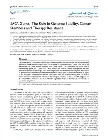
Buffer conditions (buffer, pH, ionic strength) should be optimized for every helicase assayed. ATP·MgCl2 stock solution (200 mM) is prepared using ATP disodium salt hydrate lyophilizedpowder (Sigma). Hydrate content should be checked in the accompanying analysiscertificate. Adjust the pH using potassium hydroxide and supplement with 1 mM Tris-HCl pH7.0. Add stoichiometric amount of MgCl2 prior to storage at -80 C. Fluorescence intensity of specific dyes can be reduced when placed adjacent to a guaninenucleotide. The influence of the position (5′ vs 3′ end of the reporter strand) of the fluorophore onhelicase activity should be tested. Strand displacement assays are typically accompanied with measurements of nucleotidetriphosphate (NTP) consumption to determine how energy expenditure is coupled totranslocation. For some helicases, NTP hydrolysis is dispensable for strand displacementbut is required for enzyme recycling (Liu et al., 2008). The number of NTP used pernucleotide translocation also vary among helicases. Several methods (Brune et al., 1994,Bradley and De La Cruz, 2012, Rajagopal and Lorsch, 2013, Özeş et al., 2014) andcommercial kits are available to monitor NTPase activity.3. RNA unwinding and decay assays of MTR4-exosome complexesRNA unwinding by MTR4 and RNA adaptor complexes ultimately leads to 3′ end trimming ordecay by the exosome. These processes are coupled through physical interaction of MTR4 withthe exosome via nuclear co-factors MPP6 and/or the C1D/EXOSC10 heterodimer. To analyzeRNA unwinding and decay activities of recombinant human MTR4-exosome complexes, wedesigned fluorophore-labeled tripartite substrates (Fig. 3, Table 1) that can be used for gelbased assays (Weick et al., 2018). For decay assays, the translocation strand (strand A) islabeled with a fluorescent moiety whereas for helicase assays, a DNA oligonucleotide (strand B)12
annealed to the translocation strand is conjugated with a fluorophore (Fig. 3). A short poly(A)8overhang is included for interaction with MTR4 (Jia et al., 2011). These substrates can serve astemplates for exploring conditions or modifications that alter MTR4-exosome activities. Thefollowing procedures describe substrate preparation and protocols for helicase and decayassays of MTR4-exosome complexes.3.1 Equipment C1000 Touch Thermal Cycler (Bio-Rad) Nanodrop 2000 (Thermo Fisher Scientific) ThermoMixer (Eppendorf) Typhoon FLA 9500 scanner (GE Healthcare) Xcell SureLock Mini-Cell tank (Thermo Fisher Scientific)3.2 Materials Annealing buffer (10 mM Tris pH 7.0, 100 mM potassium acetate) ATP·MgCl2 solution (200 mM ATP, 200 mM MgCl2, pH adjusted to 7.0 using KOH andsupplemented with in 1 mM Tris-HCl pH 7.0) (See Subsection 2.5 for details) 50 mM ß-mercaptoethanol (BME) DNA capture strand (see Table 1, 3 µM Capture2 in annealing buffer) Lyophilized RNA (See Table 1; synthetic oligos from IDT or Dharmacon) Assay buffer (20 mM HEPES-KOH pH 7.5, 50 mM potassium acetate, 0.5 mM magnesiumacetate, 2.5 mM DTT , 0.01% IGEPAL CA-630) Human placenta RNAse inhibitor (40 U/µl, New England Biolabs)13
Human DIS3/EXOSC10/MTR4-exosome protein complexes in assay buffer engineered withor without mutations in the DIS3 and/or EXOSC10 active sites that disrupt hydrolysis andcatalytic activity for helicase or decay assays, respectively (Weick et al., 2018) 2X Quench solution (1% w/v sodium dodecyl sulfate, 10 mM ethylenediaminetetraaceticacid (EDTA), 10% v/v glycerol, 80 units/ml Proteinase K, 0.01% w/v xylene cyanol) 0.5X TBE running buffer (44.5 mM Tris-borate, 1 mM EDTA, pH 8.3) Novex 12-well 4-20% TBE polyacrylamide gel (Thermo Fisher Scientific)3.3 Protocols3.3.1 Preparation of RNA substrates1. Resuspend and prepare oligonucleotides (StrandA1, StrandA2, StrandB1, StrandB2, andStrandC1) as in steps 1-3 of Subheading 2.4.1.2. Prepare a stock solution of 32 µM for each oligonucleotide.3. For helicase substrate, mix 10 µl of 32 µM StrandA1, 10 µl of 32 µM StrandB2, 10 µl of 32 µMStrandC1 and 10 µl annealing buffer in 0.2 ml PCR tube. For decay substrate, mix 10 µl of 32µM StrandA2, 10 µl of 32 µM StrandB1, 10 µl of 32 µM StrandC1 and 10 µl annealing buffer in0.2 ml PCR tube. Prepare a 10X gel shift unwound control that will be used as a marker forcaptured reporter strand product: mix 35.5 µl annealing buffer, 0.5 µl of 8 µM StrandB2 and 4 µl3 µM Capture2.4. Using Bio-Rad C1000 Touch Thermal Cycler, heat the RNA mixture to 95 C for 5 minfollowed by cooling to 16 C for 10 minutes and 4 C overnight. Transfer RNA mixture to amicrocentrifuge tube and store at -80 C until needed.14
5. Analyze annealed RNA substrate using native polyacrylamide gel electrophoresis. Prepare10 nM RNA substrate or fluorescent reporter strand control (StrandA2 or StrandB2) in 1X assaybuffer. Mix 10 µl 10 nM RNA substrate or 10 nM reporter strand control with 10 µl of 2X quenchsolution. Load 10 µl sample into 12-well 20% Novex TBE polyacrylamide gel. Run gel in 1X TBE(pre-chilled to 4 C) under constant voltage (200 V) for 1 hour at 4 C. Scan gel for 6FAM/fluorescein fluorescence (473 nm laser, LPB filter). More than 95% of the fluorescentstrand should be annealed.3.3.2 RNA unwinding assay of MTR4-exosome complexes1. For each reaction, mix 69 µl assay buffer, 1 µl 40 units/µl human placenta RNAse inhibitor, 10µl 100 nM RNA and 10 µl 200 nM MTR4-exosome complexes with catalytically inertribonucleases (Weick et al., 2018) in a microcentrifuge tube.2. Incubate at 20 C for 5 min.3. For sample at 0 time point, take 18 µl reaction mixture and mix with 2 µl assay buffer and 10µl quench solution.4. Transfer 54 µl reaction mixture to a fresh microcentrifuge tube.5. Prepare a fresh 10X ATP-capture solution. For 50 µl solution, mix 42.5 µl nuclease-freewater, 2.5 µl 200 mM ATP·MgCl2 solution and 5 µl 3 µM DNA capture strand.6. Initiate the reaction by adding 6 µl ATP-capture solution.7. Take 10 µl aliquot of reaction mixture at various time points and terminate the reaction byadding 5 µl quench solution.8. Incubate quenched samples at 30 C overnight to complete proteinase K digestion.9. Rinse each well of a pre-cast Novex 12-well 4-20% TBE polyacrylamide gel with water.15
10. Place the gel cassette in XCell SureLock Mini-Cell tank and fill the upper and lowerchambers with 0.5X TBE running buffer (pre-chilled to 4 C). Flush each well with 0.5X TBErunning buffer.11. Load 3.5 µl sample into each lane. For captured reporter strand marker, mix 39.5 µl assaybuffer, 0.5 µl 40 units/µl human placenta RNAse inhibitor, 5 µl 10X gel shift unwound control, 5µl 20 mM ATP·MgCl2, 50 µl quench solution. Load 3.5 µl captured reporter strand marker into awell.12. Run samples under constant voltage (200 V) for 45 min at 4 C.13. Remove the gel from the cassette and scan for 6-FAM fluorescence (473 nm laser, LPBfilter) using Typhoon FLA 9500 scanner.3.3.3 RNA decay assays of MTR4-exosome complexes1. For each reaction, mix 69 µl assay buffer, 1 µl 40 units/µl human placenta RNAse inhibitor, 10µl 100 nM RNA and 10 µl 20 mM ATP·MgCl2 of MTR4-exosome complexes in a microcentrifugetube with or without mutations in EXOSC10 that disrupt its activity to facilitate analysis of DIS3activities (Weick et al., 2018).2. Incubate at 20 C for 5 min.3. For sample at 0 time point, take 18 µl aliquot of reaction mixture and mix with 2 µl assaybuffer and 10 µl quench solution.4. Pipet 54 µl reaction mixture to a fresh microcentrifuge tube.5. Initiate the reaction by adding 6 µl 1 µM MTR4-exosome complex.6. Take 10 µl aliquot of reaction mixture at various time points and stop the reaction by adding 5µl quench solution.7. Incubate quenched samples at 30 C overnight to complete proteinase K digestion.8. Rinse each well of a pre-cast Novex 12-well 4-20% TBE polyacrylamide gel with water.16
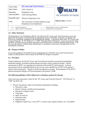
9. Place the gel cassette in XCell SureLock Mini-Cell tank and fill the upper and lower chamberswith 0.5X TBE running buffer. Flush each well with 0.5X TBE running buffer.10. Load 3.5 µl sample into each lane.11. Run samples under constant voltage (200 V) for 45 min at 22 C.12. Remove the gel from the cassette and scan for fluorescein fluorescence (473 nm laser, LPBfilter)using Typhoon FLA 9500 scanner.3.4 Notes Assay buffer can be replaced with 20 mM Tris-HCl pH 7.0, 50 mM NaCl, 0.5 mM MgCl 2, 2.5mM DTT, 0.01% IGEPAL CA-630 to avoid precipitation of SDS by potassium salt.4. Preparation of RNA-loaded human MTR4-exosome complex for structural studiesTo understand how MTR4 and other exosome-associated helicases unwind and feed RNA intothe exosome, we engineered substrates to capture the helicase during the course of RNAtranslocation. We previously reported use of a substrate with a chimeric DNA-RNA translocationstrand (Fig. 4a) to determine the cryogenic electron microscopy (cryo-EM) structure of RNAloaded human MTR4-exosome complex in the process of unwinding and delivering RNA to theexosome (Weick et al., 2018). The DNA-RNA translocation strand allows MTR4 tracking on theRNA segment of the substrate but induces stalling as the helicase reaches the DNA-RNAjunction (Fig. 4b). We designed the length of the translocation strand based on the number ofnucleotides accommodated by MTR4, exosome core, and DIS3 in reported crystal structures ofS. cerevisiae homologs (Weir et al., 2010, Zinder et al., 2016). The optimal length was refinedby performing a helicase reaction followed by a crosslinking assay (Weick et al., 2018). In this17
section, we describe the preparation and loading of a tripartite DNA-RNA chimera substate toMTR4-exosome complex for cryo-EM analysis.4.1 Equipment ÄKTA pure 25 L chromatography system (GE Healthcare) Bio-Rad ChemiDoc XRS C1000 Touch Thermal Cycler (Bio-Rad) Nanodrop 2000 (Thermo Fisher Scientific) ThermoMixer (Eppendorf) Typhoon FLA 9500 scanner (GE Healthcare) XCell SureLock Mini-Cell tank (Thermo Fisher Scientific)4.2 Materials Lyophilized RNA (see Table 1; synthetic oligos from IDT or Dharmacon) Annealing buffer (10 mM Tris-HCl pH 7.0, 100 mM potassium acetate) ATP·MgCl2 (200 mM ATP, 200 mM MgCl2, pH adjusted to 7.0 using KOH and supplementedwith in 1 mM Tris-HCl pH 7.0) (See Subsection 2.5 for details) DNA capture strand (see Table 1, 90 µM Capture2 in annealing buffer) ß-mercaptoethanol (BME) Human placenta RNAse inhibitor (New England Biolabs) Human DIS3/EXOSC10/MTR4-exosome complex with mutations that disrupt DIS3 andEXOSC10 exoribonuclease activity (Weick et. al., 2018; protein complex in 20 mM Tris-HClpH 8.0, 50 mM NaCl, 0.5 mM MgCl2, 2.5 mM BME)18
4X Lithium dodecyl sulfate (LDS) sample buffer (Thermo Fisher Scientific) Loading Buffer (20 mM Tris-HCl pH 8.0, 100 mM NaCl, 2.5 mM BME, 0.5 mM MgCl 2, 20%v/v glycerol, 10 mM EDTA, 0.2% v/v SDS, 50 units/ml Proteinase K) Lyophilized oligonucleotides (see Table 1, synthetic oligos from IDT or Dharmacon) MES running buffer (Thermo Fisher Scientific) Novex 12-well 4-20% TBE polyacrylamide gel (Thermo Fisher Scientific) NuPage 4-12% Bis-Tris polyacrylamide gel (Thermo Fisher Scientific) 2X Quench solution (20 mM Tris-HCl pH 8.0, 50 mM NaCl, 6 mM AMPPNP, 1 mM BME) Superdex S200 Increase 10/300 GL (Thermo Fisher Scientific) SYBR Gold Gel Stain (Thermo Fisher Scientific) 1X TBE running buffer (89 mM Tris-borate, 2 mM EDTA, pH 8.3)4.3 Protocol4.3.1 Substrate preparation1. Resuspend lyophilized, HPLC-purified oligonucleotides in annealing buffer (10 mM Tris pH7.0, 100 mM potassium acetate) to an estimated final concentration of 400 µM.2. Follow steps 2-3 in Section 3.3.1.3. Prepare 200 µl mixture of 100 µM StrandA3, 200 µM StrandB3, and 100 µM StrandC1.Aliquot 50 µl into a PCR 8-tube strip.4. Using a thermocycler, incubate the RNA mixture at 95 C for 5 min, 16 C for 10 minutes then4 C overnight. Pool annealed RNA soluton into a 1.7 ml microcentrifuge tube.6. Purify annealed RNA using Superdex S200 Increase 10/300 GL pre-equilibrated withannealing buffer. Representative chromatogram is shown in Fig. 4c.19
7. Analyze fractions for nucleic acid content using native PAGE. Take 1 µl aliquot and add 19 µlof 1X TBE sample buffer. Resolve samples (10 µl/lane) in Novex 4-20% TBE polyacrylamide gelusing pre-chilled 1X TBE running buffer (200 V for 1 hour at 4 C). Stain gel with 1X SYBR Golddissolved in 1X TBE for 10 minutes. Wash gel with 1X TBE for 10 minutes. Scan gel forfluorescence using Bio-Rad ChemiDoc XRS . Representative native PAGE analysis of nucleicacids in Superdex S200 Increase fractions is shown in Fig. 4d.8. Take peak fractions, determine RNA concentration, and store at -80 C until needed.4.3.2 Preparation of RNA-loaded human MTR4-exosome complex1. Mix 50 µl 9 µM human MTR4-exosome complex with 50 µl 9 µM substrate.2. Prepare fresh ATP-capture solution. For 50 µl solution, mix 20 µl nuclease-fre water, 5 µl 200µM 200 mM ATP·MgCl2 solution and 25 µl 90 µM DNA capture strand. Add 10 µl of 20 mM ATPcapture mix to initiate reaction.3. Incubate rea
human nuclear RNA exosome is composed of 9 core subunits with three S1/KH-domain . & Fraser, 2014). Kinetic data can be obtained in minutes instead of hours as is often required for shift assays where assays are followed by gel electrophoresis and . Typhoon FLA 9500 scanner (GE Healthcare) XCell SureLock Mini-Cell tank (Thermo .