Transcription
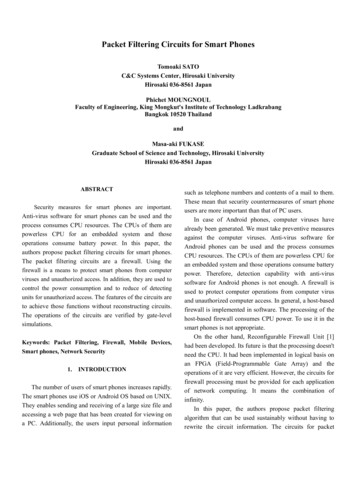
UBICOMP '14, SEPTEMBER 13 - 17, 2014, SEATTLE, WA, USABiliCam: Using Mobile Phones toMonitor Newborn JaundiceLilian de Greef1, Mayank Goel1, Min Joon Seo1, Eric C. Larson2,James W. Stout MD MPH3, James A. Taylor MD3, Shwetak N. Patel112Computer Science & Engineering, DUB GroupComputer Science & Engineering3Department of PediatricsSouthern Methodist UniversityUniversity of WashingtonDallas, TX 75205Seattle, WA 98195{ldegreef, mayankg, minjoon, shwetak, jstout, uncjat}@uw.edu, eclarson@lyle.smu.eduABSTRACTHealth sensing through smartphones has receivedconsiderable attention in recent years because of thedevices’ ubiquity and promise to lower the barrier fortracking medical conditions. In this paper, we focus onusing smartphones to monitor newborn jaundice, whichmanifests as a yellow discoloration of the skin. Although adegree of jaundice is common in healthy newborns, earlydetection of extreme jaundice is essential to preventpermanent brain damage or death. Current detectiontechniques, however, require clinical tests with bloodsamples or other specialized equipment. Consequently,newborns often depend on visual assessments of their skincolor at home, which is known to be unreliable. To this end,we present BiliCam, a low-cost system that usessmartphone cameras to assess newborn jaundice. Weevaluated BiliCam on 100 newborns, yielding a 0.85 rankorder correlation with the gold standard blood test. We alsodiscuss usability challenges and design solutions to makethe system practical.Figure 1. Parents or medical practitioners can monitor a newborn’sjaundice with their smartphones through BiliCam.monitoring [30], pulmonology [18,19], point-of-carediagnostics [14,33], and various other health applications[1]. They demonstrate how the capabilities and ubiquity ofmodern smartphones make them excellent candidates forclinical and health platforms, despite their inherent sensinglimitations. In this paper, we present the design and criticalevaluation of assessing newborn jaundice using asmartphone in a study with 100 newborns. Our prototype,BiliCam, is a smartphone-based medical device that usesthe embedded camera and a paper-based color calibrationcard to monitor newborn jaundice.Author KeywordsHealth sensing, mobile phones, neonatal jaundice, bilirubin,image processing.ACM Classification KeywordsH.5.m. Information interfaces and presentation (e.g., HCI):Miscellaneous.Jaundice is defined as the yellow discoloration of the skincaused by excess bilirubin, a chemical byproduct ofrecycling old blood cells. It is one of the most commonphysiological conditions among newborns: an estimated84% of newborns develop jaundice [5]. A moderate level ofbilirubin is normal in healthy newborns. However, if nottreated, extreme levels can be fatal or cause devastating andirreversible brain damage. Accurate medical tests to assessthis condition require a blood draw or the use of aspecialized measuring device, making them impracticaloutside of medical settings. However, bilirubin levelstypically peak well after most infants are discharged fromthe hospital. Consequently, visual assessment is the mostcommon method to monitor jaundice in a family’s home,where clinical technology is unavailable, as well as at mostINTRODUCTIONA number of smartphone-based medical devices arebecoming increasingly common for fitness [13], heart ratePermission to make digital or hard copies of all or part of this work forpersonal or classroom use is granted without fee provided that copies arenot made or distributed for profit or commercial advantage and that copiesbear this notice and the full citation on the first page. Copyrights forcomponents of this work owned by others than the author(s) must behonored. Abstracting with credit is permitted. To copy otherwise, orrepublish, to post on servers or to redistribute to lists, requires priorspecific permission and/or a fee. Request permissions fromPermissions@acm.org.UbiComp '14, September 13 – 17, 2014, Seattle, WA, USACopyright is held by the author(s). Publication rights licensed to ACM.ACM 978-1-4503-2968-2/14/09 15.00.http://dx.doi.org/10.1145/2632048.2632076331
UBICOMP '14, SEPTEMBER 13 - 17, 2014, SEATTLE, WA, USAEXPLANATION OF JAUNDICE AND TREATMENToutpatient clinics, where administering a blood test islogistically difficult. While parents and clinicians areusually able to visually identify the presence of jaundice,numerous studies show that even experienced healthcareproviders cannot accurately estimate the severity ofjaundice [26]. The importance of monitoring newbornjaundice at home under these conditions creates the needfor an accessible screening system such as BiliCam.Bilirubin is a natural product of the breakdown of expiredred blood cells, which the liver further metabolizes forexcretion. The accumulation of excess bilirubin results inthe yellow discoloration of the skin known as jaundice.Newborns tend to metabolize bilirubin slower (as theirlivers may not function at full capacity yet), have bloodcells with shorter lifespans, and have higher concentrationsof red blood cells than adults. Consequently, jaundice is oneof the most common medical conditions in newborns; up to84% of them develop jaundice during their first week of life[5]. This temporary excess of bilirubin is usually harmless.However, highly elevated concentrations of bilirubin innewborns are neurotoxic and can irreversibly damage thebrain. This potentially lethal condition, called kernicterus,can cause deafness or hearing loss, cerebral palsy, andprofound developmental delay. Fortunately, kernicterus isavoidable through early detection and treatment. Highlevels of bilirubin can be controlled through phototherapy, aprocess that involves bathing the affected newborn inspecific wavelengths of blue light that convert bilirubin intoa harmless, excretable form. For extremely high levels,excess bilirubin must be removed through exchange bloodtransfusions [26].As demonstrated by their recent popularity for healthsensing in the UbiComp community [1], smartphones offerdistinct advantages as a medical platform in terms of cost,accessibility, and computational and sensing capabilities.The programmability and Internet connectivity of thesedevices allow algorithms to adapt much more effectively.Most importantly, their ubiquity enables a multitude offamilies with newborns to use their phones as medicaldevices, helping many of them avoid the cost, anxiety, andhassle of extra hospital visits.By leveraging these inherent advantages of smartphones,BiliCam mitigates the risks in visually assessing jaundice.BiliCam uses the phone’s built-in camera to photograph anewborn. After confirming that the images are usable, thesystem uploads the relevant portions to a server, whichanalyzes the newborn’s skin to estimate the bilirubin level.It then communicates the results back to the user andrecommends a course of action. Each photograph includes acustom, low-cost color calibration card to help BiliCamadjust for different lighting conditions and apply colorcorrections. Other than the smartphone and the colorcalibration card, this non-invasive solution requires noadditional hardware.Medically Accepted Methods to Measure BilirubinTo determine whether a newborn should receivephototherapy or an exchange blood transfusion, doctors ornurses reference specialized graphs with the newborn’s age,number of weeks of gestation, and bilirubin level [26]. Onesuch graph is the Bhutani nomogram [4], like the oneshown in Figure 2, which came from an extensive study byBhutani et al. The nomogram provides a means to assess anewborn’s risk based on the percent of newborns in thestudy with given bilirubin levels and ages. Highintermediate risk is considered above the 75th percentile,and high risk above the 95th percentile. Bilirubin levels arecommonly expressed in milligrams per deciliter (mg/dl) ormicromoles of bilirubin per liter (µmol/L) [26].Our research team includes experienced pediatricians whohave helped to design and evaluate BiliCam. We conducteda clinical study to validate our smartphone-based approachwith 100 newborn participants at the University ofWashington Medical Center. We collected photographs ofnewborns with BiliCam within two hours of measuring totalserum bilirubin levels (TSB), the medical gold standard.BiliCam compares to the TSB with a rank order correlationof 0.85 and a mean error of 2.0 mg/dl. We also compareBiliCam’s results to that of a transcutaneous bilirubinometer (TcB), another non-invasive, although costly,technique that uses structured light to screen for high levelsof bilirubin. We conclude that BiliCam cannot yet replacethe TcB, but offers distinct cost and accessibilityadvantages that make it appropriate for screening newbornsfrom home.We describe the image processing and machine-learningtechniques used to infer bilirubin and show its robustnesswith a range of skin colors and lighting conditions. Intandem with several pilot deployments, these studies alsoinformed important user interface considerations and futuredesigns of BiliCam.Figure 2. A Bhutani nomogram used to assess a newborn's riskbased on bilirubin level and age, generated with risk zoneboundaries from BiliTool [23,24].Clinicians measure the blood concentration of bilirubin on acontinuous scale with either a TSB or TcB. A total serum332
SESSION: HEALTH & CHILDRENbilirubin (TSB) test directly measures the bilirubin from ablood sample. Although invasive, the TSB is the mostaccurate way to measure bilirubin and serves as the medicalgold standard. A transcutaneous bilirubinometer (TcB) is aspecialized meter for a non-invasive alternative thatindirectly measures bilirubin levels. Health practitionerstouch the end of this device to the newborn’s forehead orsternum. It emits specific wavelengths of light andmeasures the resultant reflectance and absorbance of theskin to infer bilirubin levels. TcBs are considered by themedical community to be unreliable above 14.5 mg/dl ofbilirubin, thus high measures from TcBs must be followedby a TSB [26]. In this way, TcBs are used as screeningtools. A TcB also costs several thousands of dollars andrequires frequent calibration. Although large nurseriesusually have more than one TcB, we found that TcBs arenot common in primary care clinician offices and dependon the expected number of newborn patients. Hence, thisscreening tool is not available in all clinics due to its cost.becoming a platform for medical and health applications.Prior work in this relatively new space inspired andmotivated the design of BiliCam. We divide our relatedwork into two categories: smartphone-based health sensingand visual- or camera-based health sensing.Smartphone-Based Health SensingA number of mobile health-sensing systems augment thephone’s capabilities with additional, custom hardware. Acommon example is exercise and physical activitymonitoring systems that use sensors to track movement,such as the commercially available FitBit and Nike . Worklike UbiFit leverages such hardware to provide additionalfeedback and exercise incentives through a phone’sbackground display [13]. In addition to physical activity,smartphones can measure other physiological signs likeheart rate. Poh et al. demonstrated a means of monitoringheart rate through a PPG attached to earbuds while playingmusic [30]. Wello, an upcoming specialized phone caseembedded with sensors, promises to let people measureheart rate, temperature, blood pressure, pulse oximetry, andlung function from their phone. The space stretches beyondmonitoring everyday health — phones are also becomingdiagnostic tools. For instance, Franko et al. built a methodof screening for scoliosis using a smartphone and a customplastic accessory [16]. Smartphones are also beginning toemulate standard medical tools and making them moreaccessible. For example, Mobisanté develops commerciallyavailable hardware that plugs into a phone to generate basicultrasound images, making a more portable and affordablealternative to traditional ultrasound equipment.Visual assessments are common in outpatient settings, suchas physician offices and a family’s home, where theaforementioned technology is unavailable. While bothparents and clinicians are usually able to identify thepresence of jaundice, there is ample evidence from manystudies that even experienced healthcare providers cannotaccurately estimate the severity of jaundice through visualassessement. In studies comparing visual assessment ofjaundice with TSB levels, correlation coefficients aregenerally in the 0.35 – 0.75 range with poor inter-observeragreement [26]. More concerning is evidence indicatingthat clinicians frequently underestimate the severity ofjaundice when using this method. Visual assessments haveeven proven unreliable with the aid of reference colors,such as with an icterometer [26]. Icterometers arespecialized plexiglass rulers marked with different tones ofyellow to reference when pressed against a newborn’s skin[2]. Clinical guidelines explicitly advocate against usingicterometers [26].A number of phone-based medical devices, like BiliCam,do not require external hardware and are purely softwarebased solutions on the existing platform. For instance, thereare pulmonary-focused systems that harness the built-inmicrophone. Our previous work uses the it to measure lungfunction (spirometry) in order to detect and monitor chroniclung conditions, and has achieved results akin to a clinicalspirometer [18]. Another project monitors audio signals totrack the frequency and quality of coughs, helping patientsmonitor coughing episodes and objectively report theircoughing frequency to their doctors [19]. Similarly, Chen etal. used the microphone to continually monitor nasalconditions, such as sneezing and runny nose [12].A number of studies investigate other methods to predicthigh bilirubin levels, including measuring blood bilirubin orantiglobulin in umbilical cords and end-tidal carbonmonozide measurement (ETCOc). They have so far proveneither unsuccessful or unreliable [26].Although there are brief proposals currently posted onlineabout using mobile phone cameras to measure neonataljaundice [3,28], they provide little detail and do notmention any clinical studies. We believe this is the firstwork to describe, in detail, an approach for using mobilephones to screen neonatal jaundice. Furthermore, ourmethods are evaluated in a clinical setting and compared tothe TSB and TcB measures of bilirubin.Vision- or Camera-Based Health SensingLike BiliCam, a number of recent explorations of healthapplications are vision based. The most similar work comesfrom a medical group in Thailand who investigated thefeasibility of screening newborn jaundice with a camerabased system similar to BiliCam. They found a correlationbetween color values from images of newborns, taken witha digital camera, and corresponding bilirubin levels. To doso, they manually inspected and adjusted each image inPhotoshop [20]. Overall, the use of cameras for healthsensing is becoming increasingly popular. For instance,RELATED WORKWith their growing ubiquity, on-board sensors, andcomputational power, smartphones are increasingly333
UBICOMP '14, SEPTEMBER 13 - 17, 2014, SEATTLE, WA, USAsome systems use a phone’s camera to measure heart rateanywhere and anytime by tracking a person’s finger forsubtle flushes in the skin from blood flow [21,22].Researchers have also investigated assisting rehabilitativephysical therapy using a depth camera [11] or with infra-redcameras in a touch-screen table [7]. Smartphones have beenshown to improve and automate point-of-care diagnostics,which require visually analyzing test results from blood orurine samples on specialized materials [14,33]. Othercamera-based systems directly evaluate physiologicalconditions. Pamplona et al. demonstrated a method toscreen eyes for specific impairments using an instrumentedsmartphone camera [27]. Also examining the eye using aphone camera, Bourouis et al. developed a method ofdetecting retinal cancer [8]. Other active areas of researchwith computer vision include recognizing skin cancer [37]and tracking chronic foot ulcers from diabetes [38].more than 35 weeks of gestation (i.e., full term newborns).Of the 134 newborn participants who opted into the study, atotal of 100 completed the study. Participants who requiredphototherapy prior to the follow-up became ineligible, dueto the effect of phototherapy on skin color, which is aknown issue for the TcB [34]. We also noted which bloodsamples were effected by hemolysis, a condition that affectsthe accuracy of TSB readings [10].Medical professionals collected all of the images on iPhone4S smartphones using a custom data collection app and thebuilt-in camera. We chose to use an iPhone because it hasthe most standardized hardware of the current smartphoneplatforms available. The design of this study was informedby a pilot study we ran with 40 newborn participants. Thepilot study data is not included in our evaluation of BiliCamdue to significant differences in study procedure.Data Collection TimelineDATA COLLECTIONWe structured the study to consist of two sets of imagesamples per newborn: a baseline and a follow-up. Thebaseline was taken at the UWMC within the first 24 hoursof life, during which the newborn’s bilirubin is typicallyvery low. The follow-up was taken at either study site whenthe newborn was 2.5 to 5.5 days old. Within two hours ofthe follow-up image, two medical bilirubin measurementswere taken: a TSB blood sample and a TcB. The TSBprovided ground-truth data and the TcB as a source ofcomparison. The TcB measures came from a PhilipsBiliCheck or Draeger Jaundice meter JM-103. We assigneda unique study ID to each participant to match theparticipant’s medical results and image data whilemaintaining confidentiality.To evaluate and inform the design of BiliCam, weconducted a clinical study at two sites in Seattle, theUniversity of Washington Medical Center (UWMC) andthe Roosevelt Pediatric Care Center, to create a dataset ofimage samples paired with ground-truth bilirubin levelsfrom TSB tests. We collected images within two hours ofthe TSB blood draw to ensure that bilirubin measures wereas accurate as possible.EnrollmentParents of newborns born at the UWMC gave informedconsent to participate in the study within 24 hours afterdelivery. Photo samples were taken within these first 24hours of life as a baseline and once more between 2.5 to 5.5days of life for a follow-up. We limited enrollment toEnglish-speaking parents of newborns who were born atAfter receiving the samples from the study phone, wesegmented each image to extract the pixel values of thesternum, forehead, and color patches on the colorcalibration card. The sternum and forehead are the primarylocations of interest for skin samples for several reasons.Medical practices standardize TcBs, which are also lightbased, to take readings from these two locations. Both theforehead and sternum also offer prominent, flat regions ofskin on which we expect even lighting. We expect thewhites of a newborn’s eyes to be more consistent acrossskin tones and potentially a better location of interest for avisual system. However, the eyes are closed (e.g. whilesleeping or crying) the majority of the time. Even whenopen, the whites are hard to discern, given their small sizecompared to the iris.Participant Demographics (N 100)Age at follow-up (hours) (mean, range)86 (60 – 129)Bilirubin Levels (mg/dl) (mean, range)9.9 (0.8 – 21.1)Hemolysis (n, %)19 (19%)Reported Ethnicity (n, %)American Indian/Alaska Native:6 (6%)African American/Black:15 (15%)Asian:20 (20%)Latino:9 (9%)Pacific Islander/Native Hawaiian:3 (3%)White:79 (79%)Other:2 (2%)Multiple RacesData Collection ApplicationFigure 3 shows a screenshot of our custom iPhoneapplication that medical professionals used for datacollection. For each sample, the app records the study ID,time of birth, whether the sample is a baseline or follow-up,and one or more sets of photographs and videos. For takingphotographs, it first instructs the placement of the colorcalibration card on the newborn (Figure 3B) and prompts24 (24%)Table 1. Demographic information for participants. Note thatparticipants may report multiple ethnicities.334
SESSION: HEALTH & CHILDRENA.B.magenta, yellow, and two skin tones (see Figure 4). Thecyan, magenta, and yellow were printed such that only therespective inks were used on each patch; the colors did notcontaminate each other. The cards were printed by a KonicaMinolta Bizhub PRO c6501 printer on Cougar 100lbuncoated paper. We used a new card for each sample toprevent spreading disease. When taking a sample, weaffixed a small, skin-safe adhesive to the back of each cardfor stable placement just above the newborn’s navel withoutbending or occluding the card from the user’s fingers.C.ALGORITHMWe hypothesize that the visual characteristics of anewborn’s skin can estimate his or her bilirubin levels.Considering that the collected images (and thereby anyextracted features) could vary considerably with differentlighting conditions, the images need to be color balancedbefore feature extraction. Our main goals, then, are (1) tocolor balance the images, (2) extract intensities of variousreflected wavelengths and other chromatic and achromaticproperties from the skin, and (3) estimate bilirubin levelsusing machine learning. We explain each stage in turn andshow a method outline in Figure 6. As an overview:Figure 3. Screenshots of data collection application with (A) theview for entering basic sample information, (B) instructions prior torecording images, and (C) a live feed from the camera with a “viewfinder” to align the color calibration card.the user to make sure there is a clear view of the card,sternum, and forehead. The phone then provides a live viewfrom the camera with an overlaid “view finder” to alignwith the calibration card (Figure 3C). These cues constrainthe distance of the camera from the newborn andcomfortably fit the card and newborn’s sternum within theimage. Our data collection application then captures a set ofimages. In case BiliCam needs to account for brief changesin skin color, such as subtle flushes from blood flow, itincludes a 10 second video sequence. The phone’s “flash”LED is on during the first 5 seconds and turns off for thelast. The system also takes a high-resolution photograph inthe middle of each 5-second segment, capturing one image“with flash” and one image “without flash.” color balancing is carried out using the calibration cardscaptured in each image for each skin patch, we estimate the mean red, green,and blue values, and the gradients of colors in the patch we employ various color transformations to approximateproperties such as hue, gamma, and saturation extracted properties are used as features in a stackedregression and classification algorithm, which results ina final estimate of the bilirubin valueThe system first analyzes the captured images to check forsample quality. It detects problems with the images such aspositioning issues, occlusions, or inconsistent lighting, byapplying a threshold on the standard deviation of pixelvalues for each color patch on the card. It then displays thecaptured images and recommends retaking them if anyproblems arose. Upon submitting a completed sample, thesystem uploads the sample data to a server through thephone's Internet connection. It also stores a local copy onthe phone as a backup.Color BalancingWe derive our features from the observed skin color, whichcan vary in different lighting conditions. To mitigate someof the effects of different lighting, we compute normalizedred, green, and blue values. We calculate these normalizedvalues by dividing each color channel value by the sum ofall three channel values. Normalization alone, however, isnot sufficient to counter color variations of illuminationsources (i.e. the differences in halogen, fluorescent, orincandescent bulbs that can cause images to seem more“yellow” or “warm”). Hence, we include our colorcalibration card in each image for further color balancing.In order to use it, the system first needs to identify thelocation of the card and each of its color patches.Image SegmentationAlthough full automation and segmentation is not the focusof this proof of concept for BiliCam, we developed analgorithm to segment the card which we used in automaticimage quality feedback when collecting data. Wesegmented skin patches by hand to reduce confounds.Figure 4: Design of the color calibration card used in our studies.We designed the color calibration card to take the form of abusiness card for easy manufacturing and its appropriatesize on newborns. The card has eight square patches withthe following colors: black, 50% grey, white, cyan,The data collection UI constrained the card to a specificregion on the image (Figure 3C). Hence, the algorithm can335
UBICOMP '14, SEPTEMBER 13 - 17, 2014, SEATTLE, WA, USAFeature Extractionignore the pixels outside of this region to reduce the searchspace. It then locates at least two color patches on the cardand extrapolates the rest of the card from these patches.Elevated levels of bilirubin result in a yellow discolorationof the skin. In order to better detect this subtlediscoloration, BiliCam transforms the original RGB valuesinto the YCbCr and Lab color spaces. We calculate theirmean values for each color channel, resulting in 9 features.To identify the color patches, the algorithm appliesthresholds to the image. The system takes advantage of thefact that the cyan, magenta, and yellow patches have verydistinct hues and high saturation. Hence, it converts theimage to the hue, saturation, and value (HSV) space andapplies empirically determined thresholds on the hue andsaturation channels. Performing a bit-wise ‘AND’ operationof the two thresholded images separates the patch from therest of the image. Figure 5 shows an example ofthresholding for a yellow patch in this manner. Because thatthe system is aware of the approximate size of each patch, itcan differentiate the patch from further noise in the image.This is done by using edge detection and morphologicaloperations; specifically, we use an opening operation andCanny edge detection. The algorithm then uses contourdetection to identify the patch’s boundary from the detectededges and smooths them using the Douglas-Peukeralgorithm [32].In addition to color transformations, we also calculate thechange in color across the image patch using a linear colorgradient. The gradient is calculated by running a 3 3 Sobelgradient filter across each color channel, and then averagingthe outputs inside the patch. This is performed in the R, G,and B color planes, resulting in 3 additional features.The data collection app for BiliCam captures 2 images ineach test: “with-flash” and “without-flash,” as describedearlier. We use mean color features from both images andthe color gradient features from the “with-flash” images,resulting in a total of 9 9 3 21 features. These features areused to train a custom machine learning regressionalgorithm with leave-one-out cross validation. For eachfold, the training set features are transformed to have unitvariance and zero mean (scaling). We also use principlecomponents analysis (PCA) to decrease redundancy (e.g.the redundancy between YCbCr and Lab color spaces) andreduce the dimensionality to six component features. Itlearns these transformations only from the training dataset.After the system finds two of the color patches, it calculatesthe orientation of the card. It then extrapolates the locationsof the remaining patches from these found corners.Machine Learning RegressionThe regression algorithm employs an ensemble of differentregressions. Each regression is chosen to give a slightlydifferent perspective of the feature data. First, the scaled orPCA transformed features are used in each regression toobtain separate estimates of the total bilirubin level. Thenthe outputs of each regression are combined based upon theagreement in the ensemble, resulting in a single value forthe bilirubin level. Figure 6 shows a flowchart of ourmachine learning process.We use an ensemble of five different regression algorithms.Each regression is discussed in turn. Most regressions arecarried out using the scikit-learn toolkit [29] in Python. Inorder to avoid overfitting, we use leave-one-out crossvalidation in all levels of learning. That is, no images fromthe training sets are used in the testing sets for any of theregressions.Figure 5. Segmentation of the yellow patch from a color calibrationcard.White BalancingWe experimented with a number of white balancingtechniques and most effective one to be an algorithm usedby many popular image-editing tools. It uses the observedred, green, and blue (RGB) values of the white color patchto adjust the RGB values of the skin. More precisely, given(R’, G’, B’), it computes the adjusted (R, G, B) byk-Nearest NeighborThe first regression algorithm is an encapsulated k-NearestNeighbor regression (k 7) [17]. Intuitively, this regressiontakes a more “local” estimate of the bilirubin level basedupon training points that have similar feature values. In thisregression, we have a database of known features andbilirubin values. When an unknown test vector is analyzed,the k-nearest neighbors are found around the test vector inthe database of features. The features for finding the nearestwhere (R’w, G’w, B’w) is the average observed color of thewhite patch on the color calibration card [36].336
SESSION: HEALTH & CHILDRENregularization so that it becomes more stable. Theparameters are chosen based on a grid search of the trainingset (but never the test set). As with LARS, we train tworegressions, one with scaled features and another with PCAcomponents.SVRAll the regressions up to this stage were linear regressions.In order to capture the possible non-linear relationship
excess bilirubin must be removed through exchange blood transfusions [26]. Medically Accepted Methods to Measure Bilirubin To determine whether a newborn should receive phototherapy or an exchange blood transfusion, doctors or nurses reference specialized graphs with the newborn's age, number of weeks of gestation, and bilirubin level[26].