Transcription
SURGERYHow to Surgically Remove the Third Eyelid in theStanding HorseJustin S. Harper, DVM, MS, Diplomate ACVSAuthor’s address: Texas Specialty Veterinary Services, PLLC - 111 Shadywood, Boerne, Texas78006; e-mail: jsharperdvm@yahoo.com. 2009 AAEP.1.IntroductionDiseases that can affect the nictating membrane orthird eyelid of the horse include neoplasia, traumatic lacerations, and inflammatory/infectious processes.1,2 Squamous cell carcinoma is, by far, themost reported neoplastic condition associated withthe third eyelid in the horse.1 Diagnosis oftenrequires surgical biopsy for definitive diagnosis.Most often, an excisional biopsy is taken, which combines diagnostics and treatment in the same procedure. Strontium probe brachytherapy, cryotherapy,or hyperthermia, when available, is occasionallyused after excision of the third eyelid to preventreoccurrence of neoplasia.3Horses with neoplasia of the nictating membraneare often referred to veterinary surgical centers tohave removal of the membrane performed undergeneral anesthesia. The anesthetic risks and financial costs associated with general anesthesia canbe prohibitive to some horse owners. Biopsy or removal of the third eyelid can be performed in a safe,routine fashion in the standing horse using appropriate sedation and local anesthesia. In addition toavoiding anesthetic and post-operative anestheticcomplications, standing surgery for removal of thethird eyelid is very economical.4 – 6 Standing sur-NOTES3802009 Ⲑ Vol. 55 Ⲑ AAEP PROCEEDINGSgery for removal of the third eyelid can also be safelyperformed in the field.7The purpose of this paper is to describe the equipment required and anesthetic techniques necessaryfor surgical removal of the third eyelid in the standing horse.2.Materials and MethodsSupplies and EquipmentMost surgical supplies and instrumentation required for standing surgical removal of the nictatingmembrane in equine patients are part of the routineinstruments used by equine practitioners in hospitaland field settings. Specific instruments requiredfor the procedure include Metzenbaum or Mayo scissors, Brown Adson or Graefe fixation forceps, andmosquito or Crile hemostatic tissue forceps. Although performing surgical removal of the nictatingmembrane in the standing horse is safe for the surgeon and patient when performed in stocks, thisprocedure can also be performed under appropriaterestraint, sedation, and local anesthesia in the field.TranquilizationDetomidine HCla (0.01– 0.02 mg/kg, IV) is the sedative most often recommended for performing ophthalmic procedures, because it provides more
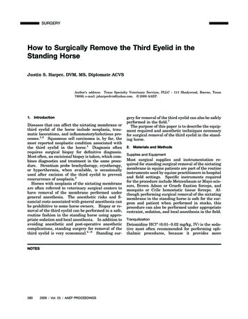
SURGERYFig. 1. Palpebral nerve block. (A) Location of the most common site for palpebral nerve block in the horse is identified by the yellowmarker. (B) Two milliliters of 2% lidocaine HCl can be injected dorsolaterally to the highest point of the zygomatic arch.profound sedation than alternatives such as xylazineb or romifidine hydrochloride.c The author,however, has also performed standing removal ofthe nictating membrane using these agents. Potential advantages of romifidine hydrochloride (40 –120 g/kg, IV) include less ataxia than that whichoccurs with other sedative agents.8 Only one doseof sedation is required for the surgical procedure.The combined use of butorphanol tartrate and thesesedatives can, in some cases, cause unwanted headmovement or exaggerated responses to stimuli.7Nerve Blocks, Topical Anesthesia, and Direct InfiltrativeTechniquesThe third eyelid is innervated by the infratrochlearnerve, a branch of the trigeminal nerve. This membrane passively extends dorsolaterally across theorbit when the eye is retracted. Anesthesia of thethird eyelid can be provided using 2% lidocaine hydrochlorided or 2% mepivicaine hydrochloride.eTo provide adequate paralysis of the eyelids andanalgesia, the author recommends performing thefollowing nerve blocks:1.The palpebral nerve, a branch of the auriculopalpebral nerve, provides motor innervation to the upper eyelid and surroundingadnexal structures. The nerve can beblocked locally where it crosses dorsolaterally to the highest point of the zygomaticarch (Fig. 1, A and B). Anesthesia of thisnerve provides paralysis of the upper eyelidand provides no sensory innervation to theregion. Thus, one could perform surgicalexcision of the third eyelid without this localblock; however, the author routinely blocks2.3.4.5.the palpebral nerve for ophthalmic examination to avoid additional movement of the surrounding structures.The frontal nerve, a branch of the supraorbital nerve, provides sensory innervation ofthe upper medial two-thirds portion of eyelid. The frontal nerve exits the skull fromthe supraorbital foramen and can be palpated proximally to medially over the supraorbital process identified as a depression(Fig. 2, A and B).The infratrochlear nerve, a branch of the trigeminal nerve, provides sensory innervationto the medial canthus of the eye. The nervecan be blocked as it courses through thetrochlear notch located at the medial canthusof the eye (Fig. 3).Some texts describe a retrobulbar nerveblock to adequately immobilize the muscularstructures of the globe and aid in surgicalremoval of the nictating membrane. It is ofthe author’s opinion and experience that thisblock is seldom warranted for successful surgical resection.Direct infiltration of the origin or ventromedial margin of the nictating membrane withanesthetic agent can be performed in addition to the palpebral, frontal, and infratrochlear nerve blocks. After previouslydescribed nerve blocks and appropriate sedation, the nictating membrane is extendeddorsolaterally. Using a 25-gauge, 0.625-inhypodermic needle, local anesthetic agent isdirectly infiltrated into the ventromedialmargin of the nictating membrane (Fig.AAEP PROCEEDINGS Ⲑ Vol. 55 Ⲑ 2009381
SURGERYFig. 2. Frontal nerve block. (A) A yellow marker identifies the location of the frontal nerve block.HCl is injected subcutaneously near the depression of the supraorbital foramen.4). This technique could be substituted forthe infratrochlear nerve block.6. Topical anesthesia of the corneal surface iswarranted and provides desensitization ofthe cornea during the surgical procedure.Topical anesthesia of the cornea can be provided by the application of several drops ofFig. 3. The infratrochlear nerve block is located near the medialcanthus of the eye. Two milliliters of 2% lidocaine HCl is injected subcutaneously.3822009 Ⲑ Vol. 55 Ⲑ AAEP PROCEEDINGS(B) One milliliter of 2% lidocaineophthalmic 0.5% proparacaine hydrochloridef solution.All previously described nerves can be anesthetizedusing 1–3 ml of anesthetic solution injected adjacent to the nerve using a 25-gauge, 0.625-in needle.Local anesthetic agents typically have onset of action in 2– 6 min and last from 30 to 120 min.9Fig. 4. Direct infiltration of 2% lidocaine HCl into ventromedialmargin of the nictating membrane.
SURGERYFig. 5. Hemostatic tissue forceps are placed across the base ofthe third eyelid, proximally to the T-shaped piece of cartilage, toserve as surgical boundaries.Surgical ProcedureAfter all previously mentioned steps have beenperformed, the patient is ready for surgical excision of the third eyelid. A twitch may be required; however, appropriate anesthetic blocksshould provide adequate anesthesia and analgesiafor the procedure.The nictating membrane is composed of conjunctival mucosa, a lacrimal gland that produces por-Fig. 6. Surgical removal of third eyelid.Surgically excised nictating membrane.tions of the aqueous component of the tear film,and a T-shaped piece of cartilage embedded withinthe tissue. When excising the third eyelid, it isrecommended to remove as much of the nictatingmembrane as possible to avoid recurrence of theoriginal disease process.The third eyelid is grasped using surgical tissueforceps, Graefe fixation forceps, or surgical glovedfingers to extend the full portion of the lid. Afterextension dorsolaterally across the globe, twocurved Mosquito or Crile hemostatic tissue forcepsare used to delineate the most ventromedial margins of the nictating membrane. Each hemostatic tissue forcep is placed across the base of thenictating membrane beyond or proximal to theT-shaped piece of cartilage (Fig. 5). The hemostatic forceps serve as surgical excision boundaries and also serve to crush conjunctiva todecrease hemorrhage from the surgical site. Subsequently, scissors (preferably curved Mayo scissors) are used to excise along the border of bothhemostatic forceps, removing the entire third eyelid (Fig. 6, A and B). After removal of the hemostatic forceps, retrobulbar adipose tissue mayoccasionally prolapse from the medial canthus region. This tissue may also be excised using scissors without any untoward effects. Suture repairof the excised margins of the third eyelid can beperformed; however, conjunctiva heals adequatelyby second intention, and the potential of iatrogenic irritation to the cornea from placement ofsuture near the globe is a good reason for notsuturing the wound. The author allows the surgical site to heal by second intention.(A) Surgical excision of nictating membrane along borders created by hemostats.AAEP PROCEEDINGS Ⲑ Vol. 55 Ⲑ 2009(B)383
SURGERYFig. 7. Completed surgical removal of nictating membrane.removal.3.(A) The eye immediately after removal.ResultsFollowing the above recommended sedative and anesthetic techniques, surgical removal or excision ofthe third eyelid can be easily accomplished in thestanding horse. This technique has successfullybeen performed by the author in 20 cases withoutany complications (Fig. 7, A and B). The mostlikely complication to be incurred while performingthis procedure in the standing horse is iatrogenicinduction of ulcerative keratitis. For this reason,the author routinely performs post-surgical flourescien ophthalmic staining of the affected eye andprescribes twice daily topical ophthalmic triple antibiotic ointment application for 5 days after surgery. Other post-operative complications includeretrobulbar adipose tissue prolapse or accumulationof mucopurulent debris in the medial canthus because of the large dead space present.104.DiscussionThe benefits of performing standing surgical excision of the third eyelid far outweigh the risks andadditional financial burden associated with generalanesthesia. Although most minor ophthalmic surgical procedures involving adnexal structures including eyelids, conjunctiva, third eyelid, and theglobe can be performed safely in the standing horse,most corneal or intraocular ophthalmic proceduresrequire general anesthesia. Standing ophthalmicsurgery of the orbital region requires appropriatesedation and the local anesthetic techniques previously discussed.3842009 Ⲑ Vol. 55 Ⲑ AAEP PROCEEDINGS(B) The eye 3 mo post-surgicalThe third eyelid provides a source of seromucinoussecretion to the precorneal tear film and redistributes the tear film across the surface of the cornea.Post-operatively, the removal of the third eyelid canlead to dry eye or keratoconjunctivitis sicca in thecanine patient; however, this is rare in the equinepatient.11 Often caused by pathology or trauma,the third eyelid is completely removed in the equinepatient. Squamous cell carcinoma is the most common neoplasm to affect the third eyelid in the horsethat requires surgical excision. Routine standingsurgical excision of the third eyelid does not seem topresent any deleterious effects to the remainingfunctional eye structures.In conclusion, standing surgical excision of thethird eyelid in the equine patient is a valuable option for equine practitioners in the field or hospitalsetting. It requires routine instruments andknowledge of general sedative and anesthesia techniques available to all equine practitioners.References and Footnotes1. Lavach JD. Large animal ophthalmology. St. Louis, MO:Mosby, 1990.2. Pusterla N, et al. Cutaneous and ocular habronemiasis inhorses: 63 cases (1988 –2002). J Am Vet Med Assoc 2003;222:978 –982.3. Gilger BC. Equine ophthalmology. St. Louis: Elsevier,2005.4. Klein L. Anesthetic complications in the horse. Vet ClinNorth Am [Equine Pract] 1990;6:665– 692.5. Parvianinen AK, Trim CM. Complications associated withanesthesia for ocular surgery: a retrospective study 1989 –1996. Equine Vet J 2000;32:555–559.
SURGERY6. Little D, Redding WR, Blikslager AT. Risk factors for reduced postoperative fecal output in horses: 37 cases (1997–1998). J Am Vet Med Assoc 2001;218:414 – 420.7. Gilger BC. How to prepare for ocular surgery in thestanding horse, in Proceedings. 48th Annual AmericanAssociation of Equine Practitioners Convention 2002;266 –271.8. Clarke KW, England GCW, Goossens L. Sedative andcardiovascular effects of romifidine, alone and in combination with butorphanol, in the horse. J Vet Anaesth 1991;18:25–29.9. Mama KR, Steffey EP. Local anesthetics. In: AdamsHR, ed. Veterinary pharmacology and therapeutics, 8th ed.Ames, IA: Iowa State University Press, 2001;353.10. Whitaker CJG, Wilkie DA. Ophthalmology. In: Rose RJ,Hodgson DR, eds. Manual of equine practice, 2nd ed. W. B.Saunders Co., 2000;427– 450.11. Carastro SM. Equine ocular anatomy and ophthalmic examination. Vet Clin North Am [Equine Pract] 2004;20:285–299.aDormosedan, Pfizer Animal Health, Exton, PA 10017.Tranquived, Vedco, St. Joseph, MO 64504.cSedivet, Boehringer Ingelheim Vetmedica, St. Joseph, 64506.dLidocaine 2% injectable, The Butler Co., Columbus, OH 43228.eCARBOCAINE-V, Pharmacia & Upjohn Company, New York,NY 10017.fAlcaine, Alcon Laboratories, Fort Worth, TX 76134.bAAEP PROCEEDINGS Ⲑ Vol. 55 Ⲑ 2009385
knowledge of general sedative and anesthesia tech-niques available to all equine practitioners. References and Footnotes 1. Lavach JD. Large animal ophthalmology. St. Louis, MO: Mosby, 1990. 2. Pusterla N, et al. Cutaneous and ocular habronemiasis in horses: 63 cases (1988-2002). J Am Vet Med Assoc 2003; 222:978-982. 3. Gilger BC. Equine .