Transcription
ViewFlex Xtra ICE CatheterPositioning Reference Manual
ViewFlex Xtra ICE CatheterThe ViewFlex Xtra ICE Catheter, which is compatible with theViewMate Z and ViewMate II ultrasound consoles, providesvisualization of cardiac structures and blood flow within theheart during electrophysiology and interventional cardiologyprocedures. This “Positioning Reference Manual” can be usedas a guide to understanding the technique of ICE imaging. Thediagrams and ultrasound images represented in the followingtext display the approximate cardiac anatomy seen during atypical ICE overview of the heart. Precise movements in catheter angulations and rotations, for a specific view, may differwith individual cardiac anatomy.Index1.2.3.4.5.6.7.8.9.10.Eustachian RidgeHome ViewAorta, Right Ventricular Outflow TractCoronary SinusIntra-Atrial Septum, Fossa OvalisMitral Valve, Left Atrial AppendageLeft Pulmonary VeinsRight Pulmonary VeinsSuperior Vena Cava, Right AtriumLeft Ventricle Long Axis
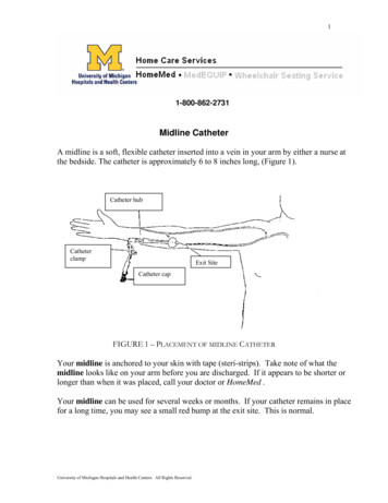
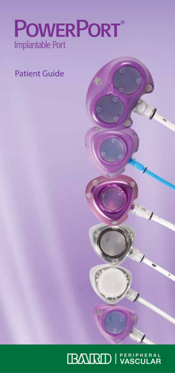
Eustachian RidgenAs the ICE catheter enters the right atrium, the eustachian ridge isthe first anatomical structure that can be visualized.EustachianRidgeLARATVOrientationAORV1
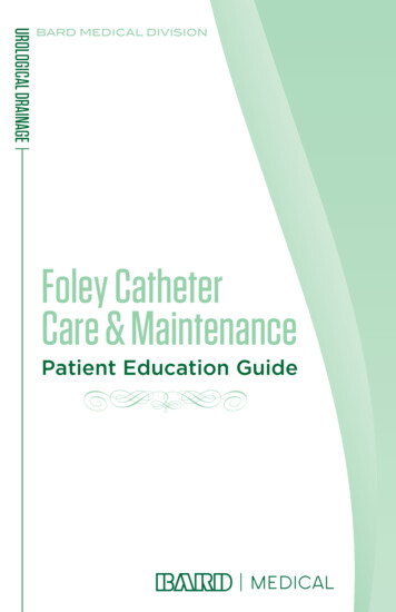
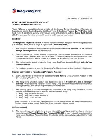
Home ViewnnnContinue advancing the catheter to the level of the mid-right atriumThe ultrasound array should be facing anteriorlyThere is no flexion on the catheterRATVOrientationRV2
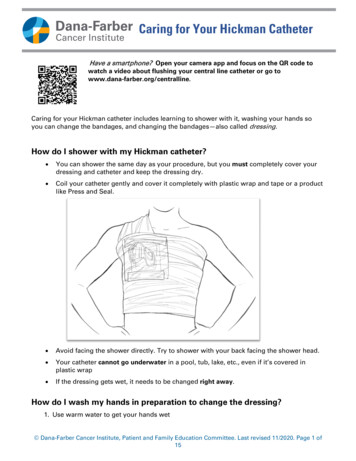
Aorta, Right Ventricular Outflow TractnnFrom the “Home View,” rotate the catheter clockwiseNo flexion on the catheter is necessaryRAAOOrientationRVOTRV3
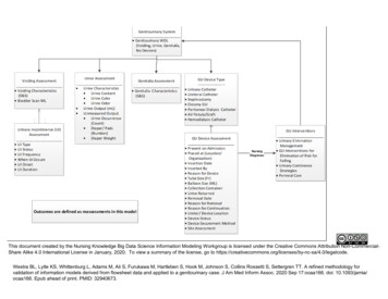
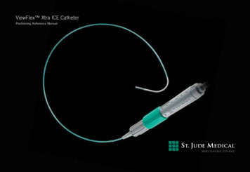
Coronary SinusnGradually rotate the catheter clockwise to visualize the ostium ofthe coronary sinusRACSOrientationLA4
Intra-atrial Septum, Fossa OvalisnnRotate the catheter clockwisePosterior flexion can be used if necessary to view more of theright atriumIntra-atrialSeptumRALAOrientation5
Mitral Valve, Left Atrial AppendagenFrom the “Intra-atrial Septum View,” gradually rotate thecatheter clockwiseRALACSOrientationMVLAALV6
Left Pulmonary VeinsnnnFrom the “Left Atrial Appendage View,” remove the posterior flexion appliedto the catheterSlightly rotate the catheter clockwiseColor Doppler can be used to identify the left pulmonary veinsLAOrientationLSPVLIPV7
Right Pulmonary VeinsnnRotate the catheter clockwise past the left pulmonary veinsAdvance the catheter slightly to obtain a better view of the rightpulmonary veinsRSPVOrientationRIPV8
Superior Vena Cava, Right AtriumnnnPlace the catheter in the mid-right atriumFlex the catheter posteriorlyIf necessary, rotate the catheter counter-clockwise to obtain a betterview of the superior vena cava and right atriumRAOrientationSVCLA9
Left Ventricle Long AxisnnnnRe-establish Home ViewOnce the tricuspid valve is clearly visible, flex the catheter anteriorly(toward tricuspid valve)Utilizing fluoroscopy and ultrasound advance the catheter past thetricuspid valve annulus into the right ventricle and then remove anterior flexionRotate the catheter clockwise until the intraventricular septum comes into viewOrientationLAAOLV10
(100057675EN)Atrial FibrillationCardiac Rhythm ManagementCardiovascularGlobal HeadquartersOne St. Jude Medical DriveSt. Paul, Minnesota 55117USA 1 651 756 2000 1 651 756 3301 FaxAtrial Fibrillation DivisionOne St. Jude Medical DriveSt. Paul, Minnesota 55117USA 1 651 756 2000 1 651 756 3301 FaxSJM Coordination Center BVBAThe Corporate VillageDa Vincilaan 11 Box F11935 ZaventemBelgium 32 2 774 68 11 32 2 772 83 84 FaxSt. Jude Medical (Hong Kong) LtdSuite 1608, 16/F Exchange Tower33 Wang Chiu RoadKowloon Bay, KowloonHong Kong SAR 852 2996 7688 852 2956 0622 FaxSt. Jude Medical Japan Co., Ltd.Shiodome City Center 15F1-5-2, Higashi-ShinbashiMinato-ku, Tokyo 105-7115JapanTel: 81 3 6255 6370Fax: 81 3 6255 6371St. Jude Medical Australia Pty Ltd.17 Orion RoadLane Cove NSW 2066Australia 61 2 9936 1200 61 2 9936 1222 FaxNeuromodulationSt. Jude Medical Brasil Ltda.Rua Itapeva, 5385º ao 8º andar01332-000 – São Paulo – SPBrazil 55 11 5080 5400 55 11 5080 5423 FaxSJMprofessional.comRx OnlyBrief Summary: Please review the Instructions for Use prior to using these devices for a complete listing of indications, contraindications, warnings, precautions,potential adverse events, and directions for use.ViewMate Z, ViewMate II, ViewFlex, ST. JUDE MEDICAL, the nine-squares symbol and MORE CONTROL. LESS RISK. are trademarks and service marks of St. JudeMedical, Inc. and its related companies. 2012 St. Jude Medical. All Rights Reserved.Item 100057675EN Rev A
ViewFlex Xtra ICE Catheter The ViewFlex Xtra ICE Catheter, which is compatible with the ViewMate Z and ViewMate II ultrasound consoles, provides visualization of cardiac structures and blood flow within the heart during electrophysiology and interventional cardiology procedures. This "Positioning Reference Manual" can be used