Transcription
Human Neutrophil Elastase Degrades SPLUNC1 andImpairs Airway Epithelial Defense against BacteriaDi Jiang1, Sally E. Wenzel2, Qun Wu1, Russell P. Bowler1, Christina Schnell1, Hong Wei Chu1*1 Department of Medicine, National Jewish Health, Denver, Colorado, United States of America, 2 Department of Medicine, University of Pittsburgh School of Medicine,Pittsburgh, Pennsylvania, United States of AmericaAbstractBackground: Acute exacerbations of chronic obstructive pulmonary disease (AECOPD) are a significant cause of mortality ofCOPD patients, and pose a huge burden on healthcare. One of the major causes of AECOPD is airway bacterial (e.g.nontypeable Haemophilus influenzae [NTHi]) infection. However, the mechanisms underlying bacterial infections duringAECOPD remain poorly understood. As neutrophilic inflammation including increased release of human neutrophil elastase(HNE) is a salient feature of AECOPD, we hypothesized that HNE impairs airway epithelial defense against NTHi by degradingairway epithelial host defense proteins such as short palate, lung, and nasal epithelium clone 1 (SPLUNC1).Methodology/Main Results: Recombinant human SPLUNC1 protein was incubated with HNE to confirm SPLUNC1degradation by HNE. To determine if HNE-mediated impairment of host defense against NTHi was SPLUNC1-dependent,SPLUNC1 protein was added to HNE-treated primary normal human airway epithelial cells. The in vivo function of SPLUNC1in NTHi defense was investigated by infecting SPLUNC1 knockout and wild-type mice intranasally with NTHi. We found that:(1) HNE directly increased NTHi load in human airway epithelial cells; (2) HNE degraded human SPLUNC1 protein; (3)Recombinant SPLUNC1 protein reduced NTHi levels in HNE-treated human airway epithelial cells; (4) NTHi levels in lungs ofSPLUNC1 knockout mice were increased compared to wild-type mice; and (5) SPLUNC1 was reduced in lungs of COPDpatients.Conclusions: Our findings suggest that SPLUNC1 degradation by neutrophil elastase may increase airway susceptibility tobacterial infections. SPLUNC1 therapy likely attenuates bacterial infections during AECOPD.Citation: Jiang D, Wenzel SE, Wu Q, Bowler RP, Schnell C, et al. (2013) Human Neutrophil Elastase Degrades SPLUNC1 and Impairs Airway Epithelial Defenseagainst Bacteria. PLoS ONE 8(5): e64689. doi:10.1371/journal.pone.0064689Editor: Samithamby Jeyaseelan, Louisiana State University, United States of AmericaReceived March 18, 2013; Accepted April 17, 2013; Published May 31, 2013Copyright: ß 2013 Jiang et al. This is an open-access article distributed under the terms of the Creative Commons Attribution License, which permitsunrestricted use, distribution, and reproduction in any medium, provided the original author and source are credited.Funding: This work was supported by grants from Flight Attendant Medical Research Institute and R01 HL088264 from the National Heart, Lung, and BloodInstitute, National Institutes of Health. The funders had no role in study design, data collection and analysis, decision to publish, or preparation of the manuscript.Competing Interests: The authors have declared that no competing interests exist.* E-mail: chuhw@njhealth.orgdegradation of surfactant proteins and induction of emphysema[8–10].Because airway epithelial cells represent the first line of lungresponses to invading bacteria, we sought to determine if HNEimpairs airway epithelial host defense functions. In the currentstudy, we focused on the effects of HNE on the function of arecently described host defense protein short palate, lung, andnasal epithelium clone 1 (SPLUNC1). A previous study suggeststhat HNE is involved in SPLUNC1 degradation [11]. However,the role of HNE-mediated SPLUNC1 degradation in host defenseagainst bacterial infection has not been investigated.SPLUNC1 is produced by large airway epithelial cells inhumans and mice [12–14]. Human and mouse SPLUNC1proteins at their precursor (non-secreted) forms have 256 and278 amino acids (AA), respectively. When secreted, the N-terminal19 AA signal peptide of SPLUNC1 precursor is cleaved to formthe mature protein. Based on its structural homology tobactericidal permeability-increasing protein (BPI, a protein mainlyfrom neutrophil granules) [15], SPLUNC1 has been predicted toexert an antimicrobial activity [16,17]. Our published data suggestthat SPLUNC1 inhibited the growth of Mycoplasma pneumoniae (Mp)IntroductionrdCOPD is the 3 leading cause of death in the US [1,2]. One ofthe major challenges in COPD healthcare is to prevent and treatacute exacerbations of COPD (AECOPD). AECOPD is mainlycaused by respiratory bacterial (e.g., nontypeable Haemophilusinfluenzae, NTHi) and viral (e.g., rhinovirus) infections [3].Recurrent respiratory infections in COPD patients suggestimpaired lung defense mechanisms. Although various mechanismshave been proposed or investigated for respiratory infectioninduced AECOPD, many research questions remain to beanswered. Further defining mechanisms underlying AECOPD isnecessary to discover more effective therapies.COPD lung including airways is inflamed, especially duringAECOPD. One of the prominent inflammatory cells in COPDlungs is neutrophils [4,5]. During their activation, neutrophilsrelease various mediators such as cytokines and proteases, whichcontribute to COPD pathogenesis including AECOPD. Amongthe proteases released from neutrophils, human neutrophil elastase(HNE) is particularly important. First, HNE levels are higher inCOPD patients than normal subjects [6,7]. Second, HNE exertsvarious deleterious effects on lung immunity and structure such asPLOS ONE www.plosone.org1May 2013 Volume 8 Issue 5 e64689
Neutrophil Elastase Dampens SPLUNC1 Functionwere collected to measure SPLUNC1 levels by using an ELISA[18,23].[18], a bacterium involved in asthma as well as COPD. Using ourrecently generated SPLUNC12/2 mice, we further confirmed thein vivo host defense function of SPLUNC1 during Mp infection[19]. Reduced SPLUNC1 protein has been reported in nasallavage fluid of ‘‘healthy’’ human smokers [20,21]. Moreover,SPLUNC1 expression is lower in brushed bronchial epithelial cellsfrom ‘‘healthy’’ smokers than healthy non-smokers [22].We hypothesized that HNE impairs human lung defensefunctions by decreasing airway epithelial SPLUNC1 levels. Ourcurrent study demonstrates that: (1) HNE directly reduced humanairway epithelial defense against NTHi; (2) HNE degraded humanSPLUNC1 protein; (3) Recombinant SPLUNC1 protein reducedNTHi levels in HNE-treated human airway epithelial cells; (4)NTHi levels in lungs of SPLUNC1 knockout mice were increasedcompared to wild-type mice; and (5) SPLUNC1 was reduced inlungs of COPD patients. Together, our findings provide a novelmechanism underlying bacterial infections in COPD patients, anda potential therapy to more effectively treat AECOPD.NTHi Infection in Cultured Well-differentiated PrimaryHuman Airway Epithelial CellsNontypeable Haemophilus influenzae (NTHi) strain 12 (kindlyprovided by Dr. Stephen J. Barenkamp, Saint Louis UniversitySchool of Medicine, Saint Louis, MO) was plated on chocolateagar plates and incubated overnight at 37uC in 5% CO2. A singlecolony was used to inoculate 10 ml of brain heart infusion broth(Remel, Lenexa, KS), and allowed to grow overnight. Thesubculture was then washed twice with PBS and the opticaldensity (O.D.) at a wavelength of 620 nm was determined toobtain the colony-forming unit (CFU) [27].To determine if HNE treatment impairs epithelial defenseagainst NTHi, cells on day 10 of ALI culture were treated at theapical surface with 50 ml of HNE (10 mg/ml) in the presence orabsence of NTHi at 103 CFU/transwell [28,29]. After 48 hrs ofinfection, apical supernatants were collected to measure NTHilevels. To quantify cell-associated NTHi, epithelial cells weredissociated by incubation with collagenase I. Briefly, cells on thetranswell membrane were treated at 37uC for 2 hrs withcollagenase I (0.25 mg/ml) supplemented with DNase I (10 mg/ml), EDTA (2 mM) and DTT (0.5 mg/ml). Thereafter, cells werescraped and transferred into a 1.5 ml tube for incubation with thecollagenase solution for additional 3 hrs at 37uC on a rocker(225 rpm) to further dissociate the cells. Thereafter, cells werespun down to remove the collagenase solution, followed by addingthe trypsin/EDTA solution for incubation at 37uC for 5 min.After removal of trypsin/EDTA solution, cell pellets wereresuspended in PBS and plated on chocolate agar plates toquantify NTHi.To test if SPLUNC1 reduction in HNE-treated cells isresponsible for increased NTHi levels, recombinant humanSPLUNC1 protein (10 mg/ml) or control protein from HEK293cells was added to the apical surface of epithelial cells after 2 hrs ofHNE treatment and NTHi infection. After 46 hrs of SPLUNC1treatment or after 48 hrs of NTHi infection, apical supernatantsand cells were harvested for NTHi quantification.MethodsEthics StatementExperimental animals used in this study were covered by aprotocol approved by Institutional Animal Care and UseCommittee (IACUC) of National Jewish Health, Denver, Colorado, USA. All experimental procedures were carried out tominimize animal discomfort, distress, and pain by following theAmerican Veterinary Medical Association Guidelines. All humanmaterials such as bronchoalveolar lavage used in this study wereapproved by Institutional Review Board (IRB) of National JewishHealth, Denver, Colorado, USA. The written informed consentwas waived by National Jewish Health IRB for de-identified organdonors without lung diseases from whom primary normal humantracheobronchial epithelial cells were obtained through theInternational Institute for the Advancement of Medicine (IIAM)(http://www.iiam.org).Generation of Recombinant Human SPLUNC1 ProteinRecombinant human SPLUNC1 protein was generated as wepreviously reported [23,24].NTHi Infection in SPLUNC1 Knockout MiceIncubation of Recombinant Human SPLUNC1 Proteinwith Human Neutrophil Elastase (HNE)SPLUNC1 knockout (KO) mouse model was used to reveal thein vivo function of SPLUNC1 against NTHi. SPLUNC1 KO miceon the C57BL/6 background were generated in our laboratoryand bred under pathogen-free housing conditions as previouslyreported [19]. SPLUNC1 KO and wild-type control mice wereintranasally inoculated with NTHi (105 colony forming unit[CFU]/mouse in 50 ml saline), and sacrificed after 24 hrs ofinfection to obtain the lung tissue for NTHi quantification. Briefly,lung tissues were homogenized and plated on chocolate agar paltesto count NTHi CFUs. All the experimental protocols wereapproved by Institutional Animal Care and Use Committee(IACUC) at National Jewish Health.HNE was purchased from the Elastin Product Company(Pacific, MO), and diluted in 0.05 M sodium acetate solutionwith 0.1 M NaCl (pH 5.0). SPLUNC1 protein at 10 mg/ml, aphysiologic dose as we reported [23], was incubated with HNE(1 mg/ml [or 0.04 U, enzymatic activity] to 50 mg/ml [2.18 U], adose range reflecting COPD lungs [25]) or 0.05 M sodium acetatesolution (vehicle, control) in 96-well plates at 37uC for 30 minutes.At the end of incubation, SPLUNC1 protein was analyzed byWestern blot.HNE Treatment in Cultured Well-differentiated PrimaryHuman Airway Epithelial CellsMeasurement of SPLUNC1 in COPD LungsPrimary normal human tracheobronchial epithelial cells wereobtained from tracheas and bronchi of deidentified organ donorsby digesting the tissue with 0.2% protease solution and thensubjected to air-liquid interface (ALI) cultures, as we reported [26].On day 6 of ALI culture, antibiotic gentamicin was removed fromboth apical surface and basolateral side. On day 10 of ALI culture,cells were treated at the apical surface with HNE (1 to 50 mg/ml)or vehicle solution. At indicated time points, apical supernatantsPLOS ONE www.plosone.orgWe obtained bronchoalveolar lavage (BAL) fluid from healthynon-smokers and smokers, as well as smokers with moderate tosevere COPD patients (Table 1) to measure SPLUNC1 protein byusing a SPLUNC1 ELISA [18,23]. Our research protocol wasapproved by the Institutional Review Board (IRB) at NationalJewish Health.2May 2013 Volume 8 Issue 5 e64689
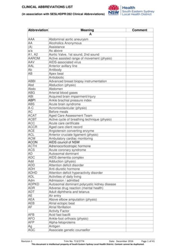
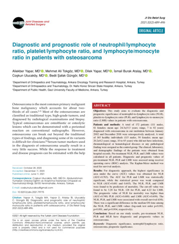
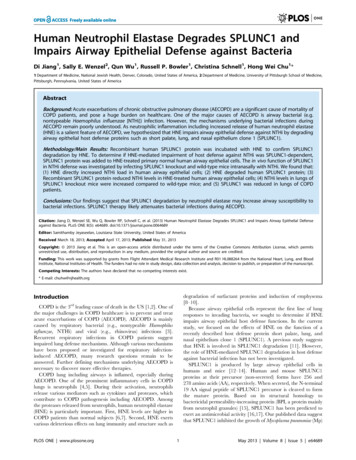
Neutrophil Elastase Dampens SPLUNC1 FunctionTable 1. Characteristics of human subjects.SubjectsNAgeSex (M/F)Smoking (pack-years) FEV1, % predictedFVC, % 5.3stHNS Healthy non-smokers; HS Healthy smokers; COPD Chronic obstructive pulmonary diseases; FEV1 forced expiratory volume in the 1 second; FVC forcedvital capacity.doi:10.1371/journal.pone.0064689.t001a quadrupole time-of-flight (QTOF) mass spectrometer to identifythe 3-kD peptide. As shown in Figure 1B, a 25 amino acid peptide(F21-T45, total mass: about 3-kD) at the N-terminus of SPLUNC1protein was identified.Statistical AnalysesOne-way analysis of variance (ANOVA) was used for multiplecomparisons, and a Turkey’s post hoc test was applied whereappropriate. P,0.05 was considered significant.HNE Degrades SPLUNC1 Protein in Cultured Welldifferentiated Human Airway Epithelial CellsResultsHNE Degrades Recombinant SPLUNC1 ProteinHaving shown the ability of HNE to degrade recombinantSPLUNC1 protein in a cell-free environment, we tested if HNEreduces SPLUNC1 protein secreted from primary normal humanairway epithelial cells that were well differentiated under air-liquidinterface culture. HNE dose-dependently decreased SPLUNC1after 4 hrs of HNE treatment (Figure 2A). HNE effects onSPLUNC1 were sustained even after 48 hrs of HNE treatment(Figure 2B).To test if HNE directly degrades SPLUNC1, recombinanthuman SPLUNC1 protein at a physiological dose (10 mg/ml) wasincubated with HNE at various doses for 30 minutes. Western blotanalysis demonstrated that HNE treatment significantly reducedintact SPLUNC1 (25-kD), and resulted in the formation of a 22kD SPLUNC1 fragment (loss of a 3-kD peptide) (Figure 1A). Wethen used liquid chromatography mass spectrometry (LC/MS) onFigure 1. Degradation of SPLUNC1 protein by human neutrophil elastase (HNE). (A) Western blot of recombinant human SPLUNC1 protein(10 mg/ml) that was incubated with HNE for 30 minutes. HNE treatment dose-dependently reduces the native SPLUNC1 protein (25 kD). A fragmentof 22-kD SPLUNC1 was seen in HNE-treated samples. (B) Mass spectrometry analysis of NHE-treated recombinant human SPLUNC1 protein revealed a25 amino acid peptide F21-T45 ne.0064689.g001PLOS ONE www.plosone.org3May 2013 Volume 8 Issue 5 e64689
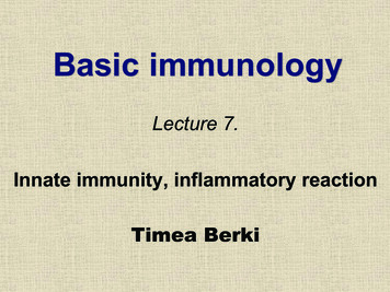
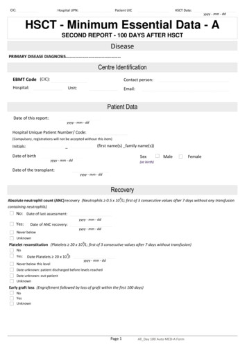
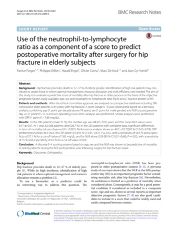
Neutrophil Elastase Dampens SPLUNC1 FunctionFigure 2. Human neutrophil elastase (HNE) decreases SPLUNC1 protein in cultured human airway epithelial cells. Primary normalhuman tracheobronchial epithelial cells (N 3) were differentiated into the mucociliary phenotype under air-liquid interface culture for 10 days. After4 (A) and 48 (B) hrs of HNE treatment, SPLUNC1 protein in apical supernatants was measured by an ELISA. Data are expressed as means 6 SEM.doi:10.1371/journal.pone.0064689.g002different donors. Although bacterial levels in HNE-treatedepithelial cells were slightly reduced by SPLUNC1, this reductionwas not statistically significant (Figure 4B). Third, we performed atime course study to observe the rescuing effect of recombinantSPLUNC1 protein on HNE-mediated SPLUNC1 reduction. Asshown in Figure 5, in HNE-treated cells, recombinant SPLUNC1protein increased SPLUNC1 levels at the apical surface for theentire 48 hrs, particularly during the first 2 hrs.HNE Dose-dependently Increases Bacterial Load in Welldifferentiated Human Airway Epithelial CellsTo determine the effects of HNE on bacterial load, apicalsurface of cultured well-differentiated normal human airwayepithelial cells was infected with NTHi for 48 hrs in the presenceor absence of HNE. NTHi levels in the apical supernatants(Figure 3A) and cells (Figure 3B) were markedly increased in aHNE dose-dependent manner.Increased Lung NTHi Load in SPLUNC1 Knockout MiceRecombinant Human SPLUNC1 Protein Reduces NTHiLoad in HNE-treated Human Airway Epithelial CellsWe have shown that SPLUNC1 in cultured human airwayepithelial cells is critical to NTHi defense, but the in vivo functionof SPLUNC1 during NTHi infection is unclear. We utilized ourSPLUNC1 knockout (SPLUNC12/2) mouse model to delineatethe in vivo function of SPLUNC1. After 24 hrs of an intranasalinoculation of NTHi, bacterial load in SPLUNC12/2 mice wasabout 30-fold higher than that in wild-type mice (Figure 6).To determine if HNE-mediated SPLUNC1 reduction isresponsible for impaired airway epithelial defense against NTHi(e.g., increased bacterial load), recombinant human SPLUNC1protein (10 mg/ml) was added 2 hrs after HNE treatment andNTHi infection. After 48 hrs of infection, NTHi levels weremeasured in apical supernatants and cells. First, in line with datashown in Figure 3, HNE increased NTHi load in both apicalsupernatants and epithelial cells. Second, recombinant SPLUNC1protein consistently reduced NTHi (43% reduction, p,0.05) inapical supernatants (Figure 4A) of epithelial cells obtained from 5Reduced SPLUNC1 in Human COPD LungsWe measured SPLUNC1 protein levels in BAL fluid of healthynon-smokers, healthy smokers and stable moderate to severeCOPD patients. As shown in Figure 7, SPLUNC1 protein wasFigure 3. Human neutrophil elastase (HNE) dose-dependently increases the load of nontypeable Haemophilus influenzae (NTHi) incultured human airway epithelial cells. Primary normal human tracheobronchial epithelial cells (N 3) were differentiated into the mucociliaryphenotype under air-liquid interface culture for 10 days, and then infected with NTHi for 48 hrs. (A) – NTHi in apical supernatants; (B) – NTHiassociated with epithelial cells. Data are expressed as means 6 SEM.doi:10.1371/journal.pone.0064689.g003PLOS ONE www.plosone.org4May 2013 Volume 8 Issue 5 e64689
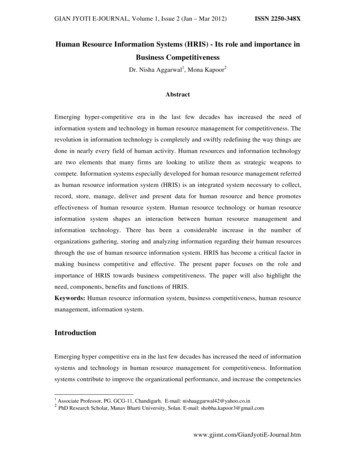
Neutrophil Elastase Dampens SPLUNC1 FunctionFigure 4. Recombinant human SPLUNC1 protein significantly reduces the load of nontypeable Haemophilus influenzae (NTHi) inapical supernatants of human neutrophil elastase (HNE)-treated human airway epithelial cells. Primary normal human tracheobronchialepithelial cells (N 5) were differentiated into the mucociliary phenotype under air-liquid interface culture for 10 days, and then infected with NTHi(103 CFU/transwell) in the presence or absence of HNE (10 mg/ml). After 2 hrs of NTHi and HNE treatment, recombinant human SPLUNC1 protein(10 mg/ml) was added to the apical surface of epithelial cells. After 46 hrs of SPLUNC1 treatment or after 48 hrs of NTHi infection, apical supernatantsand cells were harvested for NTHi quantification. (A) – NTHi in apical supernatants; (B) – NTHi associated with epithelial cells. Each connected colorline represents the data collected from cells of an individual donor. The horizontal dotted red lines indicate medians of NTHi fold changes (HNEtreatment versus vehicle solution eased lung bacterial load in SPLUNC1 knockout mice.Together, our data suggest that increased HNE, a feature ofexcessive inflammation during acute exacerbations of COPD andother chronic lung diseases, may contribute to bacterial infectionsin part through degrading host defense protein SPLUNC1.Acute exacerbations of COPD (AECOPD) pose the highest risk(i.e., mortality) to patients and the greatest costs for COPD-relatedhealthcare. Respiratory bacterial and viral infections are the majorcause of AECOPD, which commonly recruit neutrophils into theairway lumen and activate them. During neutrophil activation,they release various mediators including HNE. Although the NEknockout mouse model suggests a beneficial role of intracellularNE in host defense against bacterial (e.g., Pseudomonas aeruginosa)infection [30], the impact of released HNE on lung (airway)bacterial infections remains poorly understood. We have foundincreased bacterial (e.g., NTHi) load in HNE-treated welldifferentiated primary human airway epithelial cells. Our datasuggest a new mechanism underlying frequent bacterial infectionduring AECOPD that is characterized by excessive lungsignificantly reduced in smokers with or without COPD ascompared to healthy non-smokers.DiscussionThe current study demonstrates that human neutrophil elastase(HNE)-mediated SPLUNC1 degradation impairs airway epithelialdefense against bacterial infection. We found that HNE markedlyreduces SPLUNC1 in well-differentiated human airway epithelialcells and subsequently increases bacterial load, which can beattenuated by recombinant human SPLUNC1 protein. Moreover,we found decreased SPLUNC1 in human COPD lungs, andFigure 5. Time course study of the rescuing effects ofrecombinant human SPLUNC1 protein on SPLUNC1 proteinlevels in human neutrophil elastase (HNE)-treated humanairway epithelial cells. Primary normal human tracheobronchialepithelial cells (N 3) were differentiated into the mucociliaryphenotype under air-liquid interface culture for 10 days, and thentreated with HNE (10 mg/ml) in the presence or absence of recombinanthuman SPLUNC1 protein (10 mg/ml). At indicated time points, SPLUNC1protein in the apical supernatants was measured by using an ELISA.Data are expressed as means 6 SEM.doi:10.1371/journal.pone.0064689.g005PLOS ONE www.plosone.orgFigure 6. SPLUNC1 knockout (SPLUNC12/2) mice increase theload of nontypeable Haemophilus influenzae (NTHi) in the lung.SPLUNC12/2 and wild-type mice (n 7 or 8 mice/group) wereintranasally inoculated with NTHi (105 CFUs/mouse). After 24 hrs ofinfection, left lungs were homogenized to quantify NTHi load byculture. The horizontal lines represent means of CFUs.doi:10.1371/journal.pone.0064689.g0065May 2013 Volume 8 Issue 5 e64689
Neutrophil Elastase Dampens SPLUNC1 Functionour cell culture model. The contribution of HNE-mediatedSPLUNC1 reduction versus epithelial barrier disruption toincreased bacterial load will be investigated in our future studies.Although the current study was focused on in vitro studies, weexamined SPLUNC1 levels in COPD lungs. We found reducedSPLUNC1 levels in COPD patients as compared to healthycontrols, especially healthy non-smokers. Interestingly, SPLUNC1levels in healthy smokers were less than those in healthy nonsmokers. We realize that SPLUNC1 reduction in vivo may becaused by multiple factors. We measured HNE levels in BAL fluidof COPD patients who were stable during the BAL procedure andcontrol subjects, and did not find a significant difference of HNE.This may be explained by the fact that our patients undergoingbronchoscopy and BAL fluid procedure were stable (no acuteexacerbations). Therefore, SPLUNC1 reduction in stable COPDpatients may be caused by other factors such as cigarette smokeexposure. In a pilot study, we exposed well-differentiated humanairway epithelial cells to whole cigarette smoke or air (control) for10 minutes as we described before [33]. After 24 hrs of cigarettesmoke exposure, SPLUNC1 levels in the apical surface werereduced by 40%. Future studies should correlate SPLUNC1 levelswith HNE and bacterial load in COPD patients before, duringand after acute exacerbations. This could be done by obtainingsputum samples, as sputum collection is safer than the bronchoscopy procedure for patients during AECOPD.Our current study has several limitations. First, we only usednormal human airway epithelial cells to study the impact of HNEon SPLUNC1 degradation and bacterial infection. We willconsider COPD airway epithelial cells in our future studies todetermine if COPD cells may differ from normal cells in theirresponses to HNE and NTHi infection. Second, since NTHi wasthe only strain of bacteria examined in the current study, ourresults should be verified in other strains of bacteria involved inCOPD, including Pseudomonas aeruginosa. Third, HNE may degradeother antimicrobial substances (e.g., surfactant protein A, [8]) thatpromote host defense against bacteria. The relative contribution ofHNE-mediated reduction of SPLUNC1 versus other antimicrobialmediators should be dissected in future studies. Lastly, we realizethe variability of bacterial load data in human airway epithelialcells from different donors. This may be caused by the variation insusceptibility of different donor cells to bacterial infection. Moreexperiments from additional donors may be needed to address theunderlying mechanisms of NTHi load variation. Furthermore,in vivo (e.g., mouse) studies are warranted to determine thephysiological significance of a less than one log bacterial loadchange in cultured well-differentiated human primary airwayepithelial cells treated with recombinant SPLUNC1 protein and/or HNE.In summary, our current study provides a new mechanism toexplain increased susceptibility of COPD patients to bacterialinfections, in particular during acute exacerbations that arefeatured with excessive neutrophilic inflammation and release ofneutrophil elastase. As SPLUNC1 is an endogenous protein andunlikely has the side effects (e.g., drug resistance) of antibiotics, ithas the potential to treat bacterial infections associated withAECOPD and perhaps other lung diseases such as asthma andcystic fibrosis.Figure 7. SPLUNC1 protein is de creased in the lungs of patientswith chronic obstructive pulmonary disease (COPD). SPLUNC1protein was measured in bronchoalveolar lavage (BAL) fluid of healthynon-smokers (n 5), healthy smokers (n 9) and COPD smokers (n 4).The horizontal solid red lines indicate mmation especially neutrophils.How HNE impairs airway epithelial defense against NTHi isunclear. Our current study focuses on the effects of HNE onSPLUNC1 as recent studies suggest that SPLUNC1 exerts bothin vitro and in vivo host defense functions [18,19,31]. First, wedemonstrated that in a cell-free environment, HNE dosedependently reduced SPLUNC1 levels. By Western blot analysis,we identified a 22-kD SPLUNC1 fragment resulting from HNEtreatment, suggesting a loss of 3-kD peptide from the 25 kD nativeSPLUNC1 protein. By using mass spectrometry, we identified a 25amino acid peptide (mass: ,3 kD), suggesting a cutting site byHNE. Whether other sites of SPLUNC1 protein can be cut byHNE remains to be explored. Second, we confirmed that HNEalso reduced SPLUNC1 levels in cultured well-differentiatedprimary normal human airway epithelial cells. Interestingly, thedifferences of SPLUNC1 levels between HNE-treated and control(no HNE) cells were smaller at 48 hr than at 4 hr, which may beexplained by several factors, including continuous secretion ofSPLUNC1 protein from epithelial cells, the loss of HNE activityover time and secretion of protease inhibitors from epithelial cells.Collectively, our data indicate that HNE can degrade SPLUNC1secreted from airway epithelial cells under a neutrophilicinflammatory setting.One of the major research questions is whether HNE-mediatedSPLUNC1 reduction/degradation in airway epithelial cells isresponsible for increased bacterial load following HNE treatment.To address this, we tested the therapeutic effects of recombinanthuman SPLUNC1 protein on NTHi load in HNE-treated cells.We confirmed that SPLUNC1 protein was able to restoreepithelial defense functions by decreasing NTHi levels. Moreover,we demonstrated a critical in vivo role of SPLUNC1 in lungdefense against NTHi by using the SPLUNC1 knockout mousemodel. Together, our data suggest that SPLUNC1 protein mayserve as a novel approach to treat bacterial infections in variouslung diseases characterized by excessive neutrophilic inflammation. Although recombinant SPLUNC1 protein reduced bacterialload in HNE-treated airway epithelial cell, it did not reduce thebacteria to the normal level. This suggests that HNE may useother mechanisms to increase bacterial load. Indeed, a previousstudy demonstrated that HNE increased epithelial permeability[32]. Therefore, it is possible that HNE could increase bacterialbinding in part through disrupting epithelial barrier function inPLOS ONE www.plosone.orgAcknowledgmentsThe authors thank Stephanie R. Case, Fangkun Ning, Gongyi Zhang,Nichole Reisdorph and Ashley Gross for their technical assistance.6May 2013 Volume 8 Issue 5 e64689
Neutrophil Elastase Dampens SPLUNC1 FunctionHWC. Contributed reagents/materials/analysis tools: DJ SW QW RPBCS HWC. Wrote the paper: DJ HWC.Author ContributionsConceived and designed the experiments: DJ SW HWC. Performed theexperiments: DJ QW RPB CS HWC. Analyzed the data: DJ SW QWReferences1. Anzueto A, Sethi S, Martinez FJ (2007) Exacerbations of chronic obstructivepulmonary disease. Proc Am Thorac Soc 4: 554–564.2. Martinez FJ, Curtis JL, Albert R (2008) Role of macrolide therapy in chronicobstructive pulmonary disease. Int J Chron Obstruct Pulmon Dis 3: 331–350.3. Murphy TF, Brauer AL, Grant BJ, Sethi S (2005) Moraxella catarrhalis inchronic obstructive pulmonary disease: burden of disease and immune response.Am J Respir Crit Care Med 172: 195–199.4. Cockayne DA, Cheng DT, Waschki B, Sridhar S, Ravindran P, et al. (2012)Systemic biomarkers of neutrophilic inflammation, tissue injury and repair inCOPD patients with differing levels of disease severity. PLoS One 7: e38629.5. Bafadhel M, McCormick M, Saha S, McKenna S, Shelley M, et al. (2012)Profiling of sputum inflammatory mediators in asthma and chronic obstructivepulmonary disease. Respiration 83: 36–44.6. Boschetto P, Quintavalle S, Zeni E, Leprotti S, Potena A, et al. (2006)Association between markers of emphysema and more severe chronicobstructive pulmonary disease. Thorax 61: 1037–1042.7. Ilumets H, Rytila PH, Sovijarvi AR, Tervahartiala T, Myllarniemi M, et al.(2008) Transient elevation of neutrophil proteinases in induced sputum duringCOPD exacerbation. Scand J Clin Lab Invest 68: 618–623.8. Hirche TO, Crouch EC, Espi
Funding: This work was supported by grants from Flight Attendant Medical Research Institute and R01 HL088264 from the National Heart, Lung, and Blood Institute, National Institutes of Health. The funders had no role in study design, data collection and analysis, decision to publish, or preparation of the manuscript.