Transcription
Biotechnology Reports 22 (2019) e00324Contents lists available at ScienceDirectBiotechnology Reportsjournal homepage: www.elsevier.com/locate/btreDesign of a chromogenic substrate for elastase based on split GFPsystem—Proof of concept for colour switch sensorsAna V. Ferreira, Egipto Antunes, Artur Ribeiro, Teresa Matamá, Nuno G. Azoia1,Joana Cunha, Artur Cavaco-Paulo*Centre of Biological Engineering (CEB), University of Minho, Campus of Gualtar, 4710-057, Braga, PortugalA R T I C L E I N F OA B S T R A C TArticle history:Received 28 December 2018Received in revised form 15 February 2019Accepted 15 March 2019Recent studies have demonstrated that human neutrophil elastase (HNE) can be used as marker forinflammation/infection of chronic wounds since it was found to be present in high concentration inexudate collected from chronic wounds. Biosensors used in wound care benefit from a chromogenicsignalling due to the readiness of signal interpretation, but the most common use faint yellowchromogenic molecules such as p-nitroaniline (pNa). In addition, if to be converted into smart dressings,the colour of the detection system should not be masked by the exudate’s colour.In this work, we designed a chromogenic substrate for HNE aiming to be incorporated in a smartdressing as a colour switch sensor. The substrate was developed using the GFP-like chromoproteinultramarine (UM), following the split GFP technology. The cleavage sequence for HNE (Ala-Ala-Pro-Val)was embedded into the sensing moiety of the substrate corresponding to the 11th β-sheet. In the presenceof HNE, the 11th β-sheet is able to interact to the signalling moiety composed of the β1-β10 incompletebarrel, allowing the re-establishment of the chromophore environment and, hence, the colourproduction. Structural homology and molecular dynamics simulations were conducted to aid on thedisclosure of the structural changes that are the base of the mechanism of action of this HNE switchsubstrate. Our findings explore the possible application of GFP-like chromogenic sensors in point-of-caredevices for the evaluation of the wounds status, representing a major step in the medical field. 2019 The Authors. Published by Elsevier B.V. This is an open access article under the CC BY-NC-NDlicense ).Keywords:Human neutrophil elastase (HNE)Split GFPChromogenic GFP-like proteinsUltramarine (UM)ProteasesMolecular dynamics simulations1. IntroductionHuman neutrophil elastase (HNE) plays a critical role duringinflammation. In spite of its protective role, HNE is also implicatedin numerous disorders, including delayed chronic wound healing[1–4]. It is difficult, expensive and time consuming to accuratelyassess the wound status and there is an urgent need for a newdiagnostic tool [5]. Early detection of incipient wound infectionand chronic inflammation reduces the severity of the lesion anddecreases health care expenses [6].The progress of wound healing and the assessment of thewound status can be achieved through monitoring and detectingAbbreviations: HNE, human neutrophil elastase; UM, ultramarine protein;UM10, ultramarine engineered without the last 11th β-strand; E11, 11th β-strand ofultramarine inserted in a eglin c portion with an HNE cleavage sequence; RMSF,root-mean-square fluctuation; pNa, p-nitroaniline; pep11, synthetic peptideequivalent to free 11th β-sheet of ultramarine.* Corresponding author.E-mail address: artur@deb.uminho.pt (A. Cavaco-Paulo).1Current affiliation: CeNTI - Centre for Nanotechnology and Smart Materials,RuaFernando Mesquita 2785, 4760-034 Vila Nova de Famalicão, Portugal.HNE in wounds. Even though several approaches have beendeveloped to monitor HNE in the wound environment [2,7–12,13–18], alternative strategies are in demand as those present knownlimitations. Most of these approaches are based on fluorogenicmethods and always require an off-line detection system or animaging analysis. For example, the commercially availableNeutrophil Elastase Activity Assay Kit from BioVision presents adetection limit below 1 ng, but relies on other componentsessential for the visualization, and demands post hoc analysisand interpretation of the results. Other systems based onchromogenic approaches make use on p-nitroaniline (pNa) as achromophore (yellow) [8,10,19], being difficult the visualizationand discrimination of the signal from the wound fluids.Green fluorescent protein (GFP) is one of the most used taggingand detection molecules in several biological and molecularstudies, in some of them due to its inherent ability of selfreassembling of the β-strands [20]. GFP family of proteins folds intoa distinctive β-barrel composed of 11 β-sheets surrounding a αhelix core that restrains the chromophore group. Maturation of thechromophore is a spontaneous 3-step autocatalytic process(cyclization, oxidation and dehydration) in the Ser65-Tyr66Gly67 tripeptide (in GFP from jellyfish Aequorea victoria) 15-017X/ 2019 The Authors. Published by Elsevier B.V. This is an open access article under the CC BY-NC-ND license ).
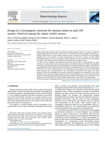
2A.V. Ferreira et al. / Biotechnology Reports 22 (2019) e00324the manifestation of colour or fluorescence does require a networkof polar interactions involving the chromophore group and severalclosely-apposed amino acids [21–25]. According to recent studies,self-reassembly of GFP and its homologous proteins is greatlydependent on C-terminus sheets (β7-11) [25]. Specifically, the 11thβ-sheet is known to protect the chromophore from the surrounding environment [26], being significantly involved on the refoldingpathways required for final chromophore maturation [27].Cabantous and colleagues [27] engineered a split GFP systeminvolving two non-fluorescent portions of GFP that when mixedtogether restored the green fluorescent signal: one portioncorresponds to GFP 1–10 fragment (amino acids 1–214, 10 βsheets) and another portion corresponds to GFP11 (amino acids215–231, 11th β-sheet). In following works, this split GFP system hasbeen extensively adapted to integrate a protease-sensitive switchon system for HIV protease, caspase-3, factor Xa, enteropeptidaseand thrombin [28] and is commercially available as a kit to monitorprotein expression (Interchim).In our work, we used the same described rationale to develop aHNE switch-on coloured system, for application on a smart wounddressing, using a non-fluorescent GFP-like chromoprotein. The twofollowing criteria had to be met regarding colour change upon HNEproteolytic activity to accomplish the intended application of thesystem: (1) easy discrimination by the human eye in the wounddressing, and (2) direct correlation between colour intensity/development time vs the amount of HNE in the wound.Considering the reddish to brownish colour of the woundenvironment (due to blood and pus) [29], we determined thatthe colour of choice for the sensor should be dark blue, dark greenor deep purple [30,31], to avoid misleading interpretations. Takingthis into consideration, we selected as subject of our workultramarine (UM), a chromogenic dark blue coloured proteinderived by genetic modifications [23,24] from Rtms5 (a naturallyoccurring GFP-like fluorescent protein) [32].We developed our strategy, inspired by the method described in[28], to convert UM into a protease switch for HNE (schematized inFig. 1). Briefly, we genetically modified UM to delete its last 11th βsheet, leading to a colourless protein composed of a barrel with 10 βsheets (UM10 protein). The sequence of the 11th β-sheet wasproduced hybridized with the stable surface-exposed α-helix fromeglin c containing the specific HNE-cleavage sequence, AlanineAlanine-Proline-Valine (AAPV) (E11 portion). As observed byCallahan and colleagues [28], we expected to restore thechromogenic environment when both polypeptides are mixedtogether in the presence of HNE. Similarly, to GFP, the released11th β-strand should interact with the incomplete β-barrel fromUM10 leading to the appearance of a blue colour (Fig. 1). To betterunderstand the phenomenon, molecular dynamics studies wereconducted to compare the structures of split UM upon HNE cleavage.The design of a colour switch sensor based on a recombinantchromogenic protein for the monitoring of HNE can revolutionizethe field of diagnostics and medical sensors, in particular in thewound care domain, since colour-based switch-on approacheswith non-fluorescent GFP-like proteins were never reportedbefore.2. Materials and methods2.1. Materials and reagentsAll reagents were analytical grade, purchased from commercialsuppliers and used as received. Standard molecular biologyprocedures were used for the gene cloning and protein recombinant expression in Escherichia coli TOP10 (Invitrogen) and E. coliBL21 DE3 (Novagen), respectively. DNA restriction enzymes NotIand NdeI and kanamycin antibiotic were purchased from ThermoFisher. Gold View for DNA staining was from UVAT Bio (Valencia,Spain). Culture media LB Broth and Terrific Broth (TB) (Autoinduction medium) were purchased from Grisp (Porto, Portugal).Isopropyl-β-D-thiogalactopyranoside (IPTG) inductor was purchased from Acros Organics (Geel, Belgium). Nickel magneticbeads for His6 tag protein purification and respective magneticseparator were purchased from Bimake (Houston, USA). Proteinmolecular weight markers, Precision Plus Protein All Blue orPrecision Plus Protein Dual Xtra Standards, were purchased fromBiorad (Portugal). Chaotropic agent urea was purchased fromlabkem (Spain). All other chemicals were purchased from SigmaAldrich (Spain), including the enzyme elastase from humanleukocytes, synthetic elastase substrate -nitroanilide (MeOSuc-AAPV-pNA),imidazole and N-Laurylsarcosine sodium salt (sarkosyl) detergent.The dialysis tubing cellulose membranes (MWCO, 1–2 kDa and 12–14 kDa) and GenElute Plasmid Miniprep Kit were also obtainedfrom Sigma-Aldrich. The ultramarine’s 11th β-strand peptide(pep11) was synthetized by JPT Peptide Technologies GmbH(Germany).Fig. 1. Schematic representation of the split UM strategy developed as sensor substrate for HNE. The GFP-like chromogenic protein ultramarine (UM) was engineered with thelast 11th β-strand removed from the 10 β-sheet-barrel, creating a colourless protein (UM10). The 11th β-strand, preceded by HNE’s cleavage site, was inserted on eglin c moiety(E11). After HNE proteolysis, the 11th β-strand is released and its proximity to the incomplete β-barrel (UM10) should regenerate UM’s blue colour. Structures were createdwith VMD 1.9.3 software. Incomplete β-barrel is presented in grey, eglin c moiety is coloured golden, 11th β-strand is coloured blue and the cleavage sequence for HNE isrepresented as a red box (For interpretation of the references to colour in this figure legend, the reader is referred to the web version of this article).
A.V. Ferreira et al. / Biotechnology Reports 22 (2019) e003242.2. InstrumentationAll absorbance and fluorescence measurements were carriedout in 96-well microplates (flat bottom, polystyrene and transparent plates, Nunc, ThermoScientific) of 300 mL capacity/well using aSynergy Mx spectrometer with the Gen5 Data Analysis Software(Bio-Tek Instruments, Inc., USA). DNA and total protein wereperformed quantified in Nanodrop 1000 (ThermoScientific).Bacterial cell lysis was accomplished with an Ultrasonic ProcessorVCX-500 W (Sonics & Materials, Inc., Connecticut, USA). Purifiedrecombinant proteins were lyophilized in a FreeZone 2.5 FreezeDry System (Labconco, Missouri, USA).3concentration of 150mU, previously determined by optimization)were recorded in a spectrophotometer for the assessment of colourchange. The split UM system was prepared by mixing UM10 (fixedat 1.5 mg/mL or 3.4 mg/mL) and E11 in a final molar ratio of 167:1 or167:5 (UM10:E11), or by mixing in the same ratios UM10 and pep11(the synthetic peptide equivalent to free 11th β-sheet). Split UMsystem was first allowed to stabilize overnight before the additionof HNE, as done by Callahan and colleagues [28]. To prevent anyinaccuracy measurements over time due to evaporation, the assaytesting the split system was evaluated at RT. In addition, to improvethe sensibility of the assay, the same assays were performed in a96-well microplate firstly blocked with a solution of 0.5% bovineserum albumin (BSA), as described in [34].2.3. Methods2.4. Molecular dynamics simulations2.3.1. Protein expression and purificationGenes of UM, UM10 (GenBank accession number BankIt2109475,MH346479) and E11 (GenBank accession number BankIt2109475,MH346480) inserted in a pET28a( ) plasmid were ordered fromGenscript (New Jersey, USA). Gene size confirmation was performedwith GenElute Plasmid Miniprep Kit and double digested with therestriction enzymes NotI and NdeI; the molecular weight of theresultant DNA fragments was confirmed in a 1% agarose gel stainedwith Gold View. All plasmids were first established in E. coli TOP10and then expressed in E. coli BL21 (DE3). The TSS method was usedfor transformations as previously described [33]. Expression andpurification conditions were established after optimization protocols performed with UM. Optimized protein expression wasaccomplished in 24 h using LB medium supplemented withkanamycin (50 mg/mL) and 180 rpm after induction with0.25 mM IPTG at a culture OD of 0.5. All proteins demanded aculture/flask ratio of 1:10 and aeration via gauze sheets. UM and E11were incubated at 30 C, whereas UM10 required a low incubationtemperature (16 C) to increase solubility. After expression, cultureswere placed at 4 C for at least 2 h to enhance chromophore folding.Then, the cells were harvested by centrifugation at 8,000g at 4 C for10 min, cell pellets were suspended in lysis buffer (20 mM NaH2PO4,500 mM NaCl, pH 7.4) and subsequently lysed by ultrasounds. Thelysate was centrifuged at 10,000g at 4 C for 30 min. Proteins werepurified using nickel magnetic beads and an imidazole gradient of10 mM to 500 mM. At every step, the fractions obtained weremonitored by SDS-PAGE with Coomassie blue staining. Removal ofimidazole from the eluted fractions was performed through dialysisagainst ultrapure water using a cellulose membrane with a MWCOof 1–2 kDa for E11 protein and 12–14 kDa for the other proteins. Thepure protein fractions were then frozen at -80 C and lyophilized.The molecular weight of all proteins obtained by SDS-PAGE was inaccordance with the theoretical size expected from the polypeptidesequences including the amino acids encoded by pET28a( ) vector.2.3.2. Assessment of the UM-based split protein as HNE proteasesubstrateHNE activity was first evaluated in the batches used in theexperiments with a control assay with the synthetic substrateMeOSuc-AAPV-pNA following the protocol supplied by themanufacturer. Briefly, the enzyme (0.5 U/mL) was mixed with25–1500 mM of substrate, to a final volume of 300 mL of reactionbuffer (100 mM HEPES with 500 mM NaCl, pH 8). A blankconsisting only in buffer and substrate was also included. Thekinetic constants were determined from the initial rates ofhydrolysis by Michaelis-Menten method. All enzymatic assayswere conducted at 37 C for 10 min in triplicate and the absorbancewas measured at 405 nm by UV–vis spectrophotometry.The lyophilized proteins were suspended immediately prior tothe assay in 100 mM Tris HCl at pH 8. The absorbance spectra ofthe UM-based split proteins prior and after addition of HNE (fixedThe molecular dynamics simulations were performed with theGromacs 4.0.7 package applying the GROMOS 54A7 united-atomforce-field [35–39], with periodic boundary conditions, at 300 Kand 1 atm.The molecular models for the UM and E11 proteins wereobtained through structure homology using the SWISS-MODELweb service (http://swissmodel.expasy.org). The X-ray model ofthe Rtms5 protein (PDB code: 3vk1) was used as template for UMwhile the NMR model of eglin c (PDB code: 1 EG L) was used for E11.The UM10, pep11 and cleaved E11 molecular structures wereobtained through manual edition of the UM and E11 models. Thesimulation pictures were rendered with VMD (Visual MolecularDynamics) software [40].2.5. Simulation systems and protocolNine different systems, composed of water and proteinsmodels, were built in this work. The proteins models were theUM, UM10, pep11, eglin c, E11, cleaved E11, UM10 with pep11,UM10 with E11 and UM10 with cleaved E11. The systems weresubjected to energy minimization, three equilibration simulations,and finally to the production run. At least to replicas of each systemwere built, and 100 ns of total production simulation werecalculated. The solvated UM, UM10, pep11, eglin c, E11 and cleavedE11 systems were simulated to equilibrate its molecular structures.The remaining UM10 with pep11, UM10 with E11 and UM10 withcleaved E11 system were simulated to unveil the possibleinteractions between these molecules, with main focus on theβ11 sequence (free or inserted in the eglin c protein) and UM10interactions. After the production runs visual inspection andseveral properties such as the temperature, pressure, and potentialenergy were evaluated to check if the system were wellequilibrated. More details about the molecular dynamics simulations is presented in supplementary information.3. Results3.1. Production of the split UM substrate for HNE detectionUltramarine (UM) protein, its incomplete version made of 10 βsheets (UM10) and the sensing substrate composed by the HNEcleavage sequence AAPV plus the sequence of the 11th β-sheetincorporated in eglin c moiety (E11) were expressed in E. coli withdistinct solubility profiles. The original UM presented a dark bluecolour and the engineered proteins UM10 and E11 were noncoloured, as expected (Fig. 2). UM and E11 proteins were mainlyobtained in the soluble fraction, but the expression of UM10 waschallenging. After verifying its low initial protein expression yield,with the same protocol used for the other two proteins, weundertook an optimization of the conditions to control the
4A.V. Ferreira et al. / Biotechnology Reports 22 (2019) e00324Fig. 2. Colour of the UM, UM10 and E11 proteins. Bacterial pellets after expressionunder optimized laboratorial set-up and in solution after purification at 1.5 mg/mLfor UM and UM10, and at 0.5 mg/mL for E11.culture’s growth rate and increase protein proper expression.Culture’s growth rate was slowed-down by reducing the incubation temperature from 30 C to 16 C to improve the chances ofcorrect folding of the protein being expressed. In addition, after celllysis, several ice-vortex cycles were performed to aid itssolubilisation, thus enhancing the correct folding of the protein.Under these optimized conditions, UM10 was expressed 50%soluble (data not shown).3.2. Effect of HNE on split UM systemPrior to the assessment of the split UM substrate in the presenceof HNE, we evaluated the protease effect in the original UM as ameasure of specificity analysis. Foremost, loss of colour was notobserved in ultramarine after incubation with HNE, even over 24 hof incubation, indicating no significant proteolytic effect of elastaseon our casting protein. However, a slight and transient increase inabsorbance was registered upon HNE addition ( 0.18 a.u.corresponding to 11% increase) (Supplementary Figure S1A).Somehow, in the first moments, the interaction of the protein withHNE led to a gain on the absorbance, but, since it returned tobaseline values, this effect was disregarded. However, by SDS-PAGEwe detected in ultramarine a small molecular weight decrease ofapproximately 1 kDa after incubation with HNE (SupplementaryFigure S1B). After analysis of the theoretical cleavage map providedby ExPASy Peptide Cutter software (SIB ExPASy BioformaticsResources Portal, available online tool) [41], we inferred, by thecorresponding fragment’s size, that it probably resulted from asmall cleavage right after the His6 tag at protein’s N-terminal(Supplementary Figure S1C). Still, this cleavage does not affect theprotein conformation nor chromophore’s arrangement, since thecolour remained blue even after several days of incubation with theenzyme (Supplementary Figure S1 and data not shown). Hence,any colour change in the split UM system as specific to the action ofHNE at the inserted cleavage site located on E11.The efficiency of the system was assayed by adding HNE topurified UM10 (fixed at 3.4 mg/mL) previously mixed with E11 inthe molar ratios 167:1 and 167:5 of UM10:E11. The syntheticpeptide equivalent to the 11th β-strand (pep11) was also included inthis assay and mixed with UM10 at the same ratios as E11.Evaluation of the interaction of the 11th β-strand and theincomplete β-barrel was made by measurement of the absorbanceover time (Fig. 3).For split UM system, a measurable increase in absorbance wasobserved for a period of 6 h after the addition of HNE ( 0.2 a.u ofabsorbance in UM10 E11 HNE mixture, corresponding to 75%increase) (Fig. 3A and Supplementary Figure S2A). However, wecould not detect by naked-eye a gain of colour of the solution. Afterthis time, the absorbance steeply decreased and then stabilized atthe 7th hour of incubation, continuing with a steady and very slowdecrease in the absorbance verified until the end of the assay.We let the solution to dry in the wells and then we observedsome purplish-blue (Supplementary Figure S3) similar to thosethat formed after evaporation of UM which evidence the formationof the UM10 E11 complex.To rule out a poor enzymatic cleavage by HNE on E11, theinteraction of UM10 with a free and readily available 11th β-sheetwas evaluated using pep11 peptide instead of E11 protein (Fig. 3Band Supplementary Figure S2B). This system followed almost thesame trend as the described for UM10 E11 HNE, although theinteraction of UM10 pep11 differed in each tested ratio, contrarilyto the observed with E11. When mixing UM10 pep11 in 167:1 ratio,the absorbance of the complex slowly increased for 42 h. Then, in30 min, it rapidly recovered the initial values and continued todrop. However, increasing the number of pep11 molecules, thedecrease in absorbance occurred in only 18 h, then it continued todrop for 24 h to much lower values than those initially recorded,and finally it stabilized in the last hours of the assay.3.3. Molecular dynamics studies for comprehension of the split UMsystemStructural homology analysis was done between UM and UM10proteins to compare both β-barrels (Supplementary Figure S4A).Despite missing the last β-sheet, UM10 showed a closelysuperposing structure with the original UM, without relevantrelaxation or constriction of the barrel’s diameter (top and bottomviews in Supplementary Figure S4). Still, as expected, thealignment of both structures showed a significant displacementof the 10th β-strand in UM10 which occupies the gap left by theabsence of the 11th β-strand.As a quantifying measure, we used the root-mean-squarefluctuation (RMSF) values of UM and UM10 since it can providephysical information regarding the residues mobility through thein silico simulations (Supplementary Figure S4B). Terminalresidues have free movement due to the absence of a constrainingforce at one end. For this reason, residues 1–7 and 217–220 in UMand 1–7 and 199–201 in UM10 were not considered in the analysisas their RMSF values are biased by the inherited freedom. Removalof the last 11th β-strand significantly affected the fluctuation ofsome residues, namely the ones located in the internal loop (49–80) that holds the chromophore group, but also in β5 (107–112) andin β6 (121–124) (Supplementary Figure S4B). In particular,chromophore interacting residues Pro59, Tyr78 and Lys80 andconserved residues Gly122 and Thr123 showed fluctuation valuesincrease by 2-fold in comparison to UM.To better understand the interaction between UM10 and E11,cleaved E11 was placed together with UM10 in the simulation boxwhich was run 4 times (Fig. 4). Two of the four replicates showedan interaction of the sequence of β11 hanging from E11 with UM10(Fig. 4C and D), although not at the original position of β11.Additionally, six replicas of the interaction of pep11 with UM10were run (Fig. 5). Four out of six showed interaction of pep11 withUM10, however only in replica 3 pep11 was located at a given timein the β11 gap of the UM10 barrel. During this simulation, pep11interacted in other faces of UM10, interspersing with searches forother sites of the barrel.4. Discussion4.1. HNE signalling occurs though transientlyThe rationale published years ago by [28] showing theadaptation of the split-GFP technology [27] into a protease switch
A.V. Ferreira et al. / Biotechnology Reports 22 (2019) e003245Fig. 3. Efficiency of (A) the split UM system in detecting HNE (150 mU) and comparison with (B) the mixture of UM10 with the synthetic peptide pep11, in molar ratios of167:1 or 167:5. Absorbance was measured at 585 nm for 24 or 44 h, respectively, and normalized against the absorbance of UM10. All assays were performed at roomtemperature. Bars representing E11 (control) and pep11 (control) are correspondent to 4.09 nmol (equivalent amount used in 167:5 ratio). Registered absorbances of UM10(control) presented as dashedlines in (A) and black dots in (B) are points referred to the secondary vertical axis.together with the need of a naked-eye detection system led us toengineer a switch-on system for HNE based on ultramarine (UM), achromogenic GFP-like protein. To produce the sensing moiety, weinserted the sequence of the 11th β-strand from UM into the stablesurface-exposed α-helix loop of eglin c and added AAPV peptide,the HNE specific cleavage sequence, upstream the β11 sequence.The signalling moiety corresponds to the incomplete UM10 βbarrel which bears the chromophore group, at this stage, unable toproduce all the required interactions for colour generation.The difficulties encountered on the production of soluble UM10should be mainly related to the absence of the last β-sheet as it isknown that hysteresis of GFP-like proteins is directly related to theC-terminal β-sheets (β7-β11) [25]. Our total extracts had a goodamount of the desired protein, but unable to fold correctly. Themodifications made to the production protocol allowed therecovery 50% of the protein in the soluble fraction.The absence of colour of UM10 (Fig. 2) is due to the inexistenceof the 11th β-sheet. The absence of the two chromophoreinteracting residues Gln209 and Glu211 from the 11th β-sheetprevents the establishment of the complete network of interactions paramount for chromophore maturation [27]. The 11th βsheet itself is significantly involved in the refolding pathways
6A.V. Ferreira et al. / Biotechnology Reports 22 (2019) e00324Fig. 4. Simulation of the interactions between UM10 signalling moiety (in grey) andcleaved E11 substrate moiety, with eglin c in golden, sequence of the 11th β-strand inblue and cleavage site in red. Conformations of the final stage resulting from foursimulations are presented separately (A–D), showing front, back and side views.Ultramarine original conformation is shown in E. All structures were generatedwith VMD 1.9.3 software. (For interpretation of the references to colour in thisfigure legend, the reader is referred to the web version of this article).required for the complete chromophore maturation and directlyprotects the chromophore from the surrounding environment [26].Being absent, β10 acquires a conformation slightly distorted.Nevertheless, the overall barrel structure of UM10 is very similar tothe original structure of UM (Supplementary Figure S4A), asobserved in the crystallography of protein GFP 1–9 (GFP lackingβ10 and β11) [42].Changes in the dynamic behaviour of UM10 were betteranalysed by the calculation of the root-mean-square fluctuation(RMSF) of residues in comparison to the original UM (Supplementary Figure S4B). A higher RMSF value is related to more flexiblemovement of a given residue during the simulation time in relationto its average position, whereas a low RMSF value indicates limitedmovement. The 2-fold increase in the fluctuation of somechromophore-interacting residues (Pro59, Tyr78 and Lys80) andconserved residues (Gly122 and Thr123) in comparison to theoriginal UM is also indicative of alterations on the conditions forthe establishment of chromophore environment.The close neighbouring of Pro59, Tyr78 and Lys80 and theelement of the chromophore triad Tyr63 (Tyr66 in GFP), whichallows the establishment of the strong interaction network thatcompose the chromophore environment is, hence, modified [43–47]. Particularly, Pro59 (Thr63 in GFP) is reported to be restrictedwithin the GFP for the establishment of carbon-proton boundswith the vicinity, instead in UM10 this residue possesses higherfluctuation and it is freely rotating [45]. Furthermore, the absentresidues Gln209 and Glu211 from the 11th β-sheet are known topromote the intramolecular complex formation of the chromophore group, being instrumental for stabilization of the keyreaction intermediates from precyclized tripeptide to the dehydrated intermediate [43,46]. In particular, Gln209 (Glu222 in GFP)is required to interact to Tyr63 for the completion of this process.We expected that chromophore environment, and hence colourproduction, could be restored by the proximity of the 11th β-strandto the incomplete β-barrel, following the same mechanism of splitGPF technology nowadays broadly implemented [27,28,34,42,48–52]. HNE cleavage on AAPV at E11 should release the N-terminal ofthe 11th β-strand, being available for interaction with theincomplete β-barrel from UM10.Our switch-on system was evaluated in vitro by measurementsof the absorbance that was expected to increase upon HNEaddition to the mixture of UM10 E11. Indeed, a 75% increase inthe absorbance was observed in the first 6 h, but the signal was lostafterwards (Fig. 3A and Suppleme
GenElute Plasmid Miniprep Kit and double digested with the restriction enzymes NotI and NdeI; the molecular weight of the resultant DNAfragmentswas confirmedina1%agarosegelstained with Gold View. All plasmids were firstestablished in E. coli TOP10 and thenexpressedinE. coli BL21(DE3). The TSS method was used