Transcription
Jt Dis Relat SurgJoint Diseases andRelated Surgery2021;32(2):489-496ORIGINAL ARTICLEDiagnostic and prognostic role of neutrophil/lymphocyteratio, platelet/lymphocyte ratio, and lymphocyte/monocyteratio in patients with osteosarcomaAliekber Yapar, MD1 , Mehmet Ali Tokgöz, MD2 , Dilek Yapar, MD3 , İsmail Burak Atalay, MD1 ,Coşkun Ulucaköy, MD1 , Bedii Şafak Güngör, MD1 Department of Orthopedics and Traumatology, Ankara Oncology Training and Research Hospital, Ankara, TurkeyDepartment of Orthopaedics and Traumatology, Dr. Nafiz Korez Sincan State Hospital, Ankara, Turkey3Department of Public Health, Gazi University Faculty of Medicine, Ankara, Turkey12Osteosarcoma is the most common primary malignantbone malignancy which accounts for about twothirds of all cases.[1,2] Most of the osteosarcomas areclassified as traditional type, high-grade tumors, anddiagnosed by radiological examinations and biopsy.Typical osteosarcomas are osteoblastic or osteolyticlesions which can be demonstrated with a periostealreaction on conventional radiographs. However,osteosarcoma can break out beyond the traditionaltypes of findings, and diagnosing some of its variantsis difficult for clinicians.[3] Serum tumor markers usedin the diagnosis of osteosarcoma usually result in avery little success. While the response to treatmentand disease prognosis can be estimated with the helpReceived: October 28, 2020Accepted: December 11, 2020Published online: June 11, 2021Correspondence: Coşkun Ulucaköy, MD. SBÜ Dr. AbdurrahmanYurtaslan Onkoloji Eğitim ve Araştırma Hastanesi Ortopedi veTravmatoloji Kliniği, 06105 Yenimahalle, Ankara, Türkiye.E-mail: coskunulucakoy@gmail.comDoi: 10.52312/jdrs.2021.79775Citation: Yapar A, Tokgöz MA, Yapar D, Atalay İB, UlucaköyC, Güngör BŞ. Diagnostic and prognostic role of neutrophil/lymphocyte ratio, platelet/lymphocyte ratio, and lymphocyte/monocyte ratio in patients with osteosarcoma. Jt Dis Relat Surg2021;32(2):489-496. 2021 All right reserved by the Turkish Joint Diseases FoundationThis is an open access article under the terms of the CreativeCommons Attribution-NonCommercial License, which permits use,distribution and reproduction in any medium, provided the originalwork is properly cited and is not used for commercial /4.0/).ABSTRACTObjectives: This study aims to evaluate the diagnostic andprognostic significance of neutrophil-to-lymphocyte ratio (NLR),platelet-to-lymphocyte ratio (PLR), and lymphocyte-to-monocyteratio (LMR) values in patients with osteosarcoma.Patients and methods: A total of 172 patients (111 males,61 females; mean age: 24.3 15.3 years; range, 7 to 82 years)diagnosed with osteosarcoma in our institution between January2002 and December 2018 were retrospectively analyzed. A totalof 165 healthy individuals (115 males, 50 females; mean age:20.2 9.2 years; range, 10 to 65 years) who did not have infectious,rheumatological or hematological diseases or any pathologicalfinding were assigned as the control group. The clinical, laboratory,and demographic findings of the patients were obtained fromhospital records. Pre-treatment NLR, PLR, and LMR values werecalculated in all patients. Diagnostic and prognostic values ofpre-treatment NLR, PLR and LMR were assessed using receiveroperating curve (ROC) analysis. The Kaplan-Meier method wasused for survival analysis.Results: For diagnostic approach, the highest significance inarea under the curve (AUC) values was obtained for NLR(AUC 0.763). The AUC for PLR and LMR was statisticallysignificant, while the statistical power was weak comparedto NLR (AUC 0.681 and 0.603). The NLR, PLR, and LMRwere found to be predictors of mortality. The cut-off value wasfound to be 3.28 for NLR, 128 for PLR, and 4.22 for LMR.The prognostic value of NLR for mortality was higher than(AUC 0.749) PLR (AUC 0.688) and LMR (AUC 0.609). TheNLR, PLR, and LMR were associated with overall survival (OS).There was a significant difference in the median OS time amongthe NLR, PLR, and LMR values (log-rank test order p 0.001,p 0.001, and p 0.004, respectively).Conclusion: Based on our study results, pre-treatment NLR,PLR and MLR have diagnostic and prognostic values inosteosarcoma.Keywords: Diagnostic significance, neutrophil/lymphocyte ratio,osteosarcoma, prognostic significance.
490of some serum markers, there is no inexpensive andwidely available marker to help in diagnosis.[4] Besides,poor prognosis and high mortality rates in metastaticosteosarcoma are similar to those of 20 years ago,despite current multimodality treatment.[5]An increasing number of evidence has shownthat, in cancer patients, host-related variables mayhelp in diagnosis with an impact on prognosis.In particular, systemic inflammatory responseand related variables are reported to have aremarkable effect.[6,7] Demonstration of the increasedpresence of inflammatory cells in the tumor localmicroenvironment has also triggered the interestin markers, such as circulating leukocyte countsand neutrophil-to-lymphocyte ratio (NLR).[8] It hasbeen shown that inflammation-based scores maycontribute to diagnostic and prognostic evaluation invarious types of cancer.[9-12]In the literature, prognostic markers calculatedon the basis of inflammatory cells are shown tohave a prognostic value in osteosarcoma patients.However, both the diagnostic and prognostic effectsof these markers on osteosarcoma have not beenfully clarified. In the present study, we aimed toinvestigate the effect of prognosis of the levels ofpre-treatment inflammatory markers (NLR, plateletto-lymphocyte ratio [PLR], and lymphocyte-tomonocyte ratio [LMR]) on patients with osteosarcomaand to compare them with the healthy controls foridentifying the diagnostic value.PATIENTS AND METHODSThis single-center, retrospective study wasconducted at Dr. Abdurrahman Yurtaslan AnkaraOncology Training and Research Hospital,Department of Orthopedics and Traumatology,between January 2002 and December 2018. A totalof 172 patients (111 males, 61 females; mean age:24.3 15.3 years; range, 7 to 82 years) diagnosed withosteosarcoma in our institution were included.A total of 165 healthy individuals (115 males,50 females; mean age: 20.2 9.2 years; range, 10 to 65year) who did not have infectious, rheumatologicalor hematological diseases or any pathologicalfinding were assigned as the control group. Thecontrol group who referred to the outpatient clinicof orthopedic and traumatology with non-specificsymptoms was similar in terms of age and sex tothe patient group. A written informed consent wasobtained from each participant and/or his/her legalguardian. The study protocol was approved bythe Dr. Abdurrahman Yurtaslan Ankara OncologyTraining and Research Hospital Ethics CommitteeJt Dis Relat Surg(date/no: 2020-06/647). The study was conducted inaccordance with the principles of the Declaration ofHelsinki.Patients with a histopathologically confirmedosteosarcoma diagnosis who had no previousanticancer treatment and who had complete medicaland follow-up records were included. Patientswith elevated C-reactive protein results, who werelost to follow-up, those with diabetes mellitus,infections disease, rheumatological diseases andother inflammatory diseases, any hematologicaldisease, previously treated with non-steroid antiinflammatory drugs, and those with missing medicalrecords were excluded from the study. Clinical anddemographic data of the patients were reviewedfrom the hospital records. Age, sex, tumor location,side, pre-treatment complete blood count results,date of diagnosis, the final follow-up date, surgicalprocedure, the status of metastasis at diagnosis,mortality, and recurrence records were reviewedfrom the patient files. The NLR and PLR werecalculated as the absolute count of neutrophilsand platelets, respectively, divided by the absolutelymphocyte count. The LMR was calculated as theabsolute count of lymphocyte divided by the absolutemonocyte count.Statistical analysisStatistical analysis was performed using theIBM SPSS for Windows, version 22.0 software(IBM Corp., Armonk, NY, USA). Descriptivestatistics are presented in number and percentagefor categorical variables and in mean standarddeviation or median (min-max) for continuousvariables. Normal distribution for continuousvariables were assessed with visual (histogramsand probability graphics) and analytic methods(Kolmogorov-Smirnov test). In the data that donot fit the normal distribution, the Mann-WhitneyU test was used for comparison analysis betweenthe two independent groups. Comparison analysesfor categorical variables between independentgroups were done by chi-square test. Diagnosticand prognostic values of pre-treatment NLR,PLR, and LMR were assessed using the receiveroperating curve (ROC) analysis. The area under theROC curve (AUC) results were considered excellentfor AUC values between 0.9-1, good for AUC valuesbetween 0.8-0.9, fair for AUC values between0.7-0.8, poor for AUC values between 0.6-0.7,and failed for AUC values between 0.5-0.6.[13,14]Following the ROC analysis results, the AUC andcut-off values, sensitivity and specificity of thesecut-offs, likelihood ratio (LHR), positive predictive
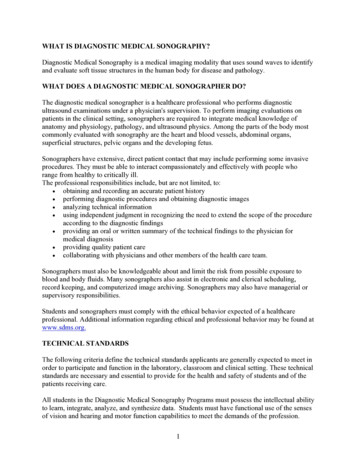
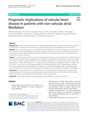
Diagnostic and prognostic role of inflammatory indexes in osteosarcomaTable IBaseline demographic characteristics of patients withosteosarcoma (n 7.49.37.67.00.60.60.6Metastasis at urvivors769644.255.8value (PPV) and negative predictive value (NPV)were calculated. Survival analyses were performedusing the Kaplan-Meier method and log-rank test.A p value of 0.05 was considered statisticallysignificant.RESULTSOf the patients with osteosarcoma, 11.6% of thepatients had metastases at the time of diagnosis,while 44.2% of the patients died during follow-up.According to the localization, tumors were mostlylocated in the femur (57%) and tibia (17.4%). Theage and sex distributions of the control and thepatient groups were similar (p 0.528 and p 0.313,respectively) (Table I).491There was no significant correlation between ageand NLR (r 0.052, p 0.337), PLR (r 0.054, p 0.320)and LMR (r 0.046, p 0.396). The NLR (p 0.544), PLR(p 0.796) and LMR (p 0.364) between the age groupsof 18 years old and 18 years old were similar. Whenit was evaluated separately for patient and controlgroups, each NLR (pcontrol 0.938, ppatients 0.243), PLR(pcontrol 0.504, ppatients 0.282), and LMR (pcontrol 0.658,ppatients 0.355) among age groups of 18 years old and 18 years old were found to be similar.The NLR and PLR values of the sarcoma groupwere significantly higher than the controls (p 0.001)(Table II). The LMR values were statisticallysignificantly lower in the sarcoma group, compared tothe controls (p 0.001). The diagnostic power of NLR,PLR, and LMR with ROC analysis was evaluated(Figure 1). Accordingly, the highest significance inAUC values was obtained for NLR (AUC 0.763).The calculated value of AUC for PLR and LMR wasstatistically significant, while the statistical powerwas weak, compared to NLR (AUC 0.681 and 0.603).The cut-off values were determined for all threeparameters, and sensitivity, specificity, LHR,PPV, and NPV values of the cut-off values werecalculated. The predictability of NLR values of 2.45 in the diagnostic approach for osteosarcomawas higher than the other two parameters(sensitivity 64.5%, specificity 78.8%, LHR 3.04,PPV 75%, and NPV 67.7) (Table III). The cut-offvalue for PLR was found to be 119 (sensitivity 60.5%,specificity 64.2%, LHR 1.69, PPV 63.4%, andNPV 60.7) for diagnostic approach.The predictability of NLR, PLR, and LMR valuesfor mortality was evaluated by the ROC analysis andthe statistical significance of AUC values obtainedfrom ROC analysis was presented in Table IV andFigure 2. The NLR, PLR, and LMR were found to beTable IIComparison of patients and control groups (n 337)Controls (n 165)n%Age (year)SexMaleFemaleNeutrophil-to-lymphocyte ratioPlatelet-to-lymphocyte ratioLymphocyte-to-monocyte ratioPatients (n 330.46-13.51 0.001*110.5363.7-296.7127.8440.51-807.8 0.001*4.811.1-9.134.190.66-20.90.001*Min: Minimum; Max: Maximum; * Mann-Whitney U test; ** Chi-square test.
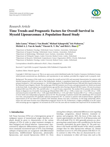
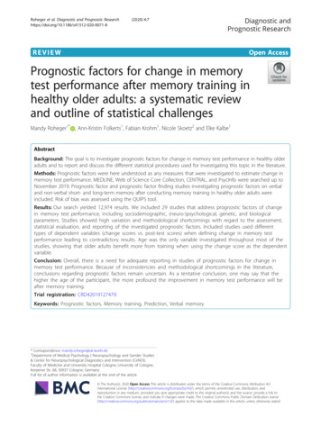
492Jt Dis Relat 0.40.20.00.40.2NLRPLRReference line0.00.20.40.60.80.01.0MLRReference line0.00.2Specificity0.40.60.81.0SpecificityFIGURE 1. Receiver operating characteristic (ROC) curves for diagnostic value of the NLR (a), PLR (a), and LMR (b). (a) Largerresults of NLR and PLR indicate more diagnostic positive test for sarcomas. (b) Smaller results of LMR indicate more diagnosticpositive test for sarcomas.NLR: Neutrophil-to-lymphocyte ratio; PLR: Platelet-to-lymphocyte ratio; LMR: Lymphocyte-to-monocyte ratio.predictors of mortality. The cut-off value was found tobe 3.28 for NLR, 128 for PLR, and 4.22 for LMR. Theprognostic value of NLR for mortality was higher than(AUC 0.749) PLR (AUC 0.688) and LMR (AUC 0.609).The median overall survival (OS) duration of theosteosarcoma patients was 46.6 (range, 22.4 to 70.82)months. While the one-year OS rate was 86.2%,the three-year OS rate decreased to 59.7% and theTable IIIStatistical analysis of various diagnostic approaches for the predictive value of pre-treatment NLR, PLR and LMR (n 337)AUC (95% CI)pCut-offSensitivity(%)Specificity(%) LHRNLR0.763 (0.711-0.814) 0.001 2.45PLR0.681 (0.624-0.738) 0.001 119LMR0.603 (0.542-0.663)0.001 60.759.960.61.5461.359.2AUC: Area under the curve; CI: Confidence interval; LHR: Positive likelihood ratio; PPV: Positive predictive value; NPV: Negative predictive value; NLR: Neutrophil-tolymphocyte ratio; PLR: Platelet-to-lymphocyte ratio; LMR: Lymphocyte-to-monocyte ratio.Table IVStatistical analysis of various prognostic approaches for the predictive value of pre-treatment NLR, PLR and LMR (n 172)AUC (95% CI)pCut-offSensitivity(%)Specificity(%) LHRPPV(%)NPV(%)NLR0.749 (0.675-0.824) 0.001 3.2861.8762.5867.171.6PLR0.688 (0.608-0.768) 0.001 12868.464.62.0160.572.1LMR0.609 (0.525-0.664)0.014 4.2263.257.31.4753.966.3AUC: Area under the curve; CI: Confidence interval; LHR: Positive likelihood ratio; PPV: Positive predictive value; NPV: Negative predictive value; NLR: Neutrophil-tolymphocyte ratio; PLR: Platelet-to-lymphocyte ratio; LMR: Lymphocyte-to-monocyte ratio.
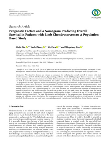
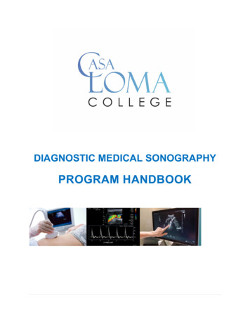
Diagnostic and prognostic role of inflammatory indexes in ivity(a)4930.40.20.40.2NLRPLRReference line0.00.00.20.40.60.8MLRReference FIGURE 2. Receiver operating characteristic (ROC) curves for prognostic value of the NLR (a), PLR (a), and LMR (b). (a) Largerresults of NLR and PLR indicate more prognostic positive test for sarcomas. (b) Smaller results of LMR indicate more diagnosticpositive test for sarcomas.NLR: Neutrophil-to-lymphocyte ratio; PLR: Platelet-to-lymphocyte ratio; LMR: Lymphocyte-to-monocyte ratio.Table VOverall survival rates (n 172)All patientsLog Rank test(p)Overall survival,monthsMedian (95% CI)1-yearsurvival rate(%)3-yearsurvival rate(%)5-yearsurvival rate(%)10-yearsurvival rate(%)-46.6 (22.4-70.82)86.259.747.239.087.3 (62.8-111.7)31.7 (22.7-40.6)92.378.07639.767.72347.835.392.4 (74.7-112.9)36.0 (29.2-42.7)89.78373.348.965.333.743.536.273.6 (44.1-103.2)33.6 (27.8-39.4)90.88273.247.561.634.340.238.2NLR 3.28 3.28 0.001PLR 128 1280.001LMR 4.22 4.220.004CI: Confidence interval; NLR: Neutrophil-to-lymphocyte ratio; PLR: Platelet-to-lymphocyte ratio; LMR: Lymphocyte-to-monocyte ratio.10-year OS rate to 39% (Figure 3a). There was asignificant difference in the median OS timeaccording to NLR, PLR, and LMR values (log-ranktest order p 0.001, p 0.001, and p 0.004, respectively)(Table V, Figures 3b-d).DISCUSSIONIn the current study, we evaluated factors showingthe systemic inflammatory response from twodifferent perspectives. Pre-treatment values ofNLR, PLR, and LMR in the peripheral blood of
494Jt Dis Relat Surg(b)1.01.00.80.80.60.6Cum survivalCum survival(a)0.40.20.40.2Survival functionCensored0.00.024.048.072.0Log rang test, p 0.001LNLR ( 3.28)HNLR ( 3.28)LNLR ( 3.28)-censoredHNLR ( 3.28)-censored0.096.0 120.0 144.0 168.00.024.0Follow-up time (months)72.096.0 120.0 144.0 168.0Follow-up time (months)(d)1.01.00.80.80.60.6Cum survivalCum survival(c)48.00.40.2Log rang test, p 0.001LPLR ( 128)HPLR ( 128)LPLR ( 128)-censoredHPLR ( 128)-censored0.00.024.048.072.096.0 120.0 144.0 168.0Follow-up time (months)0.40.2Log rang test, p 0.0040.0HLMR ( 4.22)LLMR ( 4.22)HLMR ( 4.22)-censoredLLMR ( 4.22)-censored0.024.048.072.096.0 120.0 144.0 168.0Follow-up time (months)FIGURE 3. Kaplan-Meier curves for overall survival. (a) Overall survival curve and of patients with osteosarcoma. The OSrates of patients at 1, 3, 5, and 10 years after surgery were 86.2, 59.7, 47.2 and 39%, respectively. (b) Overall survival curveof patients with osteosarcoma in different NLR groups. (c) Overall survival curve of patients with osteosarcoma in differentPLR groups. (d) Overall survival curve of patients with osteosarcoma in different LMR groups.OS: Overall survival; NLR: Neutrophil-to-lymphocyte ratio; LNLR: Low neutrophil-to-lymphocyte ratio; HNLR: High neutrophil-to-lymphocyte ratio;LPLR: Low platelet-to-lymphocyte ratio; HPLR: High platelet-to-lymphocyte ratio; LLMR: Low lymphocyte-to-monocyte ratio; HLMR: High lymphocyteto-monocyte ratio.osteosarcoma patients were found to be diagnosticand prognostic values. The prognostic value of pretreatment inflammatory markers (NLR, PLR, andLMR) in the osteosarcoma patients was mentioned inthe literature.[5,15,16] Consistent with previous studies,our study confirmed that high NLR, PLR, and lowLMR were significantly associated with poor OS.To the best of our knowledge, the current studyis the one of the premise studies showing bothdiagnostic and prognostic value of inflammatory
Diagnostic and prognostic role of inflammatory indexes in osteosarcomabased markers.[17] We demonstrated that inflammatorymarkers, particularly NLR, could be significantlysupportive in diagnosing osteosarcoma. In a patienthaving a suspicion of osteosarcoma, the clinicalsuspicion increased, when the NLR value was 2.45,while the probability of the disease progressing withpoor prognosis became stronger, if the pre-treatmentNLR was 3.28.In a study, the NLR cut-off values were found to be3.43 for OS and 3.67 for progression-free survival, andit was found that the probability of poor prognosison both parameters increased in patients with NLRvalues above the cut-off.[18] The prognostic valuecalculations in the current study are consistent withthe literature. Another important finding of thecurrent study is that the OS rate of patients with anNLR value of 3.28 was significantly lower at three(39.7%) and five (23%) years.Similar to changes in peripheral blood immunecells, changes in immune infiltrations aroundthe tumor microenvironment are also reliableand effective prognostic factors for many tumors,including bone and soft tissue sarcomas.[19] Besides,early peripheral blood lymphocyte recovery afterinitiation of chemotherapy is a reliable prognosticindicator for superior outcomes in patients withosteosarcoma.[20,21] Considering the aforementionedinformation and current study findings, the highNLR, PLR, and low LMR ratio appeared as a poorprognostic indicator, which could be due to reflectionof the inability of the host metabolism to respond tothe tumor with sufficient cellular immunity.In the literature, there are studies on the diagnosticvalue of inflammatory variables in malignancies. In astudy, the NLR was shown to have a diagnostic valuefor cartilaginous tumors, although it was not foundto be effective in distinguishing the subtypes of thetumor.[22] Another study demonstrated that NLR LMR combination was effective both in separatingglioma from acoustic neuroma and meningioma, anddifferential diagnosis of glioblastoma multiformefrom low-grade gliomas.[9] The value of inflammatorymarkers was also investigated in determining themalignancy potential of spinal tumors and the cutoff value was calculated as 3.19 for NLR and as141 for PLR value. Malignancy potential of spinalmass patients with the results above these valueshas been reported to be high.[23] Although there arestudies showing the diagnostic value of inflammatorymarkers, a study on ovarian mass reported that NLRwas an ineffective marker in predicting the malignantcharacteristics of a pelvic mass.[24] The findings of thecurrent study showed that inflammatory variables495could be supportive in diagnosing osteosarcoma suchas spinal masses, cartilaginous tumors, and gliomas.Nonetheless, there are several limitationsto this study. First, this study has a single-center,retrospective design. Second, despite its advantages inthis cohort, inflammatory markers were a non-specificpredictor for osteosarcoma and, thus, inevitably hadintrinsic weaknesses and limitations. However, thereare no specific serum markers for osteosarcoma.Non-specific findings such as NLR, PLR, and LMRcould be beneficial to support the suspicion of clinicaland radiological osteosarcoma for some patients.On the other hand, the current study has severalstrengths. First, the infection markers were evaluatedand the effects of the infection on the inflammatorymarkers were prevented by excluding the patientswho were considered to be infected. Second, bothdiagnostic and prognostic values of inflammatorymarkers were evaluated and cut-off values wereseparately specified for both. In the current study, theclinician should also pay attention to the pre-treatmenthigh NLR to evaluate the OS in bone masses withsuspected malignancy. The NLR, PLR, and LMR aresimple, low cost, easy to measure with widely usedand standardized tests, and the current study is thefirst to demonstrate the diagnostic value of these nonspecific inflammatory markers.In conclusion, our study results suggest thatpatients with a bone mass with malignant potentialhave higher levels of NLR and PLR and lower levelsof LMR than healthy individuals. Pre-treatment NLR,PLR, and MLR have diagnostic and prognostic valuesin osteosarcoma, supporting the prognostic value ofinflammatory markers in osteosarcoma. In addition,NLR may be a superior distinguishing factor to LMRand PLR. There are many different situations whichdetermine the prognosis of neoplastic diseases, andNLR may be one of them. However, the NLR valuescannot be a standalone decision maker in patients withosteosarcoma, but they can be one of the variables tobe taken into account while making a treatment plan.Further multi-center, large-scale, prospective studiesare needed to confirm our findings and investigatethe value of the combined use of these inflammatorymarkers to prolong the remission time of patientswith osteosarcoma.Declaration of conflicting interestsThe authors declared no conflicts of interest with respect tothe authorship and/or publication of this article.FundingThe authors received no financial support for the researchand/or authorship of this article.
496REFERENCES1.2.3.4.5.6.7.8.9.10.11.12.Ferguson JL, Turner SP. Bone cancer: diagnosis andtreatment principles. Am Fam Physician 2018;98:205-13.Ritter J, Bielack SS. Osteosarcoma. Ann Oncol 2010;21 Suppl7:vii320-5.Hayashi K, Tsuchiya H, Yamamoto N, Shirai T, Nishida H,Takeuchi A, et al. Diagnosis and treatment of low-gradeosteosarcoma: experience with nine cases. Int J Clin Oncol2014;19:731-8.Savitskaya YA, Rico-Martínez G, Linares-González LM,Delgado-Cedillo EA, Téllez-Gastelum R, Alfaro-RodríguezAB, et al. Serum tumor markers in pediatric osteosarcoma:a summary review. Clin Sarcoma Res 2012;2:9.Wang S, Zheng S, Hu K, Sun H, Zhang J, Rong G, et al.A predictive model to estimate the pretest probabilityof metastasis in patients with osteosarcoma. Medicine(Baltimore) 2017;96:e5909.McMillan DC. Systemic inflammation, nutritional statusand survival in patients with cancer. Curr Opin Clin NutrMetab Care 2009;12:223-6.Roxburgh CS, McMillan DC. Role of systemic inflammatoryresponse in predicting survival in patients with primaryoperable cancer. Future Oncol 2010;6:149-63.Sorbye SW, Kilvaer TK, Valkov A, Donnem T, SmelandE, Al-Shibli K, et al. Prognostic impact of peritumorallymphocyte infiltration in soft tissue sarcomas. BMC ClinPathol 2012;12:5.Zheng SH, Huang JL, Chen M, Wang BL, Ou QS, Huang SY.Diagnostic value of preoperative inflammatory markersin patients with glioma: a multicenter cohort study. JNeurosurg 2018;129:583-92.Idowu OK, Ding Q, Taktak AF, Chandrasekar CR, YinQ. Clinical implication of pretreatment neutrophilto lymphocyte ratio in soft tissue sarcoma. Biomarkers2012;17:539-44.Nikolić I, Kukulj S, Samaržija M, Jeleč V, Žarak M,Orehovec B, et al. Neutrophil-to-lymphocyte and plateletto-lymphocyte ratio help identify patients with lung cancer,but do not differentiate between lung cancer subtypes.Croat Med J 2016;57:287-92.Sierzega M, Lenart M, Rutkowska M, Surman M, MytarB, Matyja A, et al. Preoperative neutrophil-lymphocyteand lymphocyte-monocyte ratios reflect immune cellpopulation rearrangement in resectable pancreatic cancer.Ann Surg Oncol 2017;24:808-15.Jt Dis Relat Surg13. Metz CE. Basic principles of ROC analysis. Semin Nucl Med1978;8:283-98.14. Obuchowski NA. Receiver operating characteristic curvesand their use in radiology. Radiology 2003;229:3-8.15. Liu T, Fang XC, Ding Z, Sun ZG, Sun LM, Wang YL. Preoperative lymphocyte-to-monocyte ratio as a predictor ofoverall survival in patients suffering from osteosarcoma.FEBS Open Bio 2015;5:682-7.16. Yang S, Wu C, Wang L, Shan D, Chen B. Pretreatmentinflammatory indexes as prognostic predictors forsurvival in osteosarcoma patients. Int J Clin Exp Pathol2020;13:515-24.17. Atik OŞ. Is there something new and interesting in myarticle? Eklem Hastalik Cerrahisi 2019;30:69.18. Xia WK, Liu ZL, Shen D, Lin QF, Su J, Mao WD. Prognosticperformance of pre-treatment NLR and PLR in patientssuffering from osteosarcoma. World J Surg Oncol 2016;14:127.19. Buddingh EP, Kuijjer ML, Duim RA, Bürger H, AgelopoulosK, Myklebost O, et al. Tumor-infiltrating macrophagesare associated with metastasis suppression in high-gradeosteosarcoma: a rationale for treatment with macrophageactivating agents. Clin Cancer Res 2011;17:2110-9.20. Yang Q, Chen T, Yao Z, Zhang X. Prognostic value of pretreatment Naples prognostic score (NPS) in patients withosteosarcoma. World J Surg Oncol 2020;18:24.21. Moore C, Eslin D, Levy A, Roberson J, Giusti V, SutphinR. Prognostic significance of early lymphocyte recoveryin pediatric osteosarcoma. Pediatr Blood Cancer2010;55:1096-102.22. Yapar A, Ulucaköy C, Sezgin EA, Atalay İB, EkşioğluMF. Diagnostic role of neutrophil-to-lymphocyte ratioand monocyte-to-lymphocyte ratio in patients withenchondroma and low-grade chondrosarcoma. Jt Dis RelatSurg 2020;31:286-90.23. Li Y, Wang B, Zhou S, Jiang L, Yang S, Liu X, et al.Do routine blood test results help in the diagnosis ofspine tumors? A retrospective study of the significanceof pretreatment neutrophil-to-lymphocyte and platelet-tolymphocyte ratios from 503 spine tumor patients. Medicine(Baltimore) 2019;98:e14902.24. Topcu HO, Guzel AI, Ozer I, Kokanali MK, Gokturk U,Muftuoglu KH, et al. Comparison of neutrophil/lymphocyteand platelet/ lymphocyte ratios for predicting malignantpotential of suspicious ovarian masses in gynecologypractice. Asian Pac J Cancer Prev 2014;15:6239-41.
The cut-off value was found to be 3.28 for NLR, 128 for PLR, and 4.22 for LMR. . Training and Research Hospital Ethics Committee (date/no: 2020-06/647). The study was conducted in . Statistical analysis of various prognostic approaches for the predictive value of pre-treatment NLR, PLR and LMR (n 172) AUC .