Transcription
biomoleculesArticleAn Alternatively Translated Connexin 43 Isoform,GJA1-11k, Localizes to the Nucleus and Can InhibitCell Cycle ProgressionIrina Epifantseva 1 , Shaohua Xiao 1 , Rachel E. Baum 1,4 , André G. Kléber 2 , TingTing Hong 1,3and Robin M. Shaw 4, *1234*Smidt Heart Institute, Graduate Program in Biomedical Sciences, Cedars-Sinai Medical Center, Los Angeles,CA 90048, USA; iepifantseva@gmail.com (I.E.); sxiao@mednet.ucla.edu (S.X.);rachel.e.baum@utah.edu (R.E.B.); TingTing.Hong@cshs.org (T.H.)Department of Pathology, Beth Israel & Deaconess Medical Center, Harvard Medical School, Boston,MA 02115, USA; akleber@bidmc.harvard.eduDepartment of Medicine, University of California Los Angeles, Los Angeles, CA 90048, USANora Eccles Harrison Cardiovascular Research and Training Institute, University of Utah, Salt Lake City,UT 84112, USACorrespondence: Robin.Shaw@hsc.utah.edu; Tel.: (801)-587-5845Received: 7 January 2020; Accepted: 15 March 2020; Published: 20 March 2020 Abstract: Connexin 43 (Cx43) is a gap junction protein that assembles at the cell border to formintercellular gap junction (GJ) channels which allow for cell–cell communication by facilitating therapid transmission of ions and other small molecules between adjacent cells. Non-canonical roles ofCx43, and specifically its C-terminal domain, have been identified in the regulation of Cx43 trafficking,mitochondrial preconditioning, cell proliferation, and tumor formation, yet the mechanisms are stillbeing explored. It was recently identified that up to six truncated isoforms of Cx43 are endogenouslyproduced via alternative translation from internal start codons in addition to full length Cx43, all fromthe same mRNA produced by the gene GJA1. GJA1-11k, the 11kDa alternatively translated isoformof Cx43, does not have a known role in the formation of gap junction channels, and little is knownabout its function. Here, we report that over expressed GJA1-11k, unlike the other five truncatedisoforms, preferentially localizes to the nucleus in HEK293FT cells and suppresses cell growth bylimiting cell cycle progression from the G0 /G1 phase to the S phase. Furthermore, these functions areindependent of the channel-forming full-length Cx43 isoform. Understanding the apparently uniquerole of GJA1-11k and its generation in cell cycle regulation may uncover a new target for affecting cellgrowth in multiple disease models.Keywords: connexin43; trafficking; internal translation; cell cycle1. Introduction.Gap Junction (GJ) channels are formed by connexin proteins, which assemble as hexamers onthe cell membrane and dock with the hexamer in an adjacent cell, creating a channel for intercellularcommunication [1]. Connexin 43 (Cx43) is the most prevalent gap junction protein in the heartand is widely expressed in most mammalian organs and cell types [2]. Dysregulation in Cx43protein expression is associated with cardiac arrhythmias [3]. Studies have identified numerousnon-canonical roles of Cx43 that are not described by gap junction channel activity, including inischemic injury protection [4,5], cancer [6–9], wound healing [10], muscle differentiation [11], organmorphogenesis [12,13], and cell migration in embryonic development [14]. With specific regard tocancer, Cx43 can function as a tumor suppressor [6,15,16]. A reduction in Cx43 expression is observed inBiomolecules 2020, 10, 473; lecules
Biomolecules 2020, 10, 4732 of 14tumor cell lines [17], and reduced Cx43 has been proposed as a biomarker of malignant tissue [8,18–20].Few studies have been able to reconcile how the gap junction Cx43 channel function can influencethese apparently channel-independent roles of Cx43 protein.Cx43 is encoded by the GJA1 gene, which has a single coding exon [21]. Therefore, GJA1 cannotprovide protein diversity by means of alternative splicing of exons. However, it has recently been foundthat several small isoforms of Cx43 are produced by alternative translation, with methionines withinthe coding exon serving as internal translation start sites [22,23]. The alternatively translated smallisoforms lack the N-terminal portions of Cx43 upstream of the initiating start sites, while retainingthe remaining downstream C-terminus portion. There are six internal methionines and a total ofsix truncated protein isoforms can be produced from Cx43 mRNA with expression levels varyingbetween isoforms and across cell types [22]. We have previously reported that the size of the mostpredominantly expressed isoform is 20kDa, thus termed GJA1-20k. It has been identified that GJA1-20kfunctions in the forward trafficking of full-length Cx43 to the plasma membrane to form gap junctionchannels [22,24] and is necessary for actin stabilization [24]. In addition to its role as a chaperone [22,24],GJA1-20k is upregulated in response to hypoxic [25] and oxidative stress for preventing mitochondrialfragmentation [26] and to mediate ischemia preconditioning protection [24,27]. Xenopus derivedGJA1-20k has also recently been identified to localize to the cell nucleus, functioning as a transcriptionactivator of N-Cadherin [28].Nucleus localization of one or more of the smaller isoforms provides the opportunity to affectcell cycle. The overexpression of various portions of the C-terminus fragment of Cx43 (Cx43 CT) hasbeen shown to occasionally localize to the nucleus [29,30] and is implicated in the suppression of cellproliferation [29,31,32]. Now that the six endogenous truncation isoforms have been identified [22],previous studies exploring various lengths of the C-terminus can be placed in the context of proteinsthat can be endogenously generated by alternative translation.In this study, we perform an expression analysis of all six mammalian isoforms and make thesurprising observation that only mammalian GJA1-11k, and not GJA1-20k or any of the other Cx43isoforms, is preferentially enriched in the nucleus of mammalian cells. Moreover, we find that GJA1-11kcan interfere with cell growth by limiting cell cycle progression in the G0 /G1 phase.2. Materials and Methods2.1. Cell CultureHEK293FT (Thermo Scientific) cells at low passages were grown on petri dishes at 37 C ina humidified atmosphere with 5% CO2 in Dulbecco’s Modified Eagle’ Medium (DMEM, ThermoScientific) high glucose with 10% fetal bovine serum (FBS), nonessential amino acids, sodium pyruvate(Thermo Scientific), and antibiotics Mycozap-CL (Lonza). For the expression of targets, cells wereplated down 24h prior to transient transfection at a density indicated in each experiment. Transienttransfection was carried out using either LipofectamineTM 2000 (Thermo Scientific) or FuGENE HD(Promega). Transfection by both reagents consistently resulted in 75–80% transfection efficiency,assessed by Flow Cytometry. The cells were used for assay after 48 hours of transfection unlessotherwise stated in the figure legends.2.2. RNAi InterferenceChemically synthesized siRNAs (Thermo Scientific) to knockdown GJA1 mRNA (sequence 5’ to3’: GG GAG AUG AGC AGU CUG CCU UUC GU; HSS178257) and StealthTM RNAi with mediumGC content as a negative control (scramble, cat. # 12935-112) were used. HEK293FT cells at thedensity 2 106 were transfected with 100nM of either siRNA or the scramble control according to themanufacturer’s reverse transfection protocol using LipofectamineTM RNAiMAX (Thermo Scientific).The cells were manually counted at 24, 48, 72, 96, and 120 h after transfection.
Biomolecules 2020, 10, 4733 of 142.3. Molecular BiologyHuman GJA1 encoding full-length and small isoforms were obtained from Open Biosystems andcloned into pDONR/221 using Gateway BP cloning to generate entry clones (BP clonase II; ThermoScientific). Destination vectors (pDEST) encoding C-terminal V5-, HA-tagged and C-terminal NES(nuclear export signal) V5-tagged proteins were subsequently made using entry clones and GatewayLR cloning (LR clonase; Thermo Scientific). All constructs for mammalian cell expression are driven bythe cytomegalovirus (CMV) promoter and include downstream internal mutated methionine-to-leucinestart sites of Cx43 to ensure single isoform expression. Mutagenesis was carried out with QuickChangeLightning Mutagenesis Kit (Agilent) according to the manufacturer’s protocol.2.4. Immunofluorescence StainingHEK293FT cells were plated on 35-mm fibronectin-coated glass bottom culture dishes (MatTekCorporation, cat #P35G-1.0-14-C) 24 hours before transfection. The cells were transfected with2 mg of two plasmids—the negative control pcDNA3.2-GW-CAT-V5 and the positive controlpcDNA3.2-GFP-V5—to verify the transfection efficiency, wild type pcDNA-GJA1-WT-V5, and all ofthe alternatively translated isoforms of the Cx43 protein (pcDNA-GJA1-43k-V5, pcDNA-GJA1-32k-V5,pcDNA-GJA1-29k-V5, pcDNA-GJA1-26k-V5, pcDNA-GJA1-20k-V5, pcDNA-GJA1-11k-V5, pcDNA-GJA1-7k-V5) and pcDNA-GJA1-11k-NES-V5. At 36h post transfection, the cells were washed inPBS and fixed in 4% paraformaldehyde for 15 min at RT. The cells were permeabilized in 0.1% Tritonin PBS and blocked in 5% goat serum for 1 h and were then subsequently incubated with primaryantibodies for 3 h at RT: rabbit polyclonal anti-Cx43, raised against a 17-residue peptide from the C-tailof Cx43 (1:1000, Sigma), and mouse monoclonal anti-V5 (1:50, Sigma). After washing 3 times with 0.1%Tween-20 in PBS, the cells were incubated with secondary antibodies: goat anti-mouse IgG conjugatedto Alexa Fluor 488 (1:500, Thermo Scientific) and goat anti-rabbit IgG conjugated to Alexa Fluor 555(1:500, Thermo Scientific) for 1 h at RT. The nuclei were detected by Hoechst 33342, trihydrochloride,trihydrate (1 µg/mL, Thermo Scientific) staining for 5 min at RT and were then mounted to ProLongGold Antifade reagent (Thermo Scientific).2.5. Image ProcessingImages were taken using a Nikon Eclipse Ti microscope with a 100 /0.75 Plan Apo objective anda Yokogowa CSU-X1 spinning-disk confocal unit with 350, 486, 561-laser units, and an ORCA-Flash4.0 Hamamatsu camera (C11440), controlled by NIS Elements software and analyzed using AdobePhotoshop. The HEK293FT cells were imaged at z-depth increments of 0.3 µm. One plane imagewas used for quantifications in ImageJ (National Institute of Health, Bethesda, MS, USA) and thefocused image (maximum projection intensity) of z-stacks was used for the publication. Fluorescenceintensity profiles were generated and quantified by Image J in at least 20 cells per sample. The ratioof nuclear/cytoplasmic fluorescence intensity of Cx43 small isoforms normalized to the region ofinterest (ROI) detected by both antibodies (anti-V5 and anti-Cx43) was quantified. Each image wasbackground-subtracted using a rolling ball radius of 50 pixels.2.6. Cell Count and Cell Cycle AssayThe cells were plated down at the density 2 106 cells per 100mm petri dish and were exposedto serum starvation in complete media with 0.2% FBS for 48 h to ensure maximum cell cyclesynchronization without cell cycle arrest. Then, the cells were transfected in 10% serum-supplementedmedia with various plasmids of interest using FuGENE HD (Promega) according to manufacturer’sprotocol. The concentration of plasmid cDNA of each sample was normalized to the amount of proteinproduced in given cells in 48 h measured by the Western Blot signal intensity normalized to actin.On 1st and 2nd days after transfection, the cells were trypsinized and manually counted. Onday 3 after transfection, the cells were incubated with 10 µM BrdU (37 C, 45min in complete media)
Biomolecules 2020, 10, 4734 of 14and were collected for cell cycle assay and manual count. Then, the cells were fixed and stained withanti-BrdU antibodies and 7-AAD DNA dye according to FITC BrdU flow kit (BD PharmingenTM )manual instruction. The cells were excited at 488nm, and signals from 50,000 cells were acquired at585/42 and 702/64 in LSR II (BD Biosciences). The results were analyzed using FlowJo software andwere expressed as the percentage of cells in each cell cycle phase within the entire population excludingdebris and apoptotic cells. At least triplicates were used for each sample and each experiment wasconducted four times.2.7. Western Blot and Subcellular FractionationTo detect protein expression by Western Blotting, the cells were plated down in 6-well platesat a concentration 0.65 106 per well. The next day, the cells were transfected with plasmid cDNAusing LipofectamineTM 2000 (Thermo Scientific) according to the manufacturer’s instructions. Thecells were lysed in RIPA buffer containing (mM): 0.1%SDS, 50 mM Tris, pH 7.4, 150mM NaCl, 1mMEDTA, 1% Triton X-100, 1% sodium deoxycholate, 1 mM NaF, 200µM Na3 VO4 , and 1 Halt Proteaseand Phosphatase Inhibitor Cocktail (Thermo Scientific), and were ruptured by sonication beforecentrifugation at 10,000 g for 20 min at 4 C. The cell lysates were normalized for total proteinwith BCA assay (Bio-Rad DC Protein Assay). The samples with 4 NuPage sample buffer (ThermoScientific) supplemented with dithiothreitol (DTT, 400mM) were heated at 70 C for 5 min, cooled toRT, and 100 µg per lane were separated by NuPAGE Bis-Tris 4–12% gradient gel (Thermo Scientific)in MES running buffer (Thermo Scientific). The gels were transferred in 10% Methanol-containingtransfer buffer to FluoroTrans PVDF membranes (Pall), which were subsequently blocked in 5%non-fat milk (Carnation) in TNT buffer (0.1% Tween-20, 150 mM NaCl, 50mM Tris pH 8.0) for 1 h atRT. Membranes were probed overnight with primary antibodies diluted in 5% milk in TNT. Mousemonoclonal anti-Cx43 directed against C-terminal region, prepared to the last 23 amino acids of Cx43(1:500, Millipore), mouse monoclonal anti-β-actin (1:2000, Sigma-Aldrich), mouse monoclonal anti-V5(1:500, Sigma-Aldrich), and goat secondary antibodies conjugated to AlexaFluor 647 (1:500, ThermoScientific) were used in this study. The membranes were imaged with a ChemiDocMP4000 fluorescentwestern detection system (BioRad). The membranes were stripped using Re-Blot plus Strong solution(Millipore) and re-probed with β-actin to ensure equal loading and were used as a normalizationcontrol. The relative intensity of signals was quantified using BioRad software.The Nuclear Extraction kit (ab113474, Abcam) was used to obtain nuclei, and cytosolic-enrichedfractions from HEK293FT cells transfected with GJA1-11k, GJA1-43k, cDNA plasmid, or negativecontrol GFP-V5 cDNA. After reconstitution in RIPA buffer, fractions were analyzed by Western Blot inBis-Tris 10% SDS-PAGE gel (100µg protein/lane), transferred to PVDF membrane and probe to mousemonoclonal anti-Cx43 directed against the C-terminal region (1:500, Millipore), mouse monoclonalanti-V5 (1:200, Sigma-Aldrich). To confirm the enrichment of correct proteins in obtained fractions,the same blot was probed with nuclear marker mouse monoclonal anti-Histone H3 (1:2000, Abcam),cytoplasmic marker mouse monoclonal anti-GAPDH (1:5000, Abcam), membrane marker mousemonoclonal NA /K -ATPase (1:10000, Millipore), Golgi marker rabbit monoclonal anti-GMP130(1:1000, Abcam), and nuclear envelope marker rabbit monoclonal anti-Lamin B1 (1:5000, Abcam).2.8. Statistical AnalysisAll quantitative data are presented as mean SEM, with ‘n’ denoting of the number of independentexperiments. The normality of the data sets was tested using Kolmogorov-Smirnov’s test and d’Agostinoand Pearson’s test. For comparison between the two groups, an unpaired two-tail Student’s t-testwas performed. For comparison among three or more groups, one-way ANOVA (followed byTukey’s post-hoc test) or two-way ANOVA (followed by Tukey’s multiple comparison test) were usedaccordingly and analyzed in Prism 6 software (GraphPad). A p-value 0.05 was considered significant.
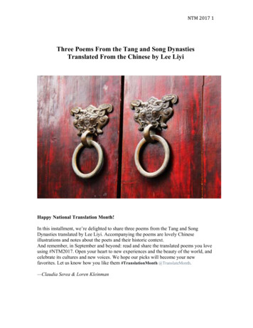
Biomolecules 2020, 10, 4735 of 143. ResultsBiomolecules 2019, 9, x FOR PEER REVIEW3.1. Alternatively Translated Cx43 Isoforms Localize to Different Subcellular Compartments3. Results5 of 14We constructed separate cDNA plasmids, each expressing an alternatively translated isoformthatcanbe generatedfrominternalmethioninestart sites(GJA1-32k,GJA1-29k, GJA1-26k, GJA1-20k,3.1. AlternativelyTranslatedCx43IsoformsLocalize to DifferentSubcellularCompartmentsGJA1-11k, GJA1-7k). For each of the plasmids, the AUGs downstream of each start codon were replacedWe constructed separate cDNA plasmids, each expressing an alternatively translated isoformby CUGs so that each plasmid only produces one Cx43 protein isoform fused with a C-terminal V5-tag.that can be generated from internal methionine start sites (GJA1-32k, GJA1-29k, GJA1-26k, GJA1-20k,We also constructed a wild-type Cx43 (GJA1-WT) plasmid with all internal AUG start sites intact and aGJA1-11k, GJA1-7k). For each of the plasmids, the AUGs downstream of each start codon wereplasmidonlyCx43(GJA1-43k),whichthe firststartcodonreplacedencodingby CUGs sothatfull-lengtheach plasmidonlyproduces oneCx43preservedprotein isoformfusedwitha C- yet mbryonickidneyHEK293FTcellsterminal V5-tag. We also constructed a wild-type Cx43 (GJA1-WT) plasmid with all internal leendogenouscompetitiontotheexogenousstart sites intact and a plasmid encoding only full-length Cx43 (GJA1-43k), which preserved the firstoverexpressionof Cx43start codon yet witheach isoforms.downstream AUG mutated to CUG (Figure S1). Human embryonic kidneyHEK293FTcells have little endogenousCx43 twoand o anti-V5)Using immunofluorescence,we usedseparate presentantibodiesC-terminusexogenousof Cx43 isoforms.totheidentifythe overexpressionsubcellular localizationof each V5-tagged isoform when exogenously introduced inUsing immunofluorescence,weexpected,used two separateantibodies(anti-Cx43and anti- on theHEK293FTcells (Figure 1A). AsGJA1-WTlocalizedas largeC-terminusplaque formationsV5) to identify the subcellular localization of each V5-tagged isoform when exogenously introducedcell–cell border. In contrast, full length GJA1-43k accumulated in perinuclear regions, indicating ain HEK293FT cells (Figure 1A). As expected, GJA1-WT localized as large plaque formations on thereduced ability to be trafficked to the cell border, consistent with our previously reported results of acell–cell border. In contrast, full length GJA1-43k accumulated in perinuclear regions, indicating asix-foldintraffickedgap junction[22].consistentGJA1-20k,a chaperonethatreporteddirectsresultsthe traffickingofreducedreductionability to beto theplaquescell border,withour cleara six-fold reduction in gap junction plaques [22]. GJA1-20k, a chaperone that directs the trafficking ofandregionsGJA1-43kand the isoformmitochondria[26]. Ofborder,note, wedid he ERfull-lengthto the cell–cellgenerallyappearsto theperinuclearandthe mitochondriaOf note,wehighlydid notdetect However,GJA1-11k[26].appearedto beenrichedthe nucleus,with minimallocalizationcell nucleus. general,However,V5GJA1-11kappearedto be highlyenrichedin thedetectionin tothethecytoplasm.Indetectionis diminishedas theisoformsgetnucleus,smaller, yet detectionisdiminishedastheisoformsnucleus to cytoplasm ratios remained consistent. GJA1-7k, the smallest isoform, was poorlygetdetected bysmaller,yet the nucleuscytoplasmratios remainedconsistent.GJA1-7k,the smallestisoform, waseitherantibody.We alsotostainednon-transfectedcellsto establishthe baselineendogenousexpressionpoorly detected by either antibody. We also stained non-transfected cells to establish the baselineof Cx43 in HEK293FT cells.endogenous expression of Cx43 in HEK293FT cells.Figure 1. Cont.
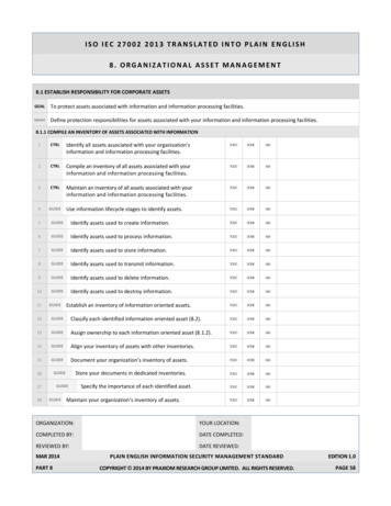
Biomolecules 2020, 10, 473Biomolecules 2019, 9, x FOR PEER REVIEW6 of 146 of 14Figure Figure1. Localizationof alternativelytranslatedCx43Cx43isoformsexpressedin HEK293FTcells.(A).(A). The fixed-cell1: Localizationof alternativelytranslatedisoformsexpressedin HEK293FTcells.The fixed-cellimmunofluorescenceof HEK293FTcells expressingshort isoformsof wasCx43detectedwas detectedimmunofluorescenceof HEK293FTcells expressingshort isoformsof Cx43by monoclonal anti-V5by monoclonalanti-V5(green)anti-Cx43and polyclonalanti-Cx43 raised(red) antibodies,raised againsta 17-residue(green) andpolyclonal(red) antibodies,against a 17-residuepeptideof C-terminal Cx43 (Sigma)peptidewithof i(blue).Thearrowheads showHoechst staining of the nuclei (blue). The arrow heads show the gap junction (plaque)formation at the cell–the gapcelljunctionthe (GJA1-WT)cell–cell borderof cellswithbar:wildtype The(GJA1-WT)border(plaque)of cells formationwith wildattypeplasmid.Scale10µm.results plasmid.are representative of fourScale bar:10µm. Theresults are (B).representativefour independent(B).The ratioofindependentexperiments.The ratio ofoffluorescentintensityexperiments.in the nucleusversuscytoplasmin 3FT cells. The data are presented as mean SEM, n 20, ****p 0.0001, by two-way ANOVA followed bypresentedas mean SEM,n 20, ****p The0.0001,by two-wayANOVA followedby Tukey’smultiple using Image J.Tukey’smultiplecomparisontest.intensitieswere measuredand normalizedto backgroundcomparison test. The intensities were measured and normalized to background using Image J.To quantify the distribution of each isoform, we calculated the ratio of the mean fluorescentTo quantifydistributionof eachisoform, (Figurewe calculatedratio ofthe meanthatfluorescentdensitythein thenucleus versuscytoplasm1B). ThistheanalysisconfirmedGJA1-11k has a twodensity in thenucleusversus detection)cytoplasm to(Figure1B). (anti-Cx43This analysisconfirmedthat GJA1-11khasFurthermore,a hment.the(anti-V5 uclearenrichment.Furthermore,nuclear localization is specific to GJA1-11k. All other isoforms capable of being thegenerated bynuclear localizationis specificto GJA1-11k.Allorotherof being density.generatedToby confirmalternativealternativetranslationhad 50%lessisoformsnucleuscapableto cytoplasmthat nucleustranslationlocalizationhad 50% or wasless notnucleusto cytoplasmdensity.To confirmthatnucleus localizationsecondaryto the V5tag interactingwithGJA1-11k,we obtainedwassimilar resultsnot secondarythe V5 fortag HA-taggedinteracting GJA1-11kwith GJA1-11k,weobtained similar results by a staining forby atostaining(FigureS2).HA-tagged GJA1-11k(FigurelocalizationS2).The nucleusof GJA1-11k was also tested by biochemistry techniques. We performedThe nucleuslocalizationofGJA1-11kwas also testedbiochemistry GJA1-11k,techniques.GJA1-43k,We performedsubcellular fractionationsof HEK293FTcells byoverexpressingand a FP-V5. Consistent with the immunofluorescence data, Western Blot analysis of cytosoliccontrol GFP-V5.Consistentwith theimmunofluorescencedata, WesternBlot analysisof cytosolicandand nuclearfractionsrevealedthat when comparedto full lengthGJA1-43k,GJA1-11kexpression isnuclear 43k,GJA1-11kexpressionismoremore enriched in the nucleus, with nearly as much protein in the nucleus as in the cytoplasm despiteenriched inathenucleus,withintranuclearnearly as muchproteinin the 2,nucleusin gether,thedespitedata ofa muchFigures 1 and 2smaller intranuclearvolume2, FigureofS3).Together,the dataof Figures1 and 2 Cx43demonstratedemonstratethat a(Figuremajor fractionGJA1-11k,unlikethe othermammalianisoforms, localizesthat a majortofractionof GJA1-11k, unlike the other mammalian Cx43 isoforms, localizes to the nucleus.the nucleus.
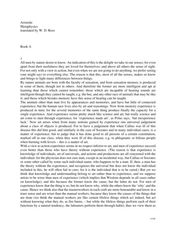
BiomoleculesBiomolecules2020,2019,10,9, x473FOR PEER REVIEWBiomolecules 2019, 9, x FOR PEER REVIEW77ofof14147 of 14Figure 2. (A). Biochemical isolation of GJA1-11k in the nucleus. Western Blot of Cx43 isoforms inFigure 2: (A). Biochemical isolation of GJA1-11k in the nucleus. Western Blot of Cx43 isoforms in subcellularsubcellularfractions (cytosolicandnuclear fractions)of HEK293FTcellsover-expressingexogenousFigure2: (A). (cytosolicBiochemicalisolationof GJA1-11kin the nucleus.WesternBlotof Cx43GJA1-11k,isoformsin subcellularfractionsand nuclearfractions)of HEK293FTcells -CT(Millipore)and (cytosolicGFP-V5 taggedplasmid fractions)cDNA, probedto To demonstratefractionsand nuclearof HEK293FTcellsCx43-CTover-expressingGJA1-43k,To iessame(Millipore)blot towasprobedusing antibodiesenriched taggedbiochemicalisolation,the probedsameblotprobedusingdifferentsubcellularand antibodies.GFP-V5plasmidcDNA,to wasmonoclonalCx43-CTantibodies.To fractiondemonstrate /K -ATPase), -ATPase),tomarkers,differentsubcellularfraction (GAPDH),markers, includingcytoplasmic(GAPDH),(NanuclearGolgi brane(Na /Kusingenrichedbiochemicalisolation,the same blotwas probedantibodiesto armarkers(Histone H3,ofLaminB1). The resultsare representativeof three(HistoneH3, LaminB1).The resultsare representativethreeindependentexperiments.(B). The densitometry markers, including cytoplasmic (GAPDH), membrane (Na /K -ATPase), Golgi (GM-130), and nuclear markersof CT-43k (11k)subcellular fractionsassessedby WesternUncutimmunoblotsare fractionsprovided inFigure S3.independentexperiments.(B). edby(Histone H3, Lamin B1). The results are representative of three independent experiments. (B). The densitometryWestern Blot. Uncut immunoblots are provided in Figure S3.of CT-43k(11k) subcellularfractionsassessed by Western Blot. Uncut immunoblots are provided in Figure S3.3.2. GJA1-11kInhibits CellProliferation3.2. GJA1-11k Inhibits Cell ProliferationThe nucleustheProliferationprimary organelle involved in cell cycle regulation. Because the C-terminus3.2. GJA1-11kInhibits isCellfragmentsareis associatedwithinhibitingcell proliferation[29,31]and GJA1-11kappearsto beThenucleusthe primaryorganelleinvolvedin cell cycleregulation.Becausethe tioninthepresenceofGJA1-11k.fragments are associated with inhibiting cell proliferation [29,31] and GJA1-11k appears to be enrichedcellsweretransfectedwith the siRNAthat dGJA1GJA1-11kto beinthe HEK293FTnucleus1 and2), weinhibitingquantifiedcell proliferationthe presenceof GJA1-11k.HEK293FTany endogenoussourceof anyCx43isoform.Note thatsilencing GJA1alone,even ls were transfected with the siRNA that silenced endogenous GJA1 mRNA, removing any endogenousbackgroundsignal,resulted in awithsignificantincreasein cellgrowthendogenouscompared to edthesiRNAthatsilencedmRNA, removingsourceofanyCx43isoform.Note hesedatasuggestevenminutequantitiesof littleany endogenoussourceof anyinCx43isoform.Note thatsilencing GJA1cellsalone,even edtonon-transfectedorscramblesiRNACx43 or its isoforms can inhibit cell proliferation.backgroundsignal,resulteda significantincreasecell minutegrowth quantitiescompared ofto Cx43non-transfectedcellscontrol(Figure3, FigureS4). inThesedata suggestthatinevenor its isoformsor scramblesiRNAcontrol (Figure 3, Figure S4). These data suggest that even minute quantities ofcaninhibit cellproliferation.Cx43 or its isoforms can inhibit cell proliferation.Figure 3: Effect of Cx43 on cell proliferation. The knockdown of endogenous Cx43 increases the cell proliferationof HEK293FT cells. For each day, data are presented as mean SEM, n 9, **p 0.01, ***p 0.001, ****p 0.0001by an unpaired two-tail Student’s t test. Western Blot analysis confirmed the knockdown of Cx43 and isrepresentative of three independent experiments.Figure3. Effectof Cx43onproliferation.cell proliferation.The knockdownof endogenousCx43 increasescellFigure3: Effectof Cx43on cellThe knockdownof endogenousCx43 increasesthe celltheproliferationproliferationHEK293FTday, dataare presentedSEM,9, ** p****p 0.01,of HEK293FTcells.ofForeach day,cells.dataForare eachpresentedas mean SEM, n as9,mean**p 0.01,***pn 0.001, 0.0001***p 0.001,****p Blotanalysisconfirmedby an unpaired two-tail Student’s t test. Western Blot analysis confirmed the knockdown of Cx43 and isthe knockdownof independentCx43 and is representativerepresentativeof threeexperiments. of three independent experiments.
Biomolecules 2020, 10, 473Biomolecules 2019, 9, x FOR PEER REVIEW8 of 148 of 14We then explored the effect of the full length Cx43 and the GJA1-11k isoforms on cell proliferation.We then exploredthewitheffectof thefull lengthCx43 GJA1-43k,and the GJA1-11kon cellWe WT,isoformsand GFP-V5as ressingGJA1-11k,GJA1-43k,GJA1-WT,andnegative control. The cells were counted over a three-day period (Figure 4). GJA1-11k significantlyGFP-V5a negativ
1 Smidt Heart Institute, Graduate Program in Biomedical Sciences, Cedars-Sinai Medical Center, Los Angeles, CA 90048, USA; iepifantseva@gmail.com (I.E.); sxiao@mednet.ucla.edu (S.X.); . Acknowledgments: We thank Flow core at Cedars-Sinai Medical Center for technical assistance. Conflicts of Interest: We have no conflict of interest to disclose.