Transcription
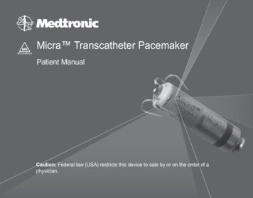
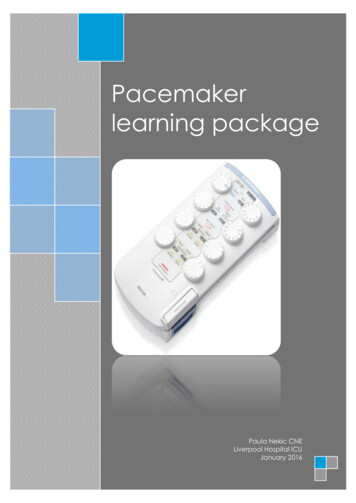
Pacemakerlearning packagePaula Nekic CNELiverpool Hospital ICUJanuary 2016
Liverpool HospitalIntensive Care: Learning PackagesPacemaker Learning PackageIntensive Care UnitCONTENTS Indications for pacing3 Anatomy4 Definitions9 Types of pacing13 How does a pacemaker work?20 Modes of pacing24 Sensitivity and output32 Thresholds35 Management37 Precautions40 Complications / troubleshooting41 Learning activities45 Reference List46LH ICU2016 Learning Package Pacemaker Learning Package2 Page
Liverpool HospitalIntensive Care: Learning PackagesPacemaker Learning PackageIntensive Care UnitINDICATIONS Sinus bradycardia AV blocks Complete heart block Overriding tachy atrial and/or ventricular arrhythmia (AF, AFlutter & SVT), Re-entrant tachycardia’s Prior to the implant of a permanent pacemaker Acute myocardial infarction complicated by heart block Temporary support of a patient after heart surgeryLH ICU2016 Learning Package Pacemaker Learning Package3 Page
Liverpool HospitalIntensive Care: Learning PackagesPacemaker Learning PackageIntensive Care UnitANATOMYFigure 1: stem-introductionThe Heart the heart is a cone-shaped, muscular organ located between the lungsbehind the sternum. The heart muscle forms the myocardium the inner lining of the myocardium is called Endocardium and the outerlayer cells is called the Epicardium. The pericardium (visceral) is the outer membranous sac with lubricatingfluid. The heart has four chambers: two upper, thin-walled atria, and two lower,thick-walled ventricles. The ventricles are the chambers that eject blood in to arteries. The functions of the atrium are to receive the incoming blood from theveinLH ICU2016 Learning Package Pacemaker Learning Package4 Page
Liverpool HospitalIntensive Care: Learning PackagesPacemaker Learning PackageIntensive Care Unit The septum is a wall dividing the right and left sides of the heart Coronary arteries are the vessels that supply blood to the heart muscle.Cardiac Valves:Cardiac valves permit blood to flow in only one direction through theheart Atrioventricular Valves:1] Tricuspid valve: separates the Right atrium from the Right ventricle2] Bicuspid valve [Mitral valve]: lies between the Left atrium and Leftventricle. Semi lunar Valves:1] Pulmonic valve: is the valve between the Right ventricle and thepulmonary artery.2] Aortic valve: is the valve between the Left ventricle and the aorta.The Heartbeat Each heartbeat is called a cardiac cycle. When the heart beats occur, the two atria contract together, then thetwo ventricles contract; then the whole heart relaxes. Systole is the contraction of heart chambers; diastole is their relaxation. The heart sounds, lub-dup, are due to the closing of the atrioventricularvalves, followed by the closing of the semi lunar valves.Conduction System The hearts cardiac conduction functions by emitting electrical currentsthat cause the myocardium to contract. The atria are the first to contract then the ventricle. There are two specialized cells in the heart that keep the synchronizationof the atria and the ventricle:1. Nodal cells2. Purkinje cellsThese cells have three characteristics that help to keep everything in time:1. Automaticity: the ability to initiate an electrical impulse2. Excitability: the ability to respond to an electrical impulseLH ICU2016 Learning Package Pacemaker Learning Package5 Page
Liverpool HospitalIntensive Care: Learning PackagesPacemaker Learning PackageIntensive Care Unit3. Conductivity: the ability to transmit an electrical impulse from one cellto another. The electrical current is first initiated in the SA node, the hearts naturalpacemaker, located at the top of the right atrium. The SA node is composed of nodal cells. In a normal resting adult heart,the SA node initiates firing at 60 to100 impulses/minute, but the rate canchange in response to the metabolic demands of the body. The impulsescause electrical stimulation and subsequent contraction of the atria. These signals then travel across the atrium to the atrioventricular node,located close to the septal leaflet of the tricuspid valve. The AV node is also made up of nodal cells. The AV node coordinatesthese incoming electrical impulses. After a slight delay (giving the atriatime to contract and complete ventricular filling), it relays the impulse tothe ventricles. The Purkinje cells then take over in the ventricle. The impulse in the ventricles is initially conducted through a bundle ofspecialized conducting tissue called the bundle of His, which then dividesinto the right bundle branch (conducting impulses to the right ventricle)and the left bundle branch (conducting impulses to the left ventricle). To transmit impulses to the left ventricle, the heart's largest chamber, theleft bundle branch divides into the left anterior and left posterior bundlebranches. Impulses travel through the bundle branches to reach the Purkinje fibers(the terminal point in the conduction system). The Purkinje cells rapidly conduct the impulses through the thick walls ofthe ventricles. At this point, the myocardial cells are stimulated, causingventricular contraction. (1)LH ICU2016 Learning Package Pacemaker Learning Package6 Page
Liverpool HospitalIntensive Care: Learning PackagesPacemaker Learning PackageIntensive Care UnitFigure 2: Cardiac conduction system. Marquette, K.H. Impulses can occur in various sites within the heart. Heart rate is determined by the Nodal and Purkinje cells with the fastestfiring rate. Normally, the SA node has the highest inherent rate, 60 to100impulses/minute. The AV node has an inherent rate of 40 to 60 impulses/minute Ventricular pacemaker sites have the lowest inherent rate, of 30 to 40impulses/minute. If the SA node malfunctions, the AV node generally takes over theheart's pacemaker function at its inherently lower rate. Should both the SA and the AV nodes fail in their pacemaker function, apacemaker site in the ventricle will fire at its inherent bradycardic rate of30 to 40 impulses/minute.It is when one or both of these nodes fail that a temporary pacemakermay be needed.Electrical activity of the heart is recorded on the ECGLH ICU2016 Learning Package Pacemaker Learning Package7 Page
Liverpool HospitalIntensive Care: Learning PackagesPacemaker Learning PackageIntensive Care UnitFigure 3: Electrical representation and cardiac cycle. Marquette, K.HLH ICU2016 Learning Package Pacemaker Learning Package8 Page
Liverpool HospitalIntensive Care: Learning PackagesPacemaker Learning PackageIntensive Care UnitDEFINITIONSThere are numerous terms used in pacing. Some of these are listed below: Action Potential:The action potential is brought on by a rapid change in membranepermeability to certain ions, with unique properties necessary for function ofthe electrical conduction system of the heart. Automaticity:Is the ability of the cardiac muscles to depolarize spontaneously, i.e. withoutexternal electrical stimulation from the nervous system. Ampere (AMP, A):Is a measure of electrical current flowing past a point in a conductor whenone volt of potential is applied across one ohm of resistance. In pacing, thesecurrents are so small that they are expressed in thousandths of amperes(milliamperes, mA) or in millionths of amperes (microamperes, μA) Amplitude:The maximum absolute value attained by an electrical waveform, voltage orcurrent. The amplitudes of pacemaker output pulses are expressed in volts(the difference in electrical potential), or in milliamperes (the measure of theelectrical current flow). AV Interval / Atrioventricular Interval, AV Delay:In a dual chamber pacemaker mode the AV Interval is the period of timebetween an atrial event (sensed or paced) and a scheduled pacedventricular event. It is typically measured in milliseconds. The AV Interval canalso be thought of as the pacemaker equivalent to the PR interval in normalconduction.LH ICU2016 Learning Package Pacemaker Learning Package9 Page
Liverpool HospitalIntensive Care: Learning PackagesPacemaker Learning PackageIntensive Care Unit Asynchronous pacing:(a not; syn together; chrono time) are those that are not together intime with the heart because they do not know what the heart is doing.Pacemakers such as AOO and VOO are asynchronous as they stimulate theheart at a fixed, preset rate independent of the electrical and/ormechanical activity of the heart. Burst Pacing:Is a technique of rapid pacing which may be used to terminate atachycardia by delivering a series of pacing pulses in which the delivery rateremains constant within each pacing attempt. This technique is used in boththe atria and the ventricles to attempt to terminate organized arrhythmias. Capture:Is initiation of depolarization of the atria and/or ventricles by an electricalstimulus delivered by an artificial pacemaker. Capture can be visualized onthe monitor by a spike before every p wave (for atrial pacing) and a spikebefore every QRS (for ventricular pacing) Demand Pacing:A pacing stimulus is delivered to the myocardium if the intrinsic rate fallsbelow the set rate on the pacemaker. Dual-Chamber Pacemaker:A pacemaker with two leads (one in the atrium and one in the ventricle) toallow pacing and/or sensing in both chambers of the heart to artificiallyrestore the natural contraction sequence of the heart. Epicardial Pacing:Is a type of temporary pacing where pacing wires are fixed directly to themyocardium (ventricular and often atrial) and are exposed through the skinon the chest wall, usually following cardiac surgery.LH ICU2016 Learning Package Pacemaker Learning Package10 P a g e
Liverpool HospitalIntensive Care: Learning PackagesPacemaker Learning PackageIntensive Care Unit Fixed Rate Pacing:A pacing stimulus is delivered to the myocardium at a programmed fixedrate regardless of the underlying rate and rhythm. This is also known asasynchronous ventricular pacing. Electromagnetic Interference (EMI):Radiated or conducted energy – either electrical or magnetic – which caninterfere with the function of the pacemaker. Fusion Beat:Is a spontaneous cardiac depolarisation which occurs coincidentally with apaced depolarisation. The paced and natural depolarisation waveformscollide or fuse, causing distortion of the surface ECG complex. Intrinsic:Inherent; belonging to or originating from the heart itself, (e.g., an intrinsicbeat refers to a naturally occurring heart beat). Inhibited:Is any pacemaker which, after sensing a spontaneous depolarization,withholds its pacing stimulus. Examples are AAI, VVI Millivolt:One one-thousandth of one volt. A unit of measure for low levels of voltage.Spontaneous, intrinsic P-waves and R-waves are measured in millivolts. Millisecond:One one-thousandth of one second. Most pacemaker timing functions, e.g.,pulse width and pacing intervals, are expressed in milliseconds. No Output:Is the absence of energy delivery to the heart. Output:The electrical stimulus or energy generated by a pulse generator andintended to trigger a depolarization in the chamber of the heart beingpaced.LH ICU2016 Learning Package Pacemaker Learning Package11 P a g e
Liverpool HospitalIntensive Care: Learning PackagesPacemaker Learning PackageIntensive Care Unit Sensitivity:Is the pacemaker’s ability to sense a patient’s intrinsic rhythm or when naturaldepolarization is occurring The sensitivity number represents the minimum size, in millivolts, of anelectrical signal that will be detected by the pacemaker The higher the sensitivity (number) the less likely the pacemaker will seethe patients rhythm Sensing Threshold:The minimum atrial or ventricular intra cardiac signal amplitude required toinhibit or trigger a demand pacemaker, expressed in millivolts. Stimulation or output Threshold:The minimum electrical stimulus needed to consistently elicit a cardiacdepolarization. It can be expressed in terms of amplitude (volts, milliamps)and pulse width (milliseconds), or energy (microjoules). Triggered:In pacemaker terms, a Triggered pacing mode is the opposite of inhibited.Upon detecting a spontaneous depolarization or other signal, a triggeredmode will deliver an electrical stimulus to the heart.Medtronic Pacing Glossary. 2005“Unless you try to dosomething beyondwhat you have alreadymastered, you willnever grow."Ronald E. OsbornLH ICU2016 Learning Package Pacemaker Learning Package12 P a g e
Liverpool HospitalIntensive Care: Learning PackagesPacemaker Learning PackageIntensive Care UnitTYPES OF PACING TRANSCUTANEOUS PACING Is only used in emergency situations due to the discomfort felt by thepatient. The electrodes are incorporated into the pads and cover a largesurface area over the skin. The pads are connected to an externalpulse generator which delivers energy through the pads to the chestwall muscles.Figure 4: Pacing pad placement for trancutaneous pacing. Lippincott’sNursing centre.com 2011 By continuously monitoring cardiac rate and rhythm and deliveringpacing impulses through the skin and chest wall muscles as needed,trancutaneous pacing causes electrical depolarization andsubsequent cardiac contraction to maintain cardiac output untilthe patient receives a transvenous pacemaker The pacing stimulus travels through chest wall, pectoralis andintercostals muscle, ribs pericardial fluid and fat before finallyreaching the heart. Therefore a much higher energy level isneeded to deliver the correct energy to the heart. One side effectof this is chest muscle contraction from the electricalstimulus. Therefore giving the patient some sedation for comfort ishigh priority Pacing or defibrillation pads must be in good contact with chestwall Once capture has been verified on monitor by a spike before everyQRS, patient’s haemodynamics must also be assessed ensuringelectrical and mechanical capture.LH ICU2016 Learning Package Pacemaker Learning Package13 P a g e
Liverpool HospitalIntensive Care: Learning PackagesPacemaker Learning PackageIntensive Care Unit See Transcutaneous pacing guideline for detailed managementinformation EPICARDIAL PACING is used post cardiac surgery and involves pacing wires being suturedto surface of heart the atrial pacing wires exit the patient’s chest on the right side andthe ventricular wires exit the chest on the left side is a form of temporary pacing for bradycardia and override pacing inatrial fibrillation These wires may only last a few days to a week. If left for longer than a week the pacing thresholds may rise and theremay not be adequate and reliable captureFigure 5: Placement of epicardial pacing wires:www.legalmedicalexhibits.comLH ICU2016 Learning Package Pacemaker Learning Package14 P a g e
Liverpool HospitalIntensive Care: Learning PackagesPacemaker Learning PackageIntensive Care Unit TRANSVENOUS PACING Transvenous pacemakers are inserted into the venous system through acatheter site such as a specially designed pulmonary artery catheter,or sheath, and deliver the stimulus via electrodes that are in directcontact with the heart tissue itself. Generally the subclavian, femoral or internal jugular veins are used toaccess the right atrium or the right ventricle.Figure 6: Transvenous pacing. Critical Care Nurse 2004Pacing powerpoint 2015 P.Nekic(CNE)LH ICU2016 Learning Package Pacemaker Learning Package15 P a g e
Liverpool HospitalIntensive Care: Learning PackagesPacemaker Learning PackageIntensive Care UnitTYPES OF PACEMAKER LEADSWith any electrical current the electrical impulse must flow between twopoles (or electrodes) in order to stimulate the heart.The pacing leads used can be either bipolar or unipolarBipolar There are two electrodes positive and negative involves a single wire with two conductors insulated from oneanother, which both run to the epicardial surfaceFigure 7: Bipolar pacing. Single chamber temporary pacing ppt 2001Unipolar Only one electrode ( negative) is in contact with the heart The positive electrode is located somewhere else on the bodyFigure 8: Unipolar pacing. Single chamber temporary pacing ppt 2001LH ICU2016 Learning Package Pacemaker Learning Package16 P a g e
Liverpool HospitalIntensive Care: Learning PackagesPacemaker Learning PackageIntensive Care UnitIn both systems the current flows from the negative terminal of the pacingbox down the pacing lead to the negative electrode and into the heart.The current is then picked up by the positive electrode and goes back up thelead to the positive terminal of the pacing box.Epicardial pacing leads: Bipolar leads are usually used post op Bipolar epicardial leads have two separate insulated wires, onepositive and negative These are sutured to the chamber of the heart that needs to be paced Both leads are in contact with the heart so either wire may be used asthe negative or pacing electrode The remaining wire is used as the ground or positive electrodeFigure 9 : Right atrial wire placement.ctsnet.org accessed 02/2012LH ICU2016 Learning Package Pacemaker Learning Package17 P a g e
Liverpool HospitalIntensive Care: Learning PackagesPacemaker Learning PackageIntensive Care UnitANATOMY OF A PACEMAKERVENTRICULAR LEADSATRIAL LEADSON/ OFFHEART RATEATRIAL OUTPUT DIALVENTRICULAR OUTPUTDIALBATTERY INDICATORLIGHTLock/ UnlockMODEFigure10: Medtronic 5392 PacemakerEnter keyAtrial and VentricularsensitivitySelection keyAV delayUpper rate limit setsmaximum ventricularpacing rate allowedwhile tracking theventriclePVARP: Post VentricularAtrial Refractory PeriodSets length of time after aventricular event duringwhich atrial sensingdoesn’t affectpacemaker timingDial to change settingsPause button to checkunderlying rhythmA.Tracking: applicable in DDD only. IfON it paces ventricle in synchronywith intrinsic atrial depolarisationsLH ICU2016 Learning Package Pacemaker Learning PackageRapid atrial pacingcan be used tointerrupt atrialtachycardia’s18 P a g e
Liverpool HospitalIntensive Care: Learning PackagesPacemaker Learning PackageIntensive Care UnitAtrial block: atrial wires to beconnected to it from patient.Block has ATRIUM written on itVentricular block: ventricularwires to be connected to itfrom patient. Block hasVENTRICLE written on itFigure 12: Pacing leads for Biotronic EDP 30 and Biotronic ReocorMode dialAV delaydialAtrialsensitivityand outputdialsRate dialAtrial Burst ratefor over-ridingtachyarrhythmiaVentricularsensitivity andoutput dialsFigure13: Biotronic Reocor pacing box.LH ICU2016 Learning Package Pacemaker Learning Package19 P a g e
Liverpool HospitalIntensive Care: Learning PackagesPacemaker Learning PackageIntensive Care UnitHow do pacemakers work? The pacemaker essentially does two things : it senses the patient’s own rhythm using a “sensing circuit”, It sends out electrical signals using an “output circuit”. If the patient’s intrinsic rhythm becomes too slow or goes awaycompletely, the electronic pacemaker senses that, and starts sendingout signals along the wires leading from the pacing box to the heartmuscle. The signals, if they’re “capturing” properly, provide a regular electricalstimulus, making the heart contract at a rate fast enough to maintainthe patient’s blood pressure. Pacemakers are not simply applied to the patient and turned on; wemust tell the pacemaker how to pace the heart. The goal of pacing the heart is to maximize cardiac output. There aretwo determinants of cardiac output; stroke volume and heart rate. Pacemakers use the sensing and output circuits and then determinethe heart rate by timing and intervalsHow do pacemakers determine heart rate?Before being able to understand pacemaker codes, it is necessary to havean understanding of pacemaker timing.INTERVALS AND TIMINGCardiac devices calculate everything in terms of intervals, from one event tothe next.The pacemaker does not keep track of how many beats the hearthas in a minute, it simply determines how long it has been since the last beathas occurred.LH ICU2016 Learning Package Pacemaker Learning Package20 P a g e
Liverpool HospitalIntensive Care: Learning PackagesPacemaker Learning PackageIntensive Care UnitIt does this by transforming the programmed minimum heart rate into a timeinterval by dividing the programmed heart rate into 60,000 (the number ofmilliseconds in one minute)The result is the number of milliseconds that the pacemaker waits beforedeciding to deliver a stimulus. (6)For Example: pacing heart rate set at 60 pacemaker waits 60,000/60 or 1000ms after the last beat before itdelivers the next stimulus if the heart has an intrinsic beat before 1000ms then pacemakerresets the lower rate timer and starts again at ure 14: Paced intervals: Single chamber temporary pacing ppt 2001INTERVAL DEFINITIONS Lower Rate interval( V-V) Lowest rate the pacemaker will pace the atrium in theabsence of intrinsic atrial activityAV Intervals Initiated by a paced atrial eventV-A Interval A pace-maker timing scheme that is based on the intervalbetween a ventricular event and the subsequent atrial event(the VA Interval). This is in contrast to an atrial escape intervalin which the interval between subsequent atrial events isfixed. (NOTE: This may make a difference when a patient hassome intrinsic AV conduction. Since a sensed ventricular beatLH ICU2016 Learning Package Pacemaker Learning Package21 P a g e
Liverpool HospitalIntensive Care: Learning PackagesPacemaker Learning PackageIntensive Care Unitcomes earlier than a paced one, and the pacemaker basesits lower rate on the atrial escape interval, the pacemakerrate from beat-to-beat could be slightly faster than theprogrammed rate. Atrial escaped interval The interval from a paced or sensed ventricular event to thenext scheduled paced atrial event.Figure 15: Intervals and timing in DDD mode. Core Pace Module 6. Single andDual Pacemaker timing ppt presentation 2003DDD ExamplesThe Four Faces of DDD Atrial and ventricular pacingAPVPAPVP– Atrial pace re-starts the lower rate timer and triggers an AV delaytimer (PAV) The PAV expires without being inhibited by a ventricular sense, resultingin a ventricular pace12On this slide, we see AV sequential or dual chamber pacing. Both the atriumand the ventricles are being paced, first the atrium and then the ventricle.Looking at the 2 complexes shown:The pacemaker begins by pacing in the atrium. 2 timers are started: The lower rate timer The AV Interval timerLH ICU2016 Learning Package Pacemaker Learning Package22 P a g e
Liverpool HospitalIntensive Care: Learning PackagesPacemaker Learning PackageIntensive Care Unit The AV Interval timer expires WITHOUT BEING INHIBITED by aventricular sense – so the pacemaker paces in the ventricle. The lower rate timer continues. If it expires without being inhibited by an atrial event (asshown above), the pacemaker paces in the atrium, and thecycle begins again.IN SUMMARYIntervals and timing can be a hard concept to understand, but fortunatelythe pacemaker calculates all these intervals for us. It is important to knowhow pacing works using its sensing, output and timing mechanisms as it helpswith troubleshooting.For example: looking at the above slide and DDD mode Why does the pacemaker’s AV interval timer expire without beinginhibited? Perhaps the patient has a heart block Perhaps the patient has delayed AV conduction — thepatient’s PR interval is longer than the PAV programmed intothe pacemaker Why does the pacemaker’s atrial escape interval expire? The patient’s sinus rate is less than the pacemaker’sprogrammed rate (i.e. sinus bradycardia)LH ICU2016 Learning Package Pacemaker Learning Package23 P a g e
Liverpool HospitalIntensive Care: Learning PackagesPacemaker Learning PackageIntensive Care UnitMODES OF PACINGOver the years pacemakers have developed from the very early deviceswhich merely delivered a timed impulse at set amplitude in a singlechamber, and paid no attention to the patient’s intrinsic rhythm.Now even temporary pacemakers are relatively sophisticated devices whichcan deliver a range of therapeutic options from the basic fixed rateasynchronous, to dual chamber demand pacing.In order to be able to easily communicate to others the potential capabilitiesand actual programming of pacemakers, it was necessary to develop acode, which is shown here, generally called the NBG code. Generally the firstthree codes are utilised.Figure 16: NBG Code.www.cardiacrhythm.coLH ICU2016 Learning Package Pacemaker Learning Package24 P a g e
Liverpool HospitalIntensive Care: Learning PackagesPacemaker Learning PackageIntensive Care UnitCODES: WHAT DOES IT ALL MEAN? Defines the chambers that are paced/sensed Defines how the pacemaker will respond to intrinsic events PACING : first letter is chamber being paced SENSING: relates to the chamber in which the pacemaker is attemptingto sense the patient’s intrinsic rhythm. RESPONSE: relates to the pacemakers response to the sensed intrinsicbeat, either triggered or inhibitedPacingChamber PacedChamber SensedAction or Responseto a Sensed EventDDDFigure 17 & 18: PSR. Pacing ppt. P.Nekic 20111st LetterChambers Paced2nd LetterChambers SensedA AtriumA AtriumV VentricleV VentricleD Dual (both Atriumand Ventricle)D DualO none3rd LetterResponse to SensingT TriggeredI Inhibit (DemandMode)D DualO none(Asynchrony)Remembering the letters PSR helps to remember the function of each letter.LH ICU2016 Learning Package Pacemaker Learning Package25 P a g e
Liverpool HospitalIntensive Care: Learning PackagesPacemaker Learning PackageIntensive Care UnitTherefore when you collate the information in the tables above you end upwith different codes and or modes of pacing. The most common modes areoutlined in the table below.MODES OF PACINGCODEChamberPaced(1st Letter)ChamberSensed(2nd Letter)AOOAtrial pacingNoneResponse tosensingTrigger orInhibited(3rd lar &atrial pacingNoneNoneAAIAtrial pacing AtrialsensingInhibitspacingA sensed P wave willcause thepacemaker towithhold atrialpacing. Absence ofintrinsic p wave willlead to atrial tspacingA sensed R wave willcause thepacemaker towithhold ventricularpacing. Absence ofintrinsic R wave willlead to ventricularpacingDDDAtrial &ventricularpacingAtrial &ventricularsensingInhibition ofpacing &trigger ofpacingimpulsesIntrinsic P & R wavescause pacemaker towithhold pacing.Pacing impulses willbe triggered whenprogrammedintervals areexceededLH ICU2016 Learning Package Pacemaker Learning PackageMEANINGAsynchronous atrialpacing occurs at aset rate, regardlessof intrinsic atrialactivity.Asynchronousventricular pacingoccurs at a set rate,regardless of intrinsicVentricular activity.Asynchronous atrial& ventricular pacingat a set rateregardless of intrinsicatrial & ventricularactivity26 P a g e
Liverpool HospitalIntensive Care: Learning PackagesPacemaker Learning PackageIntensive Care UnitWhat does it all mean? : DDDDDD MODE: The first letter is D, so both the Atrium and theVentricles are being paced. The second letter is also D which means that the pacemaker is lookingfor sensed intrinsic beats in both the Atrium and the Ventricle The third letter D means that the pacemaker can both Trigger aVentricular pace in response to a sensed Atrial event, and inhibit theAtrial and/or Ventricular pace if it senses an intrinsic event in thatchamber before its internal timing tells it to pace. An atrial sense: Inhibits the next scheduled atrial pace Re-starts the lower rate timer Triggers an AV interval (called a Sensed AV Interval or SAV)An atrial pace: Re-starts the lower rate timer Triggers an AV delay timer (the Paced AV or PAV)A ventricular sense: Inhibits the next scheduled ventricular pace Examples of DDD mode and how it worksDDD mode can have four possible outcomes for pacing depending on thepatient’s condition or underlying rhythm Atrial and ventricular pacing Atrial pace re-starts the lower rate timer (Long arrow) andtriggers an AV delay timer (PAV)( shorter arrow) The PAV expires without being inhibited by a ventricularsense, resulting in a ventricular pace Both the atrium and the ventricles are being paced, firstthe atrium and then the ventricle.The pacemaker begins by pacing in the atrium. 2 timers are started:LH ICU2016 Learning Package Pacemaker Learning Package27 P a g e
Liverpool HospitalIntensive Care: Learning PackagesPacemaker Learning PackageIntensive Care Unit The lower rate timer The AV Interval timer (we call this timer the PAV or PacedAV interval) The AV Interval timer expires WITHOUT BEING INHIBITED by aventricular sense – so the pacemaker paces in theventricle. The lower rate timer continues. If it expires without beinginhibited by an atrial event (as shown above), thepacemaker paces in the atrium, and the cycle beginsagain.Why does the pacemaker’s AV interval timer expire without being inhibited? Perhaps the patient has a heart block Perhaps the patient has delayed AV conduction — thepatient’s P-R interval is longer than the PAV programmedinto the pacemakerWhy does the pacemaker’s atrial escape interval expire? The patient’s sinus rate is less than the pacemaker’sprogrammed rate e.g. sinus bradycardia Atrial pacing and ventricular sensing Atrial pace re-starts the lower rate timer and triggers an AV delay timer(PAV) The PAV expires without being inhibited by a ventricularsense, resulting in a ventricular paceLower rate timer 1000msIn this example we see atrial pacing with an intrinsic ventricular response (VS). The pacemaker begins by pacing in the atrium. The 2 timers arestarted: The lower rate timer (longer arrow) The AV Interval timer (this is the PAV or Paced AV interval)(shorterarrow) The AV Interval timer IS INHIBITED by a ventricular sense – so the AVtimer is reset, but the lower rate timer
collide or fuse, causing distortion of the surface ECG complex. Intrinsic: Inherent; belonging to or originating from the heart itself, (e.g., an intrinsic beat refers to a naturally occurring heart beat). Inhibited: Is any pacemaker which, after sensing a spontaneous depolarization, withholds its pacing stimulus. Examples are AAI, VVI

