Transcription
Hindawi Publishing CorporationBioMed Research InternationalVolume 2013, Article ID 461230, 11 pageshttp://dx.doi.org/10.1155/2013/461230Review ArticleHeat Shock Proteins: Stimulators of Innate andAcquired ImmunityCamilo A. Colaco,1 Christopher R. Bailey,1 K. Barry Walker,2 and James Keeble31ImmunoBiology Limited, Babraham Research Campus, Cambridge CB22 3AT, UKAeras, 1405 Research Boulevard, Rockville, MD 20850, USA3NIBSC, Blanche Lane, South Mimms, Potters Bar EN6 3QG, UK2Correspondence should be addressed to Camilo A. Colaco; camilo.colaco@immbio.comReceived 11 February 2013; Accepted 9 May 2013Academic Editor: Anshu AgrawalCopyright 2013 Camilo A. Colaco et al. This is an open access article distributed under the Creative Commons AttributionLicense, which permits unrestricted use, distribution, and reproduction in any medium, provided the original work is properlycited.Adjuvants were reintroduced into modern immunology as the dirty little secret of immunologists by Janeway and thus beganthe molecular definition of innate immunity. It is now clear that the binding of pathogen-associated molecular patterns (PAMPs)by pattern recognition receptors (PRRs) on antigen presenting cells (APCs) activates the innate immune response and providesthe host with a rapid mechanism for detecting infection by pathogens and initiates adaptive immunity. Ironically, in addition toadvancing the basic science of immunology, Janeway’s revelation on induction of the adaptive system has also spurred an era ofrational vaccine design that exploits PRRs. Thus, defined PAMPs that bind to known PRRs are being specifically coupled to antigensto improve their immunogenicity. However, while PAMPs efficiently activate the innate immune response, they do not mediate thecapture of antigen that is required to elicit the specific responses of the acquired immune system. Heat shock proteins (HSPs)are molecular chaperones that are found complexed to client polypeptides and have been studied as potential cancer vaccines. Inaddition to binding PRRs and activating the innate immune response, HSPs have been shown to both induce the maturation ofAPCs and provide chaperoned polypeptides for specific triggering of the acquired immune response.1. IntroductionThe exposure of adjuvants as the immunologist’s dirty little secret by Janeway in his seminal introduction to theCold Spring Harbor Symposium on Quantitative Biology,“Approaching the Asymptote? Evolution and Revolution inImmunology” [1], resulted in a revision of the working modelof the immune system and provided a conceptual frameworkfor our current understanding of the innate immune responseand its control of adaptive immunity [1, 2]. Janeway reasonedthat as the adaptive immune system uses randomly generatedreceptors to recognise antigen, it cannot reliably distinguishbetween self and nonself. Adaptive immune cells must thus beinstructed as to the origin of an antigen by a system that candetermine whether an antigen is derived from self, infectious(i.e., microbial) nonself, or innocuous (i.e., noninfectious andnonmicrobial) nonself. He suggested that the evolutionarilyancient innate immune system might be able to providesuch instruction and proposed a mechanism by whichthe innate immune system could detect an infection and relayits conclusions to the adaptive immune system. Janeway proposed that the innate immune system would detect infectionby the use of germ-line encoded pattern recognition receptors (PRRs) to recognise conserved, microbial pathogenassociated molecular patterns (PAMPs). These PAMPs wouldbe unique to microbes and not found in eukaryotic cellsso that they would accurately signal infection. Furthermore,they would be common to a broad class of microbes so that alimited number of germ-line encoded receptors could detectall infections and be essential for the life of the microbe so thattheir detection could not be easily circumvented by mutation.Most importantly, Janeway proposed that the recognition ofinfection by PRRs on cells of the innate immune systemwould lead to the induction of signals that resulted ininitiation of the adaptive immune response. The subsequentidentification of the Toll-like receptors (TLRs) as key PRRsled to an explosion of research on innate immunity and the
2definition of a number of families of PRRs and signallingpathways that modulate inflammatory responses [2].Extension of this work into the area of vaccinologyhas suggested a classification of adjuvants into two majorfunctional groups, those being dependent and independentof binding to TLRs [3, 4]. TLR-dependent adjuvants actdirectly on dendritic cells (DCs), inducing the upregulationof cytokines, MHC class II costimulatory molecules, andpromoting DC migration to the T-cell area of the lymphnode [3, 4]. For example, peptidoglycans and other skeletalcell wall components in the Bacillus Calmette-Guérin (BCG)vaccine are recognized by TLR2 and TLR4 and help mediateprotective immunity against Mycobacterium tuberculosis [5].Conjugate vaccines against Haemophilus influenzae use theouter-membrane proteins from Neisseria to elicit effectiveadaptive responses via the triggering of TLR2 [6] and theadjuvant properties of short nucleotide sequences containingunmethylated CpG clusters mediated through TLR9 [7]. Incontrast, the mechanism of TLR-independent adjuvants likealum and the squalene-based oil-in-water emulsion MF59remains contentious [2, 8]. Alum has been shown to haveimmunostimulating activities in vivo as it results in therecruitment of monocytes, which take up antigen and migrateto the draining lymph nodes where they differentiate intofully competent inflammatory DCs [8]. Moreover, it hasbeen proposed that adsorption to alum increases antigenavailability at injection site, allowing an efficient uptake byantigen-presenting cells (APCs) [8, 9]. However, other studieshave shown that alum could also increase antigen uptakeby DCs in vitro and, in studies on alum as an adjuvant forantigens encapsulated in biopolymers, the improvement inimmunogenicity can be correlated to antigen entrapment andrelease, suggesting that, in addition to the maturation of DCs,alum may also perform an antigen delivery function [9, 10].Heat shock proteins (HSPs) are ubiquitous chaperonesthat bind and help fold nascent or denatured polypeptides[11]. HSPs have also been recognised as major immunogensin the immune response against pathogens [12, 13]. Thesestudies, as well as numerous studies on HSPs as cancervaccines, have revealed that apart from acting as immunogenic antigens themselves, HSPs can also act as adjuvants tostimulate the immunogenicity of heterologous polypeptidesto which they are either covalently or noncovalently coupled[13, 14]. Thus it can be argued that HSPs constitute a thirdfunctional group of adjuvants. This review will summarise thestudies that show that HSPs are not just stimulators of innateimmunity but can also traffic antigens into APCs facilitatingthe induction of specific acquired immune responses. In thiscontext, it is important to note that native HSPs isolatedfrom any organism will carry chaperoned polypeptides thatare specific to the source organism and can thus be useddirectly as vaccine candidates as has been demonstrated bythe development of autologous cancer vaccines [14, 15].2. Discovery of the Heat Shock Responseand HSPsThe heat shock response was first observed when the temperature of an incubator housing Drosophila was inadvertentlyBioMed Research Internationalelevated, resulting in a change to the pattern of chromosomalpuffing within the chromosomes of the salivary glands [17].Subsequently, a number of proteins were observed to beproduced within the same time frame as the appearance of thechromosome puffs and these are what subsequently becameknown as HSPs [18]. In addition to heat, these proteins werefound to be inducible upon exposure to a range of environmental stresses including oxygen deprivation, pH extremes,and nutrient deprivation [19]. This range of responses demonstrated a more general function in providing protectionagainst cellular stress, by limiting protein aggregation anddenaturation, and they are thus now more commonly referredto as stress proteins [20]. HSP synthesis occurs at 5–15 Cabove the optimal environmental temperature of that organism, depending on the organism’s growth temperature range[21]. The response is rapid (usually within 2–5 minutes afterheat shock), and the expression profile displays a temperaturerelated dynamic, in which the levels of specific HSPs changeover the range of different heat shock temperatures [21].Generally there is a transient increase in the synthesis of HSPsat low level temperature elevation, with a more sustainedresponse observed at higher temperatures, and this patternof response has been consistently observed in numerousorganisms [19–21]. For example, heat shocking BCG at 42 Cresults in the production of both HSP65 and HSP70, while at45 C, HSP70 synthesis is more pronounced [22]. At the thetranscriptional level, with BCG, the accumulation of mRNAfor HSP70 appears to peak at 45 minutes after initiation oftemperature elevation, declining after 60 minutes; whereasthe elevated mRNA expression of HSP65 mRNA did not persist after temperature elevation to 42 C [22]. The induction ofHSPs in mycobacteria can also be induced by other stresses,not the least being phagocytosed by macrophages [23]. Theheat shock response and the upregulation of levels of HSPshave been observed in all tissues and in both prokaryoticand eukaryotic organisms, indicating that it is a ubiquitousand critical biological response [21]. The early hypothesis wasthat these proteins were involved in the stress management ofthe cells by stabilisation of housekeeping proteins that werecritical for survival [20]. Thus, the initial pulsing of cells withlow temperature heat stress increased their levels of HSPsand their ability to survive a much higher thermal stress incomparison to untreated cells [21]. However, the more recentdemonstration of the constitutive nature of expression ofHSPs in all cells strongly suggests that these proteins play amore fundamental role in protein housekeeping within thecell, chaperoning the folding of nascent polypeptides andprevention of protein aggregation [19–21].Numerous studies have now revealed that HSPs arehighly conserved molecules that exhibit a high degree ofsequence homology between species [19, 20, 24]. HSPs arefound throughout the cell, but different HSP families can belocalised to specific cellular locations and can be divided intobroad families based on size (see Table 1). The HSPs involvedin protein folding can be separated into differing functionalsystems, with some overlap [20, 24, 25]. The HSP60-HSP10(GroEL-GroES) system is involved in classical protein folding [24]. The HSP70-HSP40 (DnaK-DnaJ) system stabilisespeptides in a linear, unfolded state and delivers them to
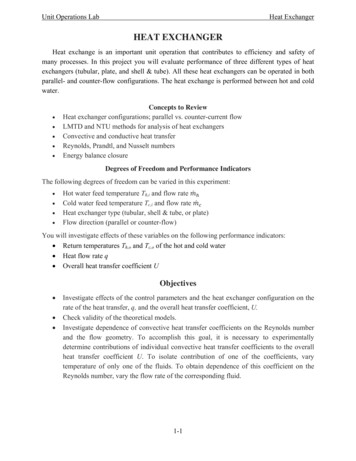
BioMed Research International3Table 1: Major prokaryotic and eukaryotic families of HSPs and their characteristics (see [15, 16]).Hsp familyCCTCalreticulinStructural featuresVaried, often large gomeric complexMonomericHSP602 stacked heptameric ringsHSP70MonomericHSP90Noncovalent homodimersHSP100Multimeric complexes withhsp70 and hsp25Small HSPsHSP40HSP47Membershsp10, GroES, hsp16, 𝛼-crystallin, hsp20, hsp25,hsp26, hsp27hsp40, DnaJ, Sis1Intracellular locationhsp47Endoplasmic reticulum (ER)CytosolCytosolTRiC (60 Kd family)Calreticulin, CalnexincytosolERCytosolhsp60, hsp65, GroELMitochondriaCytosolhsp71, hsc70 (hsp73), hsp110/SSE, DnaK, SSC1, SSQ1,MitochondriaECM10, Grp78 (BiP), Grp170ERCytosolhsc84, hsp86, HTPG, Gp96 (Grp94, endoplasmin)ERCytosolhsp104, Hsp110, Clp proteins, Hsp78Mitochondriathe HSP60-HSP10 system [25]. Small HSP family memberscan bind partially folded peptides and mediate their loadingonto one of the folding systems (e.g., HSP60-HSP10) [25].The HSP90 family are found predominantly in the cytoplasmand are thought to mediate the folding of specialised proteinssuch as steroid receptors and protein kinases [26]. Thermal tolerance, disaggregation, and unfolding of aggregatedproteins for enzymatic digestion are handled by the largerHSP100 chaperones [27]. Being involved in such a variety ofcellular processes, it is unsurprising that the majority of HSPs(HSP60, HSP70, and HSP90) are fundamental to cell survival,and mutation or deletion of the major HSP genes is oftenlethal to both cells and organisms [24–27].The major HSP families are associated with ATPase activity that is essential for their function as molecular chaperones[24, 28]. In the HSP60 system, ATP binding brings about aconformational change that exposes its peptide binding coreallowing peptides to enter the peptide binding chamber [28,29]. This is then followed by the binding of the cochaperoneHSP10, which closes the chamber and ATP hydrolysis to ADPand then energises the folding of the nascent polypeptidechain in a hydrophobic environment [29]. In the HSP70system, ATP binding brings about a conformational changein the HSP that exposes its peptide binding site, allowingpeptides to enter the binding cleft and ATP hydrolysis to ADPthen closes this cleft [28, 30]. The nascent protein can thenundergo folding without interference from other constituentsof the intracellular environment [30]. In the HSP90 (Gp96)system, in addition to ADP/ATP, peptide binding is underthe control of calcium levels that brings about the requiredconformational changes for peptide binding [31].While their role as molecular chaperones is their mostobvious biological function, their reported functions relatingto the immune system are still being elucidated. Numerousstudies have implicated HSPs in various aspects of theimmunological response to antigens, leading to the proposalthat these proteins carry out a “moonlighting” function as“chaperokines” [32–34]. These studies have shown that HSPsact both as adjuvants by triggering TLRs on cells of the innateimmune system, in particular macrophages and DCs, andalso as carriers of antigens by providing a mechanism forchaperoning polypeptides for the loading of MHC moleculesand the subsequent facilitation of induction of acquiredimmunity [32–36] (Figure 1).3. Innate ImmunityInitially HSPs were thought to be exclusively intracellularproteins that were only released into cellular environmentupon cellular injury or necrosis, but not apoptosis and,as such, they were not generally regarded as PAMPs butconsidered to be “danger associated molecular patterns”(DAMPs) [37]. DAMPs are molecules that serve as alternativeligands for PRRs but signal the presence of cellular damage, asdistinct from the presence of pathogens, thus also activatingthe innate immune response [38]. However it is now apparentthat HSPs can be actively secreted into the extracellular environment by tumour cells or released from cells undergoingnecrotic lysis in response to cytotoxic lymphocyte (CTL)or natural killer (NK) action, or viral infections [39–41].Members of HSP60, HSP70, and HSP90 (gp96) families haveall been linked with innate immune stimulation [12, 14, 36,42]. They have been observed to elicit nonspecific cytokineand chemokine secretion from cells of the mammalian innateimmune system, to upregulate costimulatory molecules, andto activate APCs in particular DCs via a number of receptors[43–45]. One of the initial HSPs to be studied for its effectson innate immunity was recombinant mycobacterial HSP65which was shown to stimulate the human monocyte cell lineTHP-1 resulting in the production of TNF-𝛼, IL-6, and IL-8[46]. In comparison to the mycobacterial HSP65, the mammalian homologue HSP60 was 10–100 times more potent atstimulating human monocytes to secrete cytokines (IL-6, IL10 TNF-𝛼, IL-12, and GM-CSF) [47, 48]. However, despiteshowing 70% amino acid homology, the two chaperonins
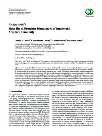
4BioMed Research D91Lox-1HSPsCytokine release(IL-12, TNF-𝛼, IL-1𝛽,GM-CSF)Chemokine release(RANTES, MIP-1𝛼, MCP-1)PeptidesSecretion ofmonokinesCD28MHC loadingCD4 T cellE.R.NucleusMHC class IIDC maturationMHC class ICD40 (B-cell activation)CD8 T cellCostimulatoryactivation signalMo/DCCD28MHC class IIAcquired immunityCD80/86 (T-cell activation)Innate immunityFigure 1: Dual role of HSPs in the activation of both innate immunity, with the induction of monokines and maturation of DCs, and acquiredimmunity, with the provision of peptides for MHC-loading and antigen specific responses.appear to mediate the innate immune responses through different cellular receptors [48–50]. For mycobacterial HSP65,signalling occurs primarily via CD14 and can be blocked bythe use of antibodies against this receptor: in contrast HSP60appears to be CD14 independent and may bind and signalvia TLR4 [49]. The domains of mycobacterial HSP65 werecloned (apical domain, intermediate domain, and equatorialdomain), and the binding to CD14 was localised to theequatorial domain [50].Mycobacterial HSP70 stimulates cytokine production inmonocytes, by interacting with both TLR2 and 4, in aCD14-dependent manner [51]. This ability to activate innateimmunity was localised to the C-terminal peptide bindingregion (aa359–610) of HSP70 which elicited production of IL12, TNF-𝛼, RANTES, and nitric oxide (NO) in THP-1 cells,whereas the N terminal nucleotide binding region of HSP70(aa 1–358) did not [52, 53]. The full mycobacterial HSP70molecule appears also to contain epitopes that inhibit DCmaturation and promote anti-inflammatory cytokines (IL10) [54]. Mycobacterial HSP70 can also interact with CD8 T-cells via CD40 to produce RANTES, MIP-1𝛼, and MIP1𝛽 [55]. Further studies revealed that only mycobacterialHSP70 but not human HSP70 induced this observation,with bacterial HSP70 (DnaK) and human HSP70 appearingto bind to different regions of CD40 on macrophages andDCs [55, 56]. It has also been reported that mycobacterialHSP70 binds to CCR5 and CD40 on human DCs, stimulatingproduction of IL-12p40 and TNF-𝛼 [57], though this hasbeen contested by other groups that dissociate the innateand acquired functions of both human and mycobacterialHSP70 (aa359–635) [58]. Thus it appears that HSP70’s ability to stimulate cells contributing to the innate immunesystem is dependent on the source of HSP, as mammalianand microbial forms appear to use different receptors [52–58]. Moreover, it appears that while mycobacterial HSP70stimulates an innate immune response, the situation withmammalian HSP70 is more variable and some members ofthis gene family may downregulate the immune responseinstead [34, 44, 54, 59]. This has led to the proposal thatthese HSPs may have a distinct role as “resolution associatedmolecular patterns” (RAMPs) that lead to the resolution ofinflammation induced by activation of the innate immuneresponse by DAMPs and PAMPs [59, 60].The eukaryotic family of HSP90/gp96 chaperones hasalso been shown to interact with TLR2 and -4 and inducethe activation of the NF-𝜅𝛽 pathway and the subsequentsecretion of IL-12 and TNF-𝛼 [61]. In addition, it has alsobeen reported that gp96 isolated from mouse liver inducedthe production of NO in both murine (Raw264.7) and human(THP-1) macrophage cell lines and that this action wasmediated by the binding of gp96 to CD36 [62, 63]. These
BioMed Research Internationalstudies also report that mixtures of IFN-𝛾 lead to a synergisticproduction of NO from these macrophage cell lines [63].There is still controversy and conflicting observationsabout the ability of mammalian HSP to stimulate the mammalian immune system [64]. However there is compellingevidence that bacterial HSPs, including mycobacterial HSP,are able to stimulate the innate immune system with datacoming from the study of domains of HSP60 [50] andHSP70 [52, 53]. A complication of initial studies was thecopurification of lipopolysaccharide (LPS) as a contaminantin preparations of recombinant HSPs. Thus Gao and Tsansuggested that the biological effect observed with humanHSP60 was as a result of LPS contamination as HSP60 with alow endotoxin activity did not result in TNF-𝛼 production inthe murine macrophage Raw 264.7 cell line [65]. However,the use of highly purified HSP60 and the stimulation ofinnate immune responses by endotoxin-free mycobacterialHSP60 show that LPS contamination does not account forall the observations reported [66]. Moreover, the chemokinestimulatory effects of mycobacterial HSP70 can be blockedby antibodies specific for CD40 but not by inhibitors ofLPS [55]. These authors also show that the effect of HSP70(but not LPS) is lost when digested with proteinase K andthe differing responses to different peptide domains of theprotein also rule out LPS contamination issues [53–55]. Thecontroversy regarding the potential contamination of HSPpreparations with PAMPs has recently been discussed indetail and supports a distinct role for HSPs in the activationof the innate immune response [64].4. Adaptive Immunity of HSPsThe first indication that HSPs could modulate the generation of adaptive immunity derived from observations incancer studies aimed at elucidating the immunogenicity ofsarcomas in genetically identical mice [42, 67]. Biochemicaldissection of chemically induced sarcomas identified gp96as the tumour rejection antigen and cloning of the geneidentified it as a member of the HSP90 family [68]. However,immunisation with gp96 elicited sarcoma-specific immunityand gp96 purified from other chemically induced tumoursor normal tissue did not elicit immunoprotection althoughno differences were observed at a protein or genetic levelfor these HSPs [67, 68]. Srivastava thus proposed that theimmunogenicity was conferred by tumour-specific peptidesassociated with the HSPs and this was supported by theobservation that a plethora of peptides could be observedbound to gp96 [42, 68].Confirmation that immunity was due to the associatedpeptides was achieved by removal of the chaperoned peptide. HSP70 purified by affinity chromatography on ADPsepharose retained its chaperoned polypeptides and providedprotection against tumour challenge, whereas purificationusing ATP-sepharose yielded HSP70 that lacked its associatedpeptides and did not provide protection [42, 69]. HSP70 has abinding pocket that was first demonstrated for the ER HSP70homolog BiP [70] and later for bacterial HSP70 [71]. Thebinding pocket interacts with peptides of 8–26 aa in length5that are rich in leucine, isoleucine, valine, phenylalanine, andtyrosine [72, 73]. Peptide binding is under the control ofATP/ADP binding to HSP70, which brings about conformational changes that expose its binding pocket [28]. In contrastto HSP70, HSP90 is found as a homodimer and has an openpeptide binding cleft that is localised between the long armsof the two monomers [74]. However, like other HSPs, peptidebinding is ATP/ADP dependent and HSP90 also functionswith cochaperones like HSP40 and HOP [74, 75]. The HSP90homologue gp96 also contains a binding pocket and, likeHSP90, it is an open binding pocket that should allowpeptides of any length to interact with it, though the presenceof a disulphide bond in this domain may also affect peptidebinding [75]. In gp96, peptide binding has also been reportedto be under the control of calcium levels that brings aboutthe required conformational changes for peptide binding, amechanism distinct from other HSP90 homologues [31, 75].The most interesting feature of the uptake of HSPchaperoned peptides by APCs is their availability for crosspresentation, which is the ability of exogenous antigens toenter endogenous loading pathway of MHC Class I moleculesand thus prime CD8 T cells [76–80]. Cross-presentation canoccur via one of two pathways, either the vacuolar/endocyticpathway (nonclassical MHC I loading) or the cytosolicpathway (classical MHC I loading) [78, 79]. In the vacuolar/endocytic pathway, antigen is taken up by the cell byphagocytosis, and formation of phagolysosomes providesthe appropriate environment for the production of peptidefragments that are then loaded onto MHC I molecules withinthis compartment: the source of MHC I molecules is believedto be from membrane recycling or from ER-phagosomefusion [78, 79]. In the cytosolic pathway, antigen is once againtaken up by the cell by phagocytosis and, once internalised,the antigen is trafficked to the cellular cytosol (through thetransmembrane protein Sec61) and enters the classical MHCI pathway of loading: this translocation to the cytosol requiresHSP90 [79, 80].The cross-presentation of peptides bound to HSPs hasbeen shown to be receptor mediated, with HSP70 and gp96binding to CD91 and HSP90/gp96 binding to Scavengerreceptor-A on APCs [43–45]. HSP70 also binds to Scavengerreceptor-A, Scavenger receptor-F1, stabilin-1, LOX-1, andSREC-1 [44, 79]. Although CD91 is found on macrophages,its distribution on DCs is low, suggesting that the scavengerreceptors and LOX-1 may be the more common receptorsinvolved in HSP-receptor-mediated cross-presentation [44,45, 81]. Thus different receptor binding and selective internalisation may account for the enhanced immunogenicityof different HSPs, and upon internalisation, the HSP boundpeptide can be taken into the vacuolar/endocytic or cytosolicpathway of cross-presentation. The factors that determinewhich pathway is taken remain unclear, but it appears to bedependent on both the nature of the bound peptide and theAPC cell type [43–45, 76–79].The ability of mycobacterial HSP70 to cross-presentchaperoned peptides onto mammalian APCs has also beeninvestigated [78, 82]. Construction of a fusion protein consisting of mycobacterial HSP70-ovalbumin (OVA) was shownto induce an antigen-specific CD8 T-cell population in
6vaccinated mice that showed cytotoxic activity against targetcells expressing recombinant OVA [82, 83]. Furthermore,Harding and colleagues have shown, in vivo, that an extendedOVA peptide, noncovalently associated with mycobacterialHSP70, could be presented via the MHC I presentationpathways of bone-marrow-derived murine macrophages andDCs to induce the secretion of IL-2 from a T-cell hybridomaspecific for OVA peptide/MHC I complex [78]. This crosspresentation was dependent on the peptide being boundto mycobacterial HSP70 and required active internalisationvia CD91 but did not involve interaction with CD40 orTLR [78, 82]. However, treatment of macrophages or Bcells with Brefeldin A, an inhibitor of ER to golgi transport and thus the cytosolic pathway of cross-presentation,did not result in a significant reduction of processing andpresentation of the fusion peptide, though a significantreduction was seen when DCs were used as APCs [83].This suggests that, in macrophages and B cells, polypeptideschaperoned by mycobacterial HSP70 are cross-presentedpredominantly via the vacuolar/endocytic pathway, whereasin DCs cross-presentation occurs via the cytosolic pathway[78, 84]. The ability of mycobacterial HSP to effect crosspresentation has also been observed in human DCs, usingan influenza A derived MHC I peptide epitopes fused tovarious HSP70 domains [34, 84]. These studies showedthat, in vivo, mycobacterial HSP70 bound peptides wereable to cross-present bound peptide, cross-prime CD8 Tcells, and generate CTL that lysed peptide-labeled targetcells: surprisingly low quantities of mycobacterial HSP70peptide complex (120 pM) were required to bring about CTLpriming, about 4 orders of magnitude lower concentrationthan that required to bring about a similar response withunchaperoned peptide [34, 84].5. HSP Cancer VaccinesThe initial work on host-derived HSPs (gp96) from tumoursas cancer vaccines has now progressed through preclinicaldevelopment into clinical trials, generating proof of concept[36, 85–87]. There have been numerous reviews on thepreclinical development studies and the reader is referred tothese for further details [14, 36, 42]. The most advanced ofthe clinical trials utilise patient-derived autologous vaccines,called Vitespen/Oncophage, which are gp96 preparationspurified from surgically removed samples of the patients’tumours using proprietary methods including affinity chromatography [42, 85]. A range of tumours including metastaticcolorectal carcinoma, metastatic melanoma, non-Hodgkinlymphoma, pancreatic adenocarcinoma, and renal cell carcinoma have been studied in clinical trials up to phase III[87]. However, while these vaccines have shown minimal sideeffects and are well tolerated, their effectiveness as therapeuticagents has been varied [87–89]. In a randomised phase IIItrial of individuals with renal cell carcinoma, administrationof isolated gp96 did not result in a statistically significantimprovement in disease outcome [88]. In contrast, whenassessed in individuals in stage IV melanoma, individualsthat were in substage M1a and M1b (those that had signs ofBioMed Research Internationalspread to other areas of the skin and lung) did show a delayin disease progression compared to a group that receivedconventional chemotherapy and/or surgery. However, in themore advanced stages of the disease, no effect was observed[89]. In groups that did show an effect, multiple vaccinationswere required ( 10), at a dose of 25 𝜇g, and it is thought thatthe disease stage can apparently modulate efficacy, and alsothe amount of available tumour tissue available to work withwill vary with disease stage [87–89]. One strategy to overcomethis limitation involves the use of tumour cells fused to DCsfor the purification of larger amounts of HSPs from thesefusion hybrids [86]. In animal models, this approach has beenshown to yield a more immunogenic vaccine than HSPs purified from tumour cells alone, and this has been ascribed to theimproved loading of peptides onto HSPs in the APCs compared to tumour cells [36, 86]. However, it should be notedthat the majority of animal studies in oncology use HSP70,not gp96 as in Vitespen, and t
Most importantly, Janeway proposed that the recognition of infection by PRRs on cells of the innate immune system would lead to the induction of signals that resulted in initiation of the adaptive immune response. e subsequent identi cation of the Toll-like receptors (TLRs) as key PRRs led to an explosion of research on innate immunity and the