Transcription
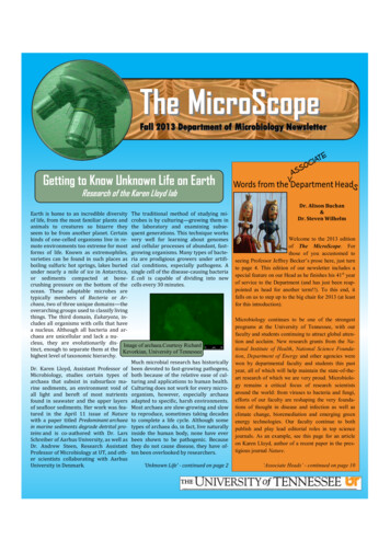
The MicroScopeFall 2013 Department of Microbiology NewsletterGetting to Know Unknown Life on EarthResearch of the Karen Lloyd labEarth is home to an incredible diversity The traditional method of studying miof life, from the most familiar plants and crobes is by culturing—growing them inanimals to creatures so bizarre they the laboratory and examining subseseem to be from another planet. Certain quent generations. This technique workskinds of one-celled organisms live in re- very well for learning about genomesmote environments too extreme for most and cellular processes of abundant, fastforms of life. Known as extremophiles, growing organisms. Many types of bactevarieties can be found in such places as ria are prodigious growers under artifiboiling sulfuric hot springs, lakes buried cial conditions, especially pathogens. Aunder nearly a mile of ice in Antarctica, single cell of the disease-causing bacteriaor sediments compacted at bone- E. coli is capable of dividing into newcrushing pressure on the bottom of the cells every 30 minutes.ocean. These adaptable microbes aretypically members of Bacteria or Archaea, two of three unique domains—theoverarching groups used to classify livingthings. The third domain, Eukaryota, includes all organisms with cells that havea nucleus. Although all bacteria and archaea are unicellular and lack a nucleus, they are evolutionarily dis- Image of archaea.Courtesy Richardtinct, enough to separate them at the Kevorkian, University of Tennesseehighest level of taxonomic hierarchy.Much microbial research has historicallyDr. Karen Lloyd, Assistant Professor of been devoted to fast-growing pathogens,Microbiology, studies certain types of both because of the relative ease of cularchaea that subsist in subsurface ma- turing and applications to human health.rine sediments, an environment void of Culturing does not work for every microall light and bereft of most nutrients organism, however, especially archaeafound in seawater and the upper layers adapted to specific, harsh environments.of seafloor sediments. Her work was fea- Most archaea are slow-growing and slowtured in the April 11 issue of Nature to reproduce, sometimes taking decadeswith a paper titled Predominant archaea to complete a life cycle. Although somein marine sediments degrade detrital pro- types of archaea do, in fact, live naturallyteins and is co-authored with Dr. Lars inside the human body, none have everSchreiber of Aarhus University, as well as been shown to be pathogenic. BecauseDr. Andrew Steen, Research Assistant they do not cause disease, they have ofProfessor of Microbiology at UT, and oth- ten been overlooked by researchers.er scientists collaborating with Aarhus‘Unknown Life’ - continued on page 2University in Denmark.Dr. Alison Buchan&Dr. Steven WilhelmWelcome to the 2013 editionof The MicroScope. Forthose of you accustomed toseeing Professor Jeffrey Becker’s prose here, just turnto page 4. This edition of our newsletter includes aspecial feature on our Head as he finishes his 41 st yearof service to the Department (and has just been reappointed as head for another term!!). To this end, itfalls on us to step up to the big chair for 2013 (at leastfor this introduction).Microbiology continues to be one of the strongestprograms at the University of Tennessee, with ourfaculty and students continuing to attract global attention and acclaim. New research grants from the National Institute of Health, National Science Foundation, Department of Energy and other agencies werewon by departmental faculty and students this pastyear, all of which will help maintain the state-of-theart research of which we are very proud. Microbiology remains a critical focus of research scientistsaround the world: from viruses to bacteria and fungi,efforts of our faculty are reshaping the very foundations of thought in disease and infection as well asclimate change, bioremediation and emerging greenenergy technologies. Our faculty continue to bothpublish and play lead editorial roles in top sciencejournals. As an example, see this page for an articleon Karen Lloyd, author of a recent paper in the prestigious journal Nature.‘Associate Heads’ - continued on page 10
‘Unknown Life’ - continued from page 1However, archaea are an integral part of many microbial ecosystems,and their ability to survive under extreme conditions makes theminteresting to scientists. Even if researchers could grow all archaeaartificially, culturing would reveal very little about ecological processes in their natural habitat. Therefore, Lloyd and the team at Aarhus needed to examine uncultured specimens of archaea living beneath the ocean floor.Lloyd says the most enjoyable aspect of the work was camaraderieamong her team members. She and the other two post-doctoral researchers cooperated to pioneer new techniques for studying uncultured microbes. Before this study, no one had ever extracted individual cells from ocean sediments, only from seawater. Since Lloyd andher colleagues had success with this process, she hopes such methods can be used in more locations around the world to expand research on archaean taxonomy and ecology.Two particular types of archaea have been found in ocean sedimentsaround the world. These are called miscellaneous crenarchaeotalgroup (MCG) and marine benthic group (MBG-D), which can be identified by unique sequences in the 16S rRNA gene. The coverage ofsampling for archaea is still sparse over much of the globe, but includes locations from every ocean. Everywhere that archaea havebeen sampled and identified, MCG and MBG-D have been present, sotheir range is assumed to be more or less worldwide. For this reason,Lloyd and team focused their study on these taxonomic groups inorder to generate results that could be applicable to benthic ecosystems globally.To study the mysterious microbes, Lloyd and colleagues collectedsediment from the bottom of a bay off the coast of Denmark and useda series of state-of-the-art techniques to amplify the genomes of individual organisms. First, individual cells were extracted from sediment, then sorted using a laser light. Next, DNA from each individualcell was extracted and amplified so the genome could be analyzed.The pain-staking cell collection and genome amplification processesrevealed four intact cells, one from the MCG group and three fromthe MBG-D group. Although amplification of DNA was uneven acrossthe genome, and therefore gave an incomplete picture, the researchers could tell enough to classify the specimens on the phylogenetictree of life. Much is still unknown about the phylogeny of archaea,but they are certainly a diverse and highly adaptable domain of organisms with over two billion years of evolutionary history. By examining the genome, the researchers determined that the MCG andMBG-D groups are only distantly related within archaea—so distant,in fact, that they are more dissimilar taxonomically than any animals—although they are both single-celled ocean-dwellers with similar metabolic functions.(From left) Dr, Karen Lloyd, Richard Kevorkian (MS student,Microbiology), and Jordan Bird (PhD student, Microbiology) withsediment cores containing archaea from an estuary in North CarolinaLloyd currently has plans to core sediments in the Baltic Sea from fardeeper than her previous study. She, along with UT MicrobiologyPhD student Jordan Bird, will delve into subsurface ecosystems asdeep as 200 meters beneath the ocean floor, a place where life hasonly recently been discovered. These extreme environments havebeen buried and cut off from the seafloor surface for hundreds ofthousands of years, so the communities of archaea and bacteria thatlive there are likely to reveal even more taxonomic lineages andphysiological processes previously unknown to science. Lloyd’s teamhas secured funding for this project from the Center for Dark EnergyBiosphere Investigations (National Science Foundation), and has already begun similar sampling in a North Carolina estuary to comThese metabolic functions are what Lloyd and her colleagues were pare archaea specimens from a slightly different environment.particularly interested in. In the genomes of all four cells, the researchers found genes coding for protein-degrading enzymes, called Lloyd is excited and optimistic for the future of her groundbreakingpeptidases, previously known only from certain kinds of bacteria. research. She says, “These organisms are almost like aliens in termsThey predicted that these enzymes, along with peptide-specific of how little we know about them, but we’ve been living with themtransport structures, were located on the outside of the cell walls. for our entire human existence and they were around way beforeThe genomes also revealed capabilities to degrade amino acids, the humans were ever here. We know almost nothing about what any ofbreakdown products of proteins, inside the cell. The extracellular them do and we’ve just skimmed the tip of the iceberg. I find it fascipeptidases are believed to be specific to the types of proteins that nating that we now have the tools to begin to determine their role inwould be found in bacteria-rich detritus on the ocean floor. This Earth’s environment.”means that when the archaea cells come in contact with proteins intheir environment, they can break them down into smaller peptidesJesse Weberand amino acids, then absorb and metabolize the amino acids forenergy to sustain the cell’s life.This finding is monumental in the field of microbial ecology. According to Lloyd, “The surprising find is extracellular peptidases. Thesemicrobes are recycling from the environment around them, not justtheir own proteins. This has never been observed before in archaeain marine sediments.” The only metabolic processes known for othermarine sedimentary archaea rely on simple carbon compounds like methane, but the discovery of MCG and MBG-D’sunique metabolism reveals an entirely new niche for archaea.2
The MicrobiologyUndergraduate ClubThe 2012-2013 school year was another productive one forthe Microbiology Undergraduate Club (MUC). When theAmerican Cancer Society held Relay for Life on UT’s campusin April, MUC raised over 2,000, more than any other teamof their size. To rally enthusiastic support from the entirecampus community, MUC members devised a fundraisingscheme targeted at faculty members, literally. For 3, anyonecould take aim at either Dr. David Golden (Food Science &Technology), Dr. Todd Reynolds, Dr. Liz Fozo, or Dr. JeffreyBecker with a water gun filled with slime. In the 3 hours thatthe professors cheerfully endured getting slimed, they raisedabout 350 from students and other faculty paying per shotof the goop gun. Other activities included swings at a microbiology-themed pinata and members participating in Relayfor Life’s 12-hour relay walk. Thanks to MUC members’ creativity, students and faculty put the “fun” in fundraising for animpressive turnout at the annual charity event.The club got its start in Fall 2011 when faculty members inthe department decided to improve interaction with students and provide them with more access to networking,career information, and research opportunities, while havingfun in the process. After announcing an initial meeting, sevDr. Jeffrey Becker and Dr. Liz Fozo place themselves in the line of fire toeral students expressed interest and the club took off fromfight cancer at the April 2013 Relay for Life at UTthere—growing to its current membership of over 40 students. Meetings are held about once per month and each arefocused on a particular theme, usually with a guest presenter. Meet- Dr. Liz Fozo is currently the faculty advisor for the club. She took oning themes cover a wide range of topics related to microbiology, this role from the club’s beginning because she wanted to interactincluding pathology, food safety, applying for graduate school, with students and see them succeed both in and outside of school.working as a physician’s assistant, etc. Speakers are from a diverse She describes her job as mainly a facilitator for the students’ ideas.array of backgrounds and credentials—whether from within the MUC members decide what they are interested in and would like tomicrobiology department, other departments at UT, or other insti- focus on at a meeting, but if they are not aware of how to pursue ittutions. Additional events include an annual barbeque social and a t- or who to talk to, Dr. Fozo is there to make it happen. She says she isshirt design contest. The winning design, based on votes from club impressed by how student-driven the club is. She applauds clubmembers, is printed on the shirts that are sold for fundraising each members for their organization, creativity, and how much they acyear. This year’s shirt features the Sunsphere, stylized as a virus, complish on their own initiative. She also admires the faculty’s endominating the Knoxville skyline under the slogan, “Microbiology: thusiasm in supporting the club. They are always happy to speak atmeetings and attend club events, even if it involves getting slimed.Watching over Knoxville since 1982.”Regarding the Relay for Life event, Dr. Fozo says, “I sent out the iniClub member and new vice president Nate Crilly is a Food Science tial email asking the department for volunteers and within twoand Technology major, but he joined the club out of interest in a minutes Dr. Becker had replied and agreed to do it. The others, too,career in virology and animal-born diseases. Crilly says, “There are agreed pretty much right away. There was no hesitation.”a lot of things that I like about the club, but my favorite thing isprobably the lectures For example, a researcher from the Body The club is gearing up for another successful year starting this fall.Farm came to one of our spring meetings to discuss her work with They hope to increase membership, plan even more events, takesoil microbes and decomposition. Earlier in the year, a graduate field trips, and continue to promote friendly fun with microbiology.student held a discussion about the microbiology of sourdoughbread. It's a really interesting way to learn more about microbiologyJesse Weberin a casual, relaxed environment.”3
The University of Tennessee Department of MicrobiologyDepartment Head Jeffrey M. Becker“I get paid for something I love to do.It’s fantastic.”These are the words of Dr. Jeffery Becker, MicrobiologyDepartment Head and Chancellor’s Professor ofMicrobiology. This year, Becker begins his third 5-yearterm as department head. The Microscope sat downwith Dr. Becker to discuss his career of nearly 40 yearsso far at UT and what he hopes for the future.What does it mean to you to be a professor?“It’s a privilege, at least I feel. I love to mentor studentstoward their graduate degrees or PhDs, and alsoundergrads. I like to introduce them to science anddiscovery, mainly discovery I would say Plus,students are always challenging me with new thoughtsand ideas. As a professor, I am constantly renewed bynew people and new ideas.”Dr. Becker and some of his students. From left: Brandon Merical,Dr. Jeffrey Becker, Jeffrey Rymer, Sarah Kauffman, Seraj UddinWhat does it mean to you to be department head?“Again, it’s a privilege. A privilege to be a part of building the department up and to be hiring great young peopleand helping them to succeed. I get to see former students establish careers and contribute to science and society.”What do you feel is the best thing about the Microbiology Department at UT?“We have had spectacular growth in the last ten years, more than double. We have a fabulous core of young facultyand almost all have major external funding and papers in strong journals. I am really excited about the growth andsuccess of both faculty and students. We have undergraduates doing great and getting into great schools andcareers. I am very proud of that.”The Jeffrey Becker Lab Then and Now199242013
Dr. Jeffrey Becker has led numerous research projects sincecoming to UT. Most notable is his work with yeast as a modelorganism to study transport and signal pathways in eukaryoticcells. The research focuses on how receptors in cell membranesrecognize external molecules and interact with transporters inthe membrane to move selected molecules into the cell. Beckeris also interested in how chemical signals travel through the celland induce particular responses to stimuli.This knowledge is crucial to development of new drugs becauseabout half of all drugs used in human medicine act on the samefamily of receptors that Becker studies in yeast. The unique proteins have been conserved in evolution since the common ancestor of humans and yeast. Allergy, pain, heart, and ulcer medications, as well as others, interact with these very same receptorsin humans.Because of the critical applications of Becker’s work to humanmedicine, his research receives long-standing funding from theNational Institute of Health (NIH). He is currently in the 35thstraight year of NIH funding.“My favorite thing about working in [Dr. Becker’s] lab isthe chance to discover something new. When you figuresomething out and realize that you are the first to knowit, that you are privy to one of the secrets of the naturalworld, it’s an incredible feeling.”-Madelyn Crawford, Undergraduate Class of 2013In fact, Becker’s lab at UT was the first to successfully clone thepeptide transport system found in nearly every living organism.This system moves peptides and amino acids, the building blocksof proteins, throughout the cell. The system is active in every aspect of a cell’s life cycle: from growth, to metabolism, to nutrientcycling, to reproduction, to death, and everything in between. Hislab is also at the forefront of research on ligand molecules. This isa family of compounds that bond to receptor proteins on the outside of a cell and initiate various cellular responses, such as thetransport of substances into the cell and along the peptidetransport chain. Many types of drugs work by using very selectiveligands to bond to specific receptors, in order to produce desiredeffects in human body cells.Dr. Becker grinning through the MicrobiologyUndergraduate Club’s “Slime a Microbiologist”fundraiser at Relay for LifeBecker is quick to point out that his work would not be possiblewithout the collaborative efforts of faculty, undergraduates, graduates, and post-doctorates at UT. He also works in close cooperationwith Dr. Fred Naider of The City University of New York. Beckeracknowledges that no one can possibly have expertise in everything, so collaboration is key to success.Becker and the rest of the faculty plan to foster and grow more of this collaborative environment in the future. Becker believes the department has plenty of untapped potential that could be realized byadding more faculty members to the team. He is seeking new hires specializing in pathogenesis, immunology, and virology inorder to more fully round out the already diverse arsenal of talent in the department. He also wants to continue attracting fantastic undergraduate and graduate students to degree programs and research, and wants elevated undergraduate participation in microbiology labs. His outlook, shared by the rest of the faculty, is one of optimism. A new building dedicated specifically to Microbiology and Biochemistry & Cellular and Molecular Biology is scheduled to be built on campus in the next five years.With the addition of this building, talented personnel, and fresh students on the horizon, Becker’s third term as departmenthead is sure to generate even further success for the Microbiology Department.Jesse Weber5
The University of Tennessee Department of MicrobiologyDepartment Hosts UT’s First Microbiology REUAt the beginning of June, a distinguished collection of ten undergraduate students traveled from universities across the country to gatheron the University of Tennessee campus. These were the participantsof UT’s inaugural Microbiology Research Experiences for Undergraduates (REU). This is a nation-wide endeavor funded by the NationalScience Foundation (NSF) to establish research programs at certainhost institutions for students seeking a Bachelor’s degree. During theREU, students work with a faculty mentor and a graduate student“big sibling” to complete a research project specially tailored to theirinterests and abilities. NSF funds well over one hundred REUs in alldisciplines of science, but only 21 nationwide involve projects specific to microbiology. UT Knoxville also hosts at least two other NSFREU sites, one in Biochemistry & Cellular and Molecular Biology andone in Materials Science & Engineering.engineering, bioinformatics, and more. Faculty mentors are not onlyfrom the Microbiology Department, but also Ecology & EvolutionaryBiology, Civil & Environmental Engineering, Biosystems Engineering &Soil Science, Earth & Planetary Sciences, along with collaboratorsfrom Oak Ridge National Lab.REU student Kristie Goughenour is attending her second REU, butthe first one specific to microbiology. Although she was acceptedinto a microbiology REU at a different university this summer, shewas impressed by the diversity of research topics available at UT’sand felt them to be a better suit for her interests. At the REU, she isresearching biofilms in drinking water systems under Dr. Qiang He,Assistant Professor of Civil & Environmental Engineering.Matthew Tuttle, from the University of Massachusetts Amherst, says UT was his topchoice out of the REUs he applied for because he wanted to focus on environmentalscience and microbial ecology, for which hethought UT offered the best opportunities.His project is studying Antarctic subglacialecosystems under Dr. Jill Mikucki, AssistantProfessor of Microbiology.The Summer 2013 UT Microbiology REU students in Neyland Stadium. From left: AndrewGarrone (Truman State University, Missouri), Matthew Tuttle (University of MassachusettsAmherst), Emily McCully (Flagler College, Florida), Jessica Stevens (University of Texas atArlington), Tanya Dilan-Perez (University of Puerto Rico at Mayaguez), Kristie Goughenour(Ohio Wesleyan University), Luis Rodriguez-Carire (University of Puerto Rico Cayey), NicolePerry (Wittenberg University, Ohio), Mariya Campbell (Georgia State University), andFransisco Lopez (Rochester Institute of Technology, New York)Last year, Microbiology Department faculty members Dr. Steven Wilhelm and Dr. Erik Zinser proposed and were awarded a summerREU grant. The first program this year received 165 applicants foronly ten positions, so faculty in the department took on the task ofselecting the most talented and qualified students from a diversearray of backgrounds and disciplines, but with a common interestfor microbiology research. According to Dr. Gary LeCleir, coordinator of this year’s REU, the selection process was daunting because somany highly qualified students applied, far more than could be accommodated in the program. The final selection included studentsfrom seven different states as well as two from Puerto Rico, with arange of majors including Microbiology, Environmental Science, Biotechnology, and others.The department aims to promote the diverse, interdisciplinary aspects of microbiology research. Available REU topics for studentsrelate to pathology, microbial ecology, oceanography, genetics, bio-6Applicants were considered based on theiracademic history, personal statement, andletters of reference. LeCleir and the rest ofthe 5-member admissions committeesought students showing potential for quality research and with interest in a sciencecareer, but also focused on students fromuniversities without much opportunity forfaculty-led student research in the field ofmicrobiology. This is in fact the main goal ofthe NSF grant: to introduce top-tier scientific research to undergraduates who mightnot otherwise have such an opportunity.The funding covers costs of the research programs, housing on campus, travel to Knoxville, and a living stipend for all the students inthe REU, at no extra cost to UT. The grant is awarded for three yearswith potential for further renewal thereafter. This year’s program isthe first of three, and hopefully many more, microbiology REUs tocome at UT.In addition to working closely with faculty and graduate students,REU participants attend weekly seminars on topics relevant to scientific research and careers. These are led by scientists from both UTand Oak Ridge National Lab and include themes such as ethics inscience, designing a project, making and presenting a poster, graduate school and career opportunities, and much more.‘NSF REU’ - continued on page 8
Taking the “Fun” Away from Pathogenic FungiTodd Reynolds’ Collaborative Effort Toward a Medical BreakthroughEvery year, well over one billion people worldwide suffer from some sort offungal infection. Most of these are relatively localized on the body and easilytreatable, like athlete’s foot and ringworm. However, serious invasive fungalinfections can be debilitating and even lethal. Even as medical technology advances, invasive types of fungal infections have become a worsening problemin the last half-century.Dr. Todd Reynolds working in his lab at UTTechnology has improved at keeping immune-compromised patients alive, butthese people are more susceptible to infections by fungi and other microbes.Healthy bodies are typically not at great risk because a normal immune system can keep most fungi in check. However, as healthcare facilities are able tosupport a growing number of immune-compromised patients, serious fungalinfections are becoming more common. A fungus that begins as an infection ofmucous layers, other soft tissue, or implanted medical devices may enter theblood stream and become systemic, taking over the deep organs. This type ofinfection causes a 20-40% mortality rate in the United States.Adding to the problem is the small number of effective antifungal drugs. There are currently three major classes of antifungals that arecommonly used to treat systemic infections, but they are not effective for all patients because of varying levels of toxicity and resistancein the body, based on the individual person being treated.Dr. Todd Reynolds, Associate Professor of Microbiology, is working to solve this problem. He and collaborator Dr. Richard Lee, medicinalchemist at St. Jude Children’s Research Hospital, have secured a National Institute of Health (NIH) grant to discover pre-therapeutic compounds for treating human fungal infections. These compounds are not drugs in themselves, but prototypes that may eventually be usedto create new drugs. Reynolds hopes to find a compound that will damage fungal cells, but not harm human cells in any way. This compound, once discovered, could become the key ingredient in a future antibiotic.The difficulty in treating fungal infections comes from the fact that human and fungal cells share many of the same components in theirconstruction, so an antibiotic compound must be extremely specific to the biochemistry of fungal cells. Reynolds believes a key to thisspecificity has been found, and he plans to figure out how to exploit it.The cell membranes of both fungal and human cells contain a particular phospholipid, an important fatty molecule, that is crucial tohealthy functioning of the cell. This phospholipid is called phosphatidylserine (PS). Although both human and fungal cells need this lipid,the difference is that the protein used to synthesize PS molecules is different in fungal cells than in human cells. If the researchers can finda compound that inhibits this particular fungal cell protein, it could be used safely inside a human body to kill fungi.Reynolds and Lee are focusing their tests on the fungus Candida albicans, because it is one of the most common causes of human fungalinfections. C. albicans is the usual culprit of female yeast infections, and it can cause thrush and systemic bloodstream infections in immune-compromised patients such as those with HIV/AIDS or immunosuppression from chemotherapy or organ transplants, respectively.C. albicans is actually naturally present in most human bodies, where it resides harmlessly on mucous membranes, but an unhealthy immune system can allow the fungus to grow out of control and become infectious. The fungus can spread through the bloodstream if surgically implanted medical devices like intravascular catheters become contaminated before or after insertion into a patient.The first step of Reynolds’ research was to show that C. albicans is not infectious when the protein that makes PS is missing from the cell.Primary tests involved destroying the gene for this protein in a sample of C. albicans, growing some of the mutant cells in laboratory cultures and injecting some into mice. As it turns out, the fungus could survive without PS in cultures, but could not grow at all in the animalhost, indicating that the PS synthesis enzyme is indeed a promising target for drug discovery.The next step is to find a compound that will inhibit this protein, so that normal C. albicans cells will lose PS in their cell membranes,thereafter behaving like mutants with PS removed, and stopping infection. To discover such a compound, a strategy was developed thattakes advantage of a particular compound called papuamide A (papA), that normally kills C. albicans. When the fungal cells are exposed topapA, the compound binds to PS molecules on the cell membrane and forms pores in the cells. This kills normal C. albicans, but mutantswithout PS or cells with PS synthesis chemically inhibited will not be killed because they have no PS for papA to bind to. Therefore, if asample of C. albicans survives treatment with papA, this indicates that it may not have PS in its cell membrane and therefore cannot remain infectious. PapA itself is not practical as a drug because it will bind to PS on human cell membranes as well, but
cell was extracted and amplified so the genome could be analyzed. The pain-staking cell collection and genome amplification processes revealed four intact cells, one from the MCG group and three from the MBG-D group. Although amplification of DNA was uneven across the genome, and therefore gave an incomplete picture, the research-