Transcription
3E x e r c i s eThe MicroscopeIf students have already had an introductory biology course in which the microscope has been introduced and used, there might be a temptation to skip this exercise. I have found that most studentsneed the review, so I recommend spending this time early in the course to make sure they are allcomfortable with the microscope, as it is used extensively throughout the laboratory manual.Time Allotment: 2 hours.Solutions:Bleach Solution, 10%Measure out 100 milliliters of household bleach. Add water to a final volume of 1 liter.Methylene Blue Solution (Loeffler’s)Weigh out 0.5 gram methylene blue. Add 1 milliliter 1% potassium hydroxide solution, and 30 milliliters absolute ethanol to 100 milliliters distilled water. Warm the water to about 50 degrees C, stirin methylene blue; filter.Physiologic Saline (Mammalian, 0.9%)Weigh out 9 grams of NaCl. Add distilled/deionized water to a final volume of 1 liter. Make freshjust prior to experiment.Laboratory MaterialsOrdering information is based on a lab size of 24 students, working in groups of 4. A list of supplyhouse addresses appears in Appendix A.24 compound microscopes, lenscleaning solution, lens paper,immersion oil24 millimeter rulers24 slides of the letter e24 slides with millimeter grids24 slides of crossed colored threads(threads should cross at a singlejunction)Filter paper or paper towels1 box of microscope slides1 box of coverslips1 box of flat-tipped toothpicks8–12 dropper bottles of physiologicsaline8–12 dropper bottles of methyleneblue stain (dilute) or iodine24 slides of cheek epithelial cells10% bleach solutionAutoclave bag, disposableAdvance Preparation1. Provide each student with a compound microscope, millimeter ruler, bottle of immersion oil, lens paper, andmillimeter grid slide. A supply of glass cleaner, such as Windex , should be available for lens cleaning.2. Have available slides of the letter e and slides of crossed colored threads. Some instructors prefer to haveslides for an entire semester available in individual boxes, which can be handed out to students. Othersprefer to keep the slides on trays to be distributed as needed.3. Set up an area for wet mount supplies, including clean microscope slides and coverslips, flat-tipped toothpicks, physiologic saline, methylene blue stain or iodine, and filter paper, or set out prepared slides ofcheek epithelial cells.12MARI1702 11 C03 pp012-017.indd 12Copyright 2014 Pearson Education, Inc.2/20/13 6:28 PM
4. Set up a disposal area containing a 1 liter beaker of 10% bleach solution and an autoclave bag. Note:Detailed instructions for treatment and disposal of materials used in labs involving human tissue andexcretions are found in the preface of this Instructor’s Guide.5. If the microscopes are binocular rather than monocular, give additional instructions on focusing.a. After the parts of the microscope have been identified, turn on the light and adjust the interpupillary distance so thata single circle of light is visible through the eyepieces. This is difficult for some students, usually because they aremoving back and forth and changing their eye position. Have each student record his/her own interpupillary distancefor later use.b. For a microscope with an adjustable left eyepiece, focus the microscope as directed, using the right eye only.c. Focus using the left eyepiece with the right eye closed. Both eyepieces should now be focused on the specimen.(Reverse the directions if the right eyepiece is adjustable.)6. The directions for perceiving depth (p. 33) are for microscopes with objective lenses that advance andretract during focusing. If the stage moves during focusing, the superior thread will come into focus firstif these directions are followed. Alter instructions if necessary.Comments and Pitfalls1. Be sure to have the students check the orientation of the letter e on the slide before putting the slide on themicroscope. If they forget to check, they will miss the point of the exercise.2. Beware of common focusing problems: dirty lenses, inverted slide, objective lens not securely in place,and wrong lens in position (oil immersion instead of high-power).3. It is difficult to use a millimeter ruler to measure the working distance of the high-power and oil immersion lenses on some microscopes. A best estimate is usually sufficient.4. Many students have difficulty with the section on determining the size of the microscope field. The directmeasurement is usually no problem, although some students measure area rather than diameter, and somestudents will have both the letter e slide and the grid on the stage at the same time. Emphasize that directmeasurement should be done using only one lens. Otherwise, measuring discrepancies cause confusion.The problem is often with the math involved. It is probably worthwhile to stop the class and work throughthe use of the formula (p. 32) when you see that most students are at this point in the exercise.5. Clarify what is meant by “detail observed” in the chart on p. 31.6. Students may forget safety precautions when preparing the wet mount. Emphasize the importance of following directions for safe disposal of toothpicks and proper cleanup of glassware.7. Many students forget to adjust the iris diaphragm and may end up using the light at its highest intensity,which is hard on the bulb. Remind students that the iris diaphragm should be adjusted so that the fieldis just filled with light when observed with the ocular lens removed. In practice, it may be necessary toadjust the iris diaphragm for best contrast, although some resolution may be lost.Answers to Pre-Lab Quiz (p. 27)1.2.3.4.5.d, stageb, The slide should be almost in focus when changing to higher magnifications.350 c, with special lens paper and cleanerfalseCopyright 2014 Pearson Education, Inc.MARI1702 11 C03 pp012-017.indd 13Exercise 3132/20/13 6:28 PM
Answers to Activity QuestionsActivity 2: Viewing Objects Through the Microscope (pp. 29–31)5. Answers will vary depending on the lenses used. Working distance decreases as lens power increases. Thee appears upside down and backwards.6. The image moves toward you. The image moves to the right.7. and 8. Grains begin to appear and are very visible with the high-power lens.The image is much larger.The entire e is visible with the low-power lens, but less than 1/4 of the letter is probably visible with thehigh-power lens.The field is smaller.The object must be centered so that it falls into the field of the higher-power lens.The light to the field is reduced as the iris diaphragm is closed.The light intensity often must be increased when changing to a higher magnification, as the lens has asmaller diameter and therefore lets in less light. In practice, if the microscope does not have a variablelight intensity adjustment, the iris diaphragm should be adjusted to obtain the best contrast.9. Grains are very visible. Yes.The working distance is less than that of the high-power lens.It is desirable to begin focusing with a low-power lens because the field is larger, making it easier to findthe specimen on the slide, and the working distance is larger, reducing the chance of hitting the slide withthe lens.Activity 3: Estimating the Diameter of the Microscope Field (pp. 32–33)3. Answers depend on the field diameter of lenses used. For lenses with field diameters of 1.8 millimeters,0.45 millimeter, and 0.18 millimeter, respectively, the estimated lengths are about 1.2 millimeters,0.14 millimeter, and 0.18 millimeter.4. No. The entire length of the object cannot be seen in one field. The estimate should be made with a lowerpower objective lens.Activity 4: Perceiving Depth (p. 33)2. When the stage descends, the first clearly focused thread is the bottom thread; the last clearly focusedthread is the top one.Answers depend on the order of the threads on the particular slides used.Activity 5: Preparing and Observing a Wet Mount (pp. 33–34)8. Most of the cells are separated from each other rather than in a continuous sheet.10. A cheek epithelial cell is about 80–100 micrometers (μ) (0.08–0.1 millimeter) in diameter.They are more similar to those in Figure 3.5 and easier to measure because they are in a continuous sheet.14Exercise 3MARI1702 11 C03 pp012-017.indd 14Copyright 2014 Pearson Education, Inc.2/20/13 6:28 PM
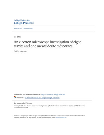
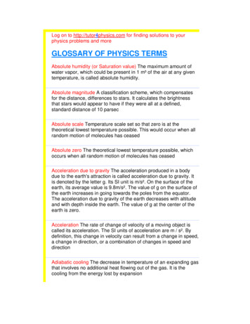
NameLab Time/DateE x e r c i s eS h ee tThe Microscope3Care and Structure of the Compound MicroscopeOcular lensesRe v i e w1. Label all indicated parts of the microscope.HeadArmRotating nosepieceObjective lensesPower switchStageLight controlMechanical stageIris diaphragm obSubstage lightBase2. Explain the proper technique for transporting the microscope.Carry with two hands—one supporting the base, the other holding the arm. Copyright 2014 Pearson Education, Inc.MARI1702 11 C03 pp012-017.indd 15152/20/13 6:28 PM
3. The following statements are true or false. If true, write T on the answer blank. If false, correct the statement by writing onthe blank the proper word or phrase to replace the one that is underlined.with grit-free lens paper1. The microscope lens may be cleaned with any soft tissue.low-power or scanning2. The microscope should be stored with the oil immersion lens in position over the stage.T3. When beginning to focus, use the lowest-power lens.away from4. When focusing, always focus toward the specimen.T5. A coverslip should always be used with wet mounts and the high-power and oil lenses. 4. Match the microscope structures in column B with the statements in column A that identify or describe them.Column BColumn Aj1. platform on which the slide rests for viewingd2. used to increase the amount of light passing throughthe specimene, i3. secure(s) the slide to the stageb4. delivers a concentrated beam of light to the specimenc5. used for precise focusing once initial focusing has been donef6. carries the objective lenses; rotates so that the different objective lenses can be brought into position over the specimena. coarse adjustment knobb. condenserc. fine adjustment knobd. iris diaphragme. mechanical stagef. nosepieceg. objective lensesh. oculari. spring clipsj. stage5. Define the following terms.virtual image: An image that is erect and appears to be where it is not resolution: Ability to discriminate two closely situated objects as separate Viewing Objects Through the Microscope6. Complete, or respond to, the following statements:16working distance1. The distance from the bottom of the objective lens to the specimen is called the .to the left2. Assume there is an object on the left side of the field that you want to bring to the center (thatis, toward the apparent right). In what direction would you move your slide?field3. The area of the specimen seen when looking through the microscope is the .954. If a microscope has a 103 ocular and the total magnification at a particular time is 9503, theobjective lens in use at that time is 3.increases contrast5. Why should the light be dimmed when looking at living (nearly transparent) cells?parfocal6. If, after focusing in low power, only the fine adjustment need be used to focus the specimen atthe higher powers, the microscope is said to be .0.757. If, when using a 103 ocular and a 153 objective, the field size is 1.5 mm, the approximate fieldsize with a 303 objective is mm.Review Sheet 3MARI1702 11 C03 pp012-017.indd 16Copyright 2014 Pearson Education, Inc.2/20/13 6:28 PM
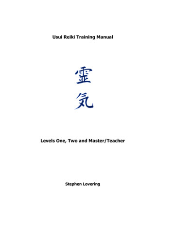
0.48. If the size of the high-power field is 1.2 mm, an object that occupies approximately a third of thatfield has an estimated diameter of mm.7. You have been asked to prepare a slide with the letter k on it (as shown below). In the circle below, draw the k as seen in thelow-power field.kk8. Figure out the magnification of fields 1 and 3, and the field size of 2. (Hint: Use your ruler.) Note that the numbers for thefield sizes below are too large to represent the typical compound microscope lens system, but the relationships depicted areaccurate.2.5 mm5 mm0.5 mm2.1.5033.500 310039. Say you are observing an object in the low-power field. When you switch to high power, it is no longer in your field of view.Why might this occur? The field decreases proportionately as magnification increases. Therefore, unless the object is centered at low power, it might be outside the higher-power field. What should be done initially to prevent this from happening? Center the object that you wish to view. 10. Do the following factors increase or decrease as one moves to higher magnifications with the microscope?resolution: increases (to a point)amount of light needed: increases working distance: decreasesdepth of field: decreases 11. A student has the high-dry lens in position and appears to be intently observing the specimen. The instructor, noting a w orkingdistance of about 1 cm, knows the student isn’t actually seeing the specimen.How so? The working distance for the h.p. lens is closer to 1 mm. 12. Describe the proper procedure for preparing a wet mount.Place the specimen on the slide with a medicine dropper or place a drop of water or saline on the slide. Mix specimen into drop using a toothpick. If staining, add a drop of stain and mix with a toothpick. Hold a coverslip with forceps so that the coverslip touches one side of the specimen drop, and then slowly and carefully lower the angled coverslip onto the specimen. 13. Indicate the probable cause of the following situations arising during use of a microscope.a.Only half of the field is illuminated: The lens is not correctly rotated into place. b.Field does not change as mechanical stage is moved: The slide is not correctly positioned in the clamp on the mechanical stage and does not move when the mechanical stage moves. Copyright 2014 Pearson Education, Inc.MARI1702 11 C03 pp012-017.indd 17Review Sheet 3172/20/13 6:28 PM
Answers to Activity Questions Activity 2: Viewing Objects Through the Microscope (pp. 29–31) 5. Answers will vary depending on the lenses used. Working distance decreases as lens power increases. The e appears upside down and backwards. 6. The image moves toward you. The image moves to the right. 7. and 8. Grains begin to appear and are very visible with the high-power lens.File Size: 338KBPage Count: 6Explore furtherLabeling the Parts of the Microscopes Free Printouts .www.microscopeworld.comthe microscope Flashcards Quizletquizlet.comParts of the Microscope with Labeling (also Free Printouts .laboratoryinfo.comRecommended to you based on what's popular Feedback