Transcription
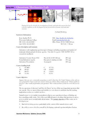
The MicroscopeMicroscope BasicsThe microscope must always be handled properly. You must observe the followingrules for its transport, cleaning, use, and storage:* Transport in an upright position with one hand on the arm and the other supporting thebase. Set it down carefully at your work station. Do not drag it across the table.* Use only special lens paper to clean the lenses. Clean all lenses before and after use.Slides should also be cleaned.* Always begin the focusing process with the 4x or 10x objective lens in position,changing to the higher-power lenses as necessary.* The coarse adjustment knob may be used with the 4x or 10x lens, but use only thefine adjustment with 40x or 100x.* Adjust lighting appropriately. Turn off the light when not in use.* Always use a cover slip with temporary (wet mount) preparations.* When you put the microscope away, remove the slide from the stage, and rotate thelowest-power objective lens into position. Wrap the cord around the clips on the back,not around the base.* Never remove or loosen any parts from the microscope.* Inform your instructor of any mechanical problems.Activity 1:Identifying the Parts of a Microscope1. Obtain a microscope and bring it to the laboratory bench. (Use the proper transporttechnique!)Compare your microscope with the figure on the following page and identify thefollowing microscope parts:Base: Supports the microscope.Substage light: Located in the base, the light passes directly upward through themicroscope.Stage: The platform the slide rests on while being viewed. The stage has a hole in it topermit light to pass through both it and the specimen. The mechanical stage permitsprecise movement of the specimen.Condenser: Concentrates the light on the specimen. The condenser has a heightadjustment knob that raises and lowers the condenser to vary light delivery. Generally,the best position for the condenser is close to the inferior surface of the stage.1
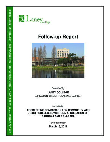
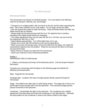
ocular lensesheadarmrotating nosepiecewith objective lensesmechanical stagestagemechanical stage controlscondensercoarse adjustment knobiris diaphragmfine adjustment knobsubstage lightbaselight controlIris diaphragm dial: Dial attached to the condenser that regulates the amount of lightpassing through the condenser. The iris diaphragm permits the best possible contrastwhen viewing the specimen.Coarse adjustment knob: Used to focus on the specimen when on 4x or 10x.Fine adjustment knob: Used for precise focusing once coarse focusing has beencompleted. Use only this knob when on 40x or 100x.Head or body tube: Supports the objective lens system, and the ocular lenses.Arm: Vertical portion of the microscope connecting the base and the head.Ocular (or eyepiece): There are two lenses at the superior end of the head, throughwhich observations are made. An ocular lens has a magnification of 10x. If your2
microscope has a pointer, it is attached to the right ocular and can be positioned byrotating the ocular lensNose piece: Has four objective lenses and permits sequential positioning of theselenses over the light beam passing through the hole in the stage. Use the nose piece tochange the objective lenses.Objective lenses: Adjustable lens system that permits the use of a scanning lens, alow-power lens, a high-power lens, or an oil immersion lens. The objective lenses havedifferent magnifying and resolving powers.2. Look at the objective lenses carefully. The shortest lens is the scanning lens, andhas magnification of 4x. The low power lens is 10x. The high-power objective lens is40x. The oil immersion objective lens is usually the longest of the objective lenses andhas a magnifying power of 100x. Record the magnification of each objective lens ofyour microscope in the first row of the summary chart.3. Rotate the lowest power objective lens until it clicks into position, and turn the coarseadjustment knob about 180 degrees. Notice how far the stage (or objective lens) travelsduring this adjustment. Move the fine adjustment knob 180 degrees, noting again thedistance that the stage (or objective lens) moves.SUMMARY CHARTScanningLow PowerHigh PowerOil ImmersionMagnification ofobjective µmmmµmWorking distanceDetail observed(draw ordescribe)Field size(diameter)3
Magnification and ResolutionThe microscope is designed to magnify specimens. Your microscope is called acompound microscope because it uses two lenses to magnify the specimen. Theobjective lens magnifies the specimen to produce a real image that is projected to theocular. This real image is magnified by the ocular lens to produce the virtual imageseen by your eye.The total magnification of any specimen being viewed is equal to the power of theocular lens multiplied by the power of the objective lens. If the ocular lens magnifies10x and the objective lens magnifies 50x, the total magnification is 500x (10 x 50).* Determine the total magnification with each of the objectives on your microscope andrecord in the chart.With a compound light microscope such as the one you are using, the level ofmagnification is almost limitless, but the resolution (resolving power) is not. Resolutionrefers to the ability to discriminate two close objects as separate. The human eye canresolve objects about 100 µm apart, but the compound microscope has a resolution of0.2 µm under ideal conditions. Objects closer than 0.2 µm are seen as a single fusedimage. Resolving power is determined by the amount and physical properties of thevisible light that enters the microscope. In general, the more light delivered to theobjective lens, the greater the resolution. The size of the objective lens aperture(opening) decreases with increasing magnification, allowing less light to enter theobjective. You will likely need to increase the light intensity at the higher magnifications.Activity 2:Viewing Objects Through the Microscope1. Obtain a millimeter ruler and a letter "e" slide. Adjust the condenser to its highestposition and switch on the light source of your microscope.2. Place the slide on the stage (in the slide holder) so that the letter e is centered overthe light beam passing through the stage.3. With your lowest power objective lens in position over the stage, use the coarseadjustment knob to bring the objective lens and stage as close together as possible.4. Look through the ocular lens and adjust the light for comfort using the iris diaphragm.Now use the coarse adjustment knob to focus slowly away from the e until it is asclearly focused as possible. Complete the focusing with the fine adjustment knob.5. Sketch the letter e in the space on the summary chart just as it appears in the field(the area you see through the microscope).4
How far is the bottom of the objective lens from the specimen? This is the workingdistance. Use a millimeter ruler to make this measurement (an estimate is fine).Record the detail observed and the working distance in the summary chart.How has the apparent orientation of the e changed (compared to what you see lookingat the slide with the naked eye)?6. Move the slide slowly away from you on the stage as you view it through the ocularlens. In what direction does the image move?Move the slide to the left. In what direction does the image move?7. Most laboratory microscopes are parfocal. This means the slide should be in focus(or nearly so) at the higher magnifications once you have properly focused. Withouttouching the focusing knobs, increase the magnification by rotating the next highermagnification lens into position over the stage. Make sure it clicks into position. Usingthe fine adjustment only, sharpen the focus. Notice the decrease in working distance.On high power or oil immersion, focusing with the coarse adjustment knob could drivethe objective lens through the slide, breaking the slide and possibly damaging the lens.Sketch the letter e in the summary chart. What new details can you observe?Record the detail observed, and working distance in the summary chart.Is the image larger or smaller than on the previous magnification?Approximately how much of the letter e is visible now?Is the field larger or smaller?5
Why is it necessary to center your object (or the portion of the slide you wish to view)before changing to a higher power?Move the iris diaphragm lever while observing the field. What happens?Is it more desirable to increase or decrease the light when changing to a highermagnification? Why?8. Repeat the steps given in direction #7 using the high-power objective lens.Record the detail observed, and working distance in the summary chart.9. We will not use the oil immersion lens today, but you will learn to use it later in thesemester. Never click an oil immersion lens into place without using oil properly. Youshould fill in as much of the summary chart as you can right now. What do you think theworking distance should be on oil immersion? What do you think you would observe?6
Size of the Microscope FieldBy this time you should know that the size of the microscope field decreases withincreasing magnification. For future microscope work, it will be useful to determine thediameter of each of the microscope fields. This information will allow you to make anestimate of the size of the objects you view in any field. For example, if you havecalculated the field diameter to be 4 mm and the object being observed extends acrosshalf this diameter, you can estimate the length of the object to be approximately 2 mm.Microscopic specimens are usually measured in micrometers (µm) and millimeters(mm), both units of the metric system.Activity 3:Determining the Size of the Microscope Field1. We will do the first part of this activity as a class. You will learn to measure the sizeof the field of view on 4x, then calculate it for the other objective lenses. The ultimategoal is to be able to estimate the size of an object in your field of view. If you arewaiting for other groups to finish, you may take a quick break (make sure you are backin time to do field size) or move on to one of the other activities scheduled for today.7
2. Estimate the length (longest dimension) of the following microscopic objects. Baseyour calculations on the field sizes you have determined for your microscope.a. Object seen in low-power field:approximate length: mmb. Object seen in high-power field:approximate length: mmor µmc. Object seen in oil immersion field:approximate length: µm8
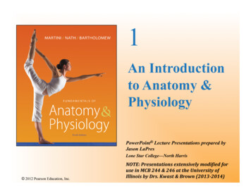
3. If an object viewed with the oil immersion lens looked as it does in the field depictedbelow, could you determine its approximate size from this view? If not then how couldyou determine it?Perceiving DepthAny microscopic specimen has depth as well as length and width. You will rarely view atissue slide with just one layer of cells. Normally you can see two or three cellthicknesses. In microscope work the depth of field (the depth of the specimen clearly infocus) is greater at lower magnifications. In other words, you can clearly see morelayers of cells on lower magnifications. On high magnifications, you can only focus onone layer at a time, and the other layers may appear blurred. Keep this in mind whenworking with the microscope.9
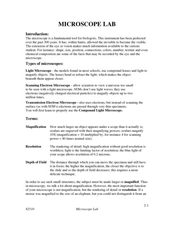
Use this diagram to help you find the following organs on the torso models:heart, kidneys, lungs, trachea, brain, esophagus, blood vessels, adrenal glands, liver,stomach, small and large intestine, pancreas, gall bladder, ureters, bladder, spleen10
1. Obtain a microscope and bring it to the laboratory bench. (Use the proper transport technique!) Compare your microscope with the figure on the following page and identify the following microscope parts: Base: Supports the microscope. Substage light: Located in the base, the light passes directly upward through the microscope.