Transcription
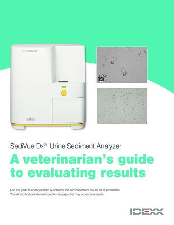
SediVue Dx Urine Sediment AnalyzerA veterinarian’s guideto evaluating resultsUse this guide to understand the quantitative and semiquantitative results for all parameters.You will also find definitions of specific messages that may accompany results.
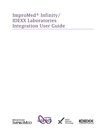
Element type: Blood cellsParameterImage tagRed blood cells(RBCs)White blood cells(WBCs)Red blood cellsRBCWBCResultsNone detected 1/HPFThe element has notSome rare features havebeen detected orbeen found in the sample;there are not enoughhowever, the quantityrecognizable featuresis below the clinicalto classify.reporting threshold.Quantitativenumericalresult/HPFWhite blood cells 50/HPF
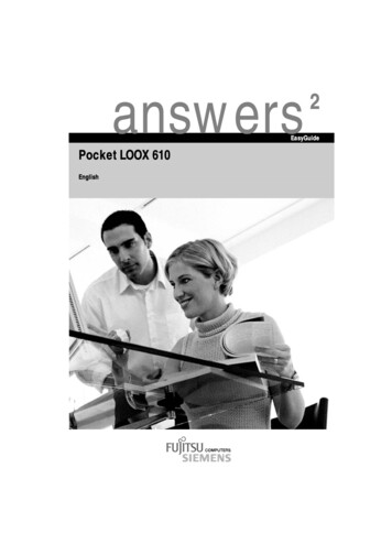
Element type: BacteriaParameterImage tagRodsN/A*CocciResultsNone detectedSuspect presencePresentThe element has not beenSome recognizable featuresThere is high confidencedetected or there are notof an element (cocci, rods,that bacteria are present inenough recognizablecasts) are present; however, thethe sample.features to classify.quantity and detail is insufficientto report as “present.”*To avoid blocking your visual interpretation, the analyzer classifies and counts all bacteria without tagging them.NOTE: Bacteria results may be confounded by other debris and artifacts in the sample (e.g., sperm, crystalline debris).RodsCocci
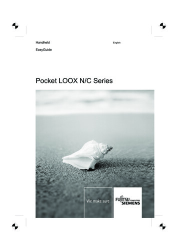
Element type: Epithelial cellsParameterImage tagSquamoussqEPIResultsNone detectedThe element has notbeen detected orthere are not enoughNonsquamousnsEPIrecognizable featuresto classify.Squamous 1/HPFSome rare featureshave been found inthe sample; however,1–2/HPF 3–5/HPF 6–10/HPF 10/HPFthe quantity is belowthe clinical reportingthreshold.Nonsquamous
Element type: CastsParameterImage tagHyalineHYANonhyaline(e.g., granular, waxy)HyalinenhCSTResultsNone detectedSuspect presenceThe element has not beenSome recognizable features of andetected or there are notelement (cocci, rods, casts) areenough recognizable fea-present; however, the quantity andtures to classify.detail is insufficient to report.Nonhyaline 1/LPF
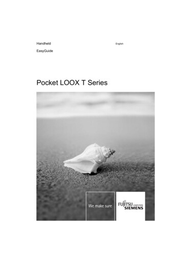
Element type: CrystalsParameterImage tagUnclassified(all other crystals)Calcium nclassifiedResultsCRYCaOxDiNone detectedThe element has notSTRbeen detected orthere are not enoughrecognizable featuresAmmBito classify. 1/HPFSome rare featureshave been found inthe sample; however, 1–5/HPF 6–20/HPF 21–50/HPF 50/HPFthe quantity is belowthe clinical reportingthreshold.BILICalcium oxalatedihydrateAmmonium biurateBilirubinStruvite
Automated messagesbased on specific resultsYour SediVue Dx Urine Sediment Analyzer may include sample messages in the patient report. Thesemessages are generated based on numerical results and are intended to provide you with further insight and guide you withrecommended next steps. Information about common messages is included in this guide. For a complete list, consult the SediVue DxUrine Sediment Analyzer Operator’s Guide at idexx.com/library.When a bacteria result is“suspect presence”Analyzer messageConfirm with one of the following: image review; air-dried, stained cytological preparation(“dry prep”); or urine culture.Some recognizable features of an element (cocci, rods, casts) are present; however, the quantity or detailsDefinitionare insufficient to report as “present.” Debris commonly found in canine and feline urine can be confusedwith bacteria.If images do not provide visual confirmation of bacteria, but patient has clinical signs or there are other cluespresent (white blood cells, etc.), it is recommended to perform a dry prep to confirm.GuidanceIf bacteria are obvious in the images or the result is “present,” a dry prep is not necessary, but you can followup with a culture and sensitivity (MIC) test if you want to further classify the bacteria as well as test for sensitivity to certain antibiotics.
When samples are crowdedwith overlapping elementsAnalyzer messageDefinitionImages crowded. Review images and follow the guidance below to determine next steps.When a urine sample is crowded and the edges of the elements overlap, the SediVue Dxconvolutional neural network may have difficulty discerning the elements from one another.When images provide clinical insight: No dilution needed, add comments to patient record.GuidanceModerate amount of cells or crystalline material: Dilute 1:5 with 0.9% normal saline and rerun.Marked amount of cells or crystalline material: Dilute 1:10 with 0.9% normal saline and rerun.Note: You will only be charged for the first run from the same patient ID within a 24-hour period.When image focus and qualitycannot be verified by the analyzerAnalyzer messageReview images to confirm results.DefinitionThe convolutional neural network cannot verify the quality of focus in the images.The sample may have very little urine sediment (e.g., “normal”) or contain air bubbles, or theanalyzer is dirty.GuidanceIf the expected results do not align with the image review, rerun the sample.If the message appears with several consecutive samples, the analyzer may require cleaning.Note: You will only be charged for the first run from the same patient ID within a 24-hour period.
CrystalsCrystals can come in a variety of different shapes, sizes, and presentations. Urine pH, specific gravity, sample preparation and handling,and drugs can all play a part in crystal formation. Crystals in small numbers (e.g., struvites) may be normal for some dogs, but others(e.g., cystine) may signify disease processes. The following Smart Flags are designed to provide further clinical insight into the presenceof crystalluria.Analyzer messageCrystalline debris detected.Crystalline debris can be abundant and variable in size and presentation in some samples. Due tobackground density, the presence of large amounts of crystalline debris can affect the identification ofDefinitionother formed elements in the sample. This flag is displayed when crystalline debris has been detectedby the algorithm. The neural network algorithm has been trained to exclude crystalline debris from theunclassified crystal (CRY) category.GuidanceWhen this flag is present, users will be notified so they can be more discerning about the bacteria result, asvery small particles of debris can resemble bacteria.
Urine protein:creatinine (UPC) ratioAnalyzer messageConsider evaluation of urine protein:creatinine ratio.When this message appears, the urine chemical results indicate the presence of protein. A UPC ratio canDefinitionbe used to quantify protein loss in the urine as it is unaffected by urine volume or concentration. It has beenincorporated into the International Renal Interest Society (IRIS) Guidelines on Staging and Treatment ofChronic Kidney Disease (CKD) as an important monitoring tool at all stages.A UPC ratio should be performed after urinalysis with urine sediment examination. It is notGuidancerecommended for use if there is an active urine sediment, as inflammatory conditions in the urinary tract willincrease protein and negate the usefulness of the ratio.Analyzer messageDefinitionGuidanceRecommend reevaluating proteinuria after resolution of active urine sediment.When this message appears, the urine chemical results indicate the presence of protein in addition to anactive urine sediment (RBCs and/or WBCs and possibly bacteria).First, resolve the infection. Once the urine sediment becomes inactive, consider running aUPC ratio to quantify protein loss.
Image tagsThe SediVue Dx Urine Sediment Analyzer identifies, classifies, and counts all elements found in the sampleand also applies an image tag. Image tags are abbreviated labels for the reported parameters that appear on the formedelements in the reported sample images.Image tags are not available: When the sample is flagged for dilution. When the sample is from an invalidated species or fluid. For all bacteria results.**Tagging of bacteria can be overwhelming and block the visual interpretation of the image. Image tags can be toggled on and off.Guidance for reviewing imagesReviewing images will validate the numerical data provided and supplement the SediVue Dx analysis.Tap on an image to make it full screen.Reverse the contrast to see details such as cell nuclei.View one imageZoom in up to 200% to see smaller elements.at a timeTurn image tags on or off.Select an image to add to the patient record or print an area of interest.Use the large arrows to scroll through images.Seventy high-resolution images are taken for every sample run.View the first3–12 images firstImages have been prioritized based on the absence or presence of formed elementsand the clinical significance of each element found.Highest scoring images are displayed first.Select View all images to easily scroll through all 70 images.Classified elements will be tagged (labeled). (Image tags are not available for “none detected” andRefer to imagebacteria results.) Image tags can be turned on or off on the image viewer.tags, when presentWhether a tag is present or not, all formed elements are classified by the analyzer and reflected inthe result.For Cornerstone Software users, three prioritized images are automatically saved to the patient recordSave images toand transmitted to VetConnect PLUS. You may select up to three additional images to save to the patientthe patient recordrecord and VetConnect PLUS.For all other practice management software, the first image will be included with the results PDF.Add comments tothe patient recordInclude comments about findings you consider noteworthy.From the patient results screen, tap Add comments. Your comments will be automatically uploaded toVetConnect PLUS and, if applicable, your practice management software.
Bacteria resultsBacteria result(rods/cocci)DefinitionThe element has not beenNone detecteddetected or there are notenough recognizablefeatures to classify.Possible reason for validationPatient has clinical signs ora history of persistent urinarytract infections.Recommended next stepsIf visual review of images is negative andpatient has no clinical signs or history,bacteriuria is unlikely. No further action isnecessary.Differentiate bacteria from debris.Review visually; if confirmed, diagnose andmanage based on your interpretation. If, however, visual review is inconclusiveSome recognizable featuresof an element (cocci, rods,Suspectcasts) are present; however,presencethe quantity and detail isinsufficient to report asand white blood cells,Crystalline or amorphous debrisred blood cells, clinical signs,is common in canine and felineand/or history of urinary tract infection aresamples (especially free catch).present, confirm presence of bacteria witha dry prep.“present.” If, however, visual review is inconclusiveand there are no supporting clues present(e.g., active urine sediment, history), thepresence of bacteria is unlikely.There is high confidence thatPresentbacteria are present in thesample.Bacteria results may beIf visual review of images is confirmatory and/confounded by other debrisor the patient has clinical signs or history,and artifacts in the samplebacteriuria is likely. No further action is(e.g., sperm, crystalline debris).necessary.
Urinalysis versus cultureUnderstanding possible discordant resultsBacteria result(rods/cocci)Culture resultCauses for discordant resultsBacterial growth was inhibited or prevented by: Antibiotic treatment at time of sample collection. Exposure of sample to extreme temperatures. Extremes of urine pH ( 4 or 9). High quantity of white blood cells in urine.Microscopy results misidentified amorphous or crystalline debris as bacteria.*PresentNo growthFalse identification of cocci was due to random motion of small colloidal particles(Brownian motion).*Stain used on urine sample prior to in-house microscopy was contaminatedby bacteria.Anaerobic bacteria were identified by the analyzer but cannot be grown inaerobic cultures (rare).*Particularly with unstained urine sediment examinations.Bacteria colony counts are too low to be visualized on urine sediment analysisbecause of: Very dilute urine.None detectedPositive Incomplete or unsuccessful antibiotic therapy. Localized pyelonephritis.In cases where clinical history is suggestive of urinary tract infection or an activeurine sediment is present, urine culture should be considered even in the absenceof bacteriuria on urinalysis.Dry prep basicsWhen?A dry prep should only be considered when you are unable to visually confirm absence orpresence and: The patient has clinical signs or previous history. There are other supporting results in the sample (e.g., white blood cells, red blood cells).A dry prep (air-dried, stained cytological slide) is a quick, effective way to:Why? Validate the absence or presence of bacteria. Distinguish bacteria from amorphous or crystalline debris.How?You can do a dry prep in 5 minutes or less!Watch a short video to learn how.
Access more training and resources online today!Visit idexx.com/uaresources 2019 IDEXX Laboratories, Inc. All rights reserved. 2164848-01All /TM marks are owned by IDEXX Laboratories, Inc. or its affiliates in the United States and/or other countries.The IDEXX Privacy Policy is available at idexx.com.
For all other practice management software, the first image will be included with the results PDF. Add comments to the patient record Include comments about findings you consider noteworthy. From the patient results screen, tap Add comments. Your comments will be automatically uploaded to