Transcription
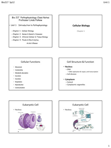
Bio217 Sp12Unit 1Bio 217 Pathophysiology Class NotesProfessor Linda Falkow Unit 1: Introduction to PathophysiologyCellular Biology– Chapter 1: Cellular BiologyChapter 1– Chapter 2: Genes & Genetic Diseases– Chapter 3: Altered Cellular & Tissue Biology– Chapter 4: Fluids & Electrolytes,Acids & BasesCellular FunctionsCell Structure & Function Nucleus Movement Conductivity Metabolic absorption Secretion Excretion– DNA DNA replication & repair, and transcription– Cell division Cytoplasm Respiration Reproduction– Cytosol– Cytoplasmic organelles CommunicationEukaryotic Cell NucleusEukaryotic Cell Nucleus1
Bio217 Sp12Unit 1Eukaryotic Cell CytoplasmOrganelles Ribosomes– Protein synthesis Endoplasmic reticulum– Rough ER – site of protein synthesis– Smooth ER - site of lipid synthesis Golgi complex– Proteins from the endoplasmic reticulum arepackaged in the Golgi complexEukaryotic OrganellesPlasma Membrane Lysosomes– Contain enzymes– Role in autodigestion as a result of cellular injury Peroxisomes– Contain oxidative enzymes– Produce H2O2 Mitochondria– Participates in oxidative phosphorylation(ATP production) Cytoskeleton– “Bones and muscles” of the cell– Microtubules and MicrofilamentsPlasma Membrane Lipids Hydrophilic and hydrophobic Phospholipids, glycolipids, and cholesterol Carbohydrates Glycoproteins Proteins– Integral, peripheral, transmembrane– FunctionsMembrane Fluidity(Fluid Mosaic Model) Plasma membrane protein functions2
Bio217 Sp12Unit 1Adenosine Triphosphate (ATP) Created from the chemical energycontained in organic molecules Used in synthesis of organic molecules,muscle contraction, and active transport Stores and transfers energyMembrane Transport Passive transportCellular Energy1. Digestion2. Glycolysis &oxidation3. Citric Acid CycleMembrane TransportPassive DiffusionFacilitated Diffusion– Diffusion (simple and facilitated) Concentration gradient [high low]– Filtration Hydrostatic pressure– Osmosis Movement of water– Tonicity Isotonic, hypertonic, and hypotonicMembrane TransportActive Transport(Na-K Pump) Active transport– Active transport pumps (Na/K Pump)– Transport by vesicle formation Endocytosis– Pinocytosis– Phagocytosis– Receptor mediated Exocytosis3
Bio217 Sp12Unit 1The Cell CycleMembrane Transport– Interphase(G1 phase, S phase, G2 phase)plus MitosisG1- cell activities andcentrioles replicateS - DNA replicatesG2 – protein synthesis,preparation for mitosisExocytosisInfluences on the Cell Cycle Cellular division rates– Complete cell cycle 12-24 hours– Mitosis 1 hour Growth factors (cytokines)– PDGF ( CT and neuroglial cells)– EGF ( epidermal cells)– IGF-I ( fat cells and CT)Types of Tissue Epithelial tissue– Simple vs. stratified squamous– Transitional– Cuboidal– Simple vs. stratified columnar– Pseudostratified ciliated Connective tissue– Dense regular or irregular– Loose and dense connective tissue– Elastic and reticular connective– Cartilage, bone, vascular, and adiposeTypes of Tissue Muscle tissue– Smooth– Striated (skeletal)– Cardiac Nerve tissue– Neurons conduct nerve impulses– Neuroglia provide support activitiesConcept Check 1. What are the main parts of the eukaryoticcell?A.B.C.D.Lipids, CHO, proteinsMinerals and waterOrganellesCytoplasm and membrane-bound nucleus4
Bio217 Sp12Unit 12. Which of the following cellular functionsdoes NOT occur in all cell types?A.B.C.D.RespirationExcretionMetabolic absorptionSecretion 4. Which can transport substances againstthe concentration gradient?A.B.C.D.Active transportOsmosisFacilitated diffusionDialysis3. Which of the following statementsregarding transport across membranes isTRUE?A. Diffusion requires a significant energy input.B. Mediated transport involves a receptor witha high degree of specificity.C. Active transport moves molecules down aconcentration gradient.D. Facilitated transport expends energy.5. Anaphasea. centrioles separate6. Chromatinb. single stranded DNA innon-dividing cellc. site of protein synthesis7. Metaphase8. Mitochondria d. chromatids separate9. Ribosomee. powerhouse of cell10. Prophasef. chomatid pairs align5
Bio217 Sp12Unit 1DNAGenes and Genetic DiseasesChapter 2 Pentose sugar (deoxyribose) Phosphate molecule Four nitrogenous bases– Pyrimidines: Cytosine and Thymine– Purines: Guanine and Adenine Double helix model NucleotideProteins One or more polypeptidesMutation Any inherited alteration of genetic material– Chromosome aberrations– Base pair substitution One base pair is substituted for another Composed of amino acids– 20 amino acids– Directed by sequence of bases (codons) Frameshift mutation– Insertion or deletion of one or more base pairs– Causes a change in the entire “reading frame” Mutagens– Increase frequency of mutations– Radiation and chemicalsMutationTranscription- DNA serves as template for formation of mRNA6
Bio217 Sp12Unit 1Translation- Process by which RNA directs thesynthesis of a polypeptideChromosomes Somatic cells (2n)– Contain 46 chromosomes (23 pairs) Gametes (n)– Contain 23 chromosomes Meiosis Formation of haploid cells from diploid cells Mitosis Forms somatic cellsChromosomes AutosomesKaryotype Ordered display of chromosomes (metaphase)– The first 22 of the 23 pairs ofchromosomes in males and females Sex chromosomes– Remaining pair of chromosomes– In females (XX)– In males (XY)Chromosome Aberrations Euploid cells have a multiple of the normalnumber of chromosomes (“eu” Gk. good )– n and 2n cells are euploid forms Polyploid - when a euploid cell has morethan the diploid number Triploidy - three copies of each chromosome(3n 69) Tetraploidy: - four copies of each (4n 92 total) Triploid or tetraploid fetuses never surviveChromosome Aberrations Aneuploidy– Somatic cell that does not contain a multiple of23 chromosomes– Cell containing three copies of onechromosome is trisomic (trisomy)– Monosomy is one copy of any chromosome,often lethal “It is better to have extra than less”7
Bio217 Sp12Unit 1Chromosome AberrationsNondisjunction Disjunction– Normal separation of chromosomesduring cell division Nondisjunction– Usually the cause of aneuploidy– Failure of homologous chromosomes toseparate normally during cell divisionAutosomal AneuploidyDown Syndrome Down syndrome (Trisomy 21)– Best known example of aneuploidy– 1:800 live births– Mentally retarded, low nasal bridge,epicanthal folds (eyes), protruding tongue,poor muscle tone– Risk increases with maternal age 35Sex Chromosome Aneuploidy Turner syndrome (45, X)– Females with only one X chromosome– Characteristics Absence of ovaries (sterile)Short stature ( 4'7")Webbing of the neckEdemaUnderdeveloped breasts; wide nipplesHigh number of aborted fetuses havethis karyotype X is usually inherited from motherSex Chromosome Aneuploidy Klinefelter syndrome (47, XXY)– Individuals with at least two Xs andone Y chromosome– Characteristics Male appearance Develop female-like breasts Small testes Sparse body hair Long limbs8
Bio217 Sp12Unit 1Abnormalities inChromosome Structure Chromosome breakage– If breakage does occur, physiologicmechanisms usually repair the break,but often heals so that chromosome structureis altered– Clastogens - harmful agents such as:Abnormalities inChromosome Structure Breakage or loss of DNA Cri du chat syndrome– “Cry of the cat”– Deletion of short arm of chromosome 5– Low birthweight, metal retardation, andmicrocephaly– ionizing radiation– chemicals– virusesGenetics HomozygousGenetics Genotype (“what they have”)– Loci on a pair of chromosomes haveidentical genes– Example O blood type (OO) Heterozygous– Loci on a pair of chromosomes havedifferent genes– Example AB blood type (A and B genes on pair of loci)– The genetic makeup of an organism Phenotype (“what they demonstrate”)– The observable, detectable, or outwardappearance Example– Individual with the A blood type could be AA orAO.Genotype Phenotype GeneticsGenetics If two alleles are found together, the allelethat is observable is dominant, and theone whose effects are hidden is recessive Carrier– A carrier is one that has a disease gene butis phenotypically normal– For a person to demonstrate a recessivedisease, the pair of recessive genes mustbe inherited– Example homozygous heterozygous homozygous Alleles can be codominant Ss sickle cell anemia carrier ss demonstrates sickle cell disease9
Bio217 Sp12Unit 1Single-Gene Disorders Autosomal dominant disorder– Abnormal allele is dominant, normal allele isrecessive, and the genes exist on a pair ofautosomes Huntington’s disease (Hh)– Dementia, uncontrollable movements– Evident after age 40– results in deathAutosomal Recessive Cystic fibrosis– Defect on Ch 7– Cl ion transport defect salt imbalance thick mucus– Affect respiratory and digestive systems– Individuals live to age 30Sex-Linked DisordersExpressivity- variation in phenotype Examples:– von Recklinghausen’s disease(Neurofibromatosis type 1)– Autosomal dominant– Long arm of chromosome 17– Disease varies from spots (caf-au-lait) onthe skin to malignant neurofibromas,scoliosis, gliomas, neuromas, etc.Consanguinity Mating of two related individuals inbred offspring Dramatically increases the recurrencerisk of recessive disordersSex-Linked Disorders The Y chromosome contains only a fewdozen genes, so most sex-linked traitsare located on the X chromosome(X-linked) Male-pattern baldness Sex-linked (X-linked) disorders are usuallyexpressed by males because femaleshave another X chromosome to maskthe abnormal gene Color-blindness– Affects 8% of males; 0.5% females– Unable to distinguish red from green– Occurs mostly in men (autosomal dominant)– Can occur in women (autosomal recessive)10
Bio217 Sp12Unit 1Sex-Linked DisordersConcept Check1. Which of the following statements is TRUE?A.B.C.D.2. Which term best describes an allele with anobservable siveCellular Adaptation Physiologic (normal) vs. pathogenic(diseased states)– Atrophy– Hypertrophy– Hyperplasia– Metaplasia– DysplasiaProtein synthesis takes place in the cytoplasm.DNA is replicated in the cytoplasm.RNA is double-stranded.RNA contains the same bases as DNA.Altered Cellular andTissue BiologyChapter 3Cellular AdaptationAtrophy – decrease in cell sizeHypertrophy – increase in cell sizeHyperplasia – increase in cellnumbers (incr. in cell div.)Metaplasia – one cell typeconverts to another cell typeDysplasia – abnormal changes incells (vary in size, shape, etc.)11
Bio217 Sp12Unit 1Cellular AdaptationCellular Injury Reversible IrreversibleCellular Injury (Hypoxic Injury due to ischemia)Cellular Injury Mechanisms Hypoxic injury– Ischemia - blood flow– Anoxia - lack of O2 ( due to blood clot)– Cellular responses Decrease in ATP, causing failure of Na-K pump andsodium-calcium exchange Cellular swelling Vacuolation (formation of vacuoles)– If O2 restored Reperfusion injury free radicalsCellular InjuryCellular Injury Mechanisms Chemical injury (poisons)– Carbon tetrachloride (CCl4) - forms free radicals by liver– Lead -- environmental or occupational sources-- affects nervous system, hemopoiesis, kidneys– Carbon monoxide (CO)-- causes hypoxic injury due toincreased affinity for Hb– Ethanol (C2H5OH)-- nutritional deficiencies & liver disorders-- conversion to acetaldehyde fat deposits, liver enlargementand necrosis, CNS depressant– Mercury (Hg) -- dental amalgams, fish, vaccines-- controversy over health effects– Social or street drugs - THC, C10H15N (meth), cocaine, opiates12
Bio217 Sp12Unit 1Unintentional and Intentional Injuries Blunt force injuries– Application of mechanical energy to thebody resulting in the tearing, shearing,or crushing of tissues– Contusion (bruise) vs.hematoma (collection of blood)– Abrasion (scrape)– Laceration (tear in skin or tissue)– Fractures (bones break)Unintentional andIntentional InjuriesUnintentional andIntentional Injuries Sharp injuries– Incised wounds– Stab wounds– Puncture wounds– Chopping woundsUnintentional andIntentional InjuriesStab WoundsAvulsed LacerationPatterned AbrasionUnintentional and Intentional Injuries Asphyxial injuries– Caused by failure of cells to receive or use oxygen Suffocation–Choking asphyxiation Strangulation–Hanging, ligature, and manual strangulationInfectious Injury Pathogenicity of a microorganism Disease-producing potential– Invasion and destruction– Toxin production– Production of hypersensitivity reactions Chemical asphyxiants–Cyanide and hydrogen sulfide Drowning–Cerebral hypoxia unconsiousness13
Bio217 Sp12Unit 1Immunologic andInflammatory Injury Phagocytic cells Immune and inflammatory substances– Histamine, antibodies, lymphokines,complement, and proteases Membrane alterations K leakage from cellManifestations of Cellular Injury Cellular accumulations (infiltrations)– Water– Lipids and carbohydrates– Glycogen– Proteins– Pigments Melanin, hemoproteins, bilirubin– Calcium– UrateOncosis (Hydropic Degeneration)- swelling (degeneration by water)Calcium InfiltrationManifestations of Cellular InjuryCellular Death Necrosis– Cellular changes after local cell death and theprocess of cellular autodigestion (self-digestion)4 types of Necrosis:– Coagulative– Liquefactive– Caseous– FattyGangrenous necrosis is large area of tissue death,not a separate type of cell death.14
Bio217 Sp12Unit 1Cellular DeathNecrosis Coagulative necrosis – result of hypoxia due to ischemiaStages of NecrosisPyknosis – nucleus shrinks andbecome dense mass of geneticmaterial– Kidneys, heart, and adrenal glands– Proteins denature coagulationKaryolysis – nucleusfragments “nuclear dust”Necrosis Liquefactive necrosis– Due to ischemic injury to brain cells (neuronsand neuroglia)– Hydrolytic enzymes digest cells– Caused by bacterial infection Staphylococci, streptococci, and Escherichia coliCoagulative Necrosis ofMyocardium of LV (heart):note anemic infarctand necrosis ofpapillary muscleNecrosis Caseous necrosis– Tuberculous pulmonary infection– Combination of coagulative andliquefactive necrosisGranuloma with CentralCaseous Necrosis:typical of pulmonary TBLiquefactive necrosisof brainNecrosis Fat necrosis– Breast, pancreas, and other abdominal organs– Due to action of lipasesNecrosis Gangrenous necrosis– Death of tissue from severe hypoxic injury Dry vs. wet gangrene– Gas gangrene ClostridiumFat Necrosis of Pancreas- note necrotic adipocytes15
Bio217 Sp12Unit 1Gangrenous NecrosisApoptosis Programmed cellular death Physiologic (normal tissue turnover)vs. pathologic (exogenous injury)Necrosis vs. ApoptosisConcept Check1. Which of the following is the most common causeof cellular injury?A.B.C.D.Free radical-induced injuryChemical injuryHypoxiaDysplasia2. Which cell adaptation is observed in the cervix?A. HyperplasiaC. DysplasiaB. HypertrophyD. Metaplasia3. Which of the following terms best describesdeath of a cell from hypoxia, generally as aresult of ischemia in the lower extremities?A.B.C.D.Coagulative necrosisLiquefactive necrosisFat necrosisGangrenous necrosis4. Cellular swelling is:A. IrreversibleB. Occurs early in all cell injuriesC. Low intracellular Na is commonD. None of the aboveMatch the manifestation with the characteristic:5. Necrosis due to Clostridia6. Rigidity of muscles afterdeath7. Increased cell number8. Necrosis from lysosomalrelease9. Replacement of one cellwith another10. Normal & pathologic cellself-destructiona. Rigor mortisb. Gas gangrenec. Hyperplasiad. Metaplasiae. Liquefactionf. Apoptosis16
Bio217 Sp12Unit 1Distribution of Body FluidsFluids and Electrolytes,Acids and BasesChapter 4Net Filtration Forces favoring filtration– Capillary hydrostatic pressure (bloodpressure)– Interstitial colloid osmotic pressure(water-pulling) Forces favoring reabsorption– Plasma oncotic pressure (water-pulling) Blood colloid osmotic pressure– Interstitial hydrostatic pressureEdema Total body water (TBW) 60% of body wt.– Intracellular fluid (ICF) 2/3 of TBW Fluid within cells– Extracellular fluid (ECF) 1/3 of TBW Interstitial fluid - intercellular and not in vessels Intravascular fluid - plasma Other ECF: Lymph, synovial, intestinal, CSF, sweat,urine, pleural, peritoneal, pericardial, andintraocular fluidsEdema Accumulation of fluid within the interstitial spaces Causes:– Increase in capillary hydrostatic pressuredue to venous obstruction or salt & water retention( thrombophlebitis, CHF)– Decrease in plasma colloid osmotic pressuredue to decreased plasma proteins (kidney disease,hemorrhage, cirrhosis of liver)– Increases in capillary permeability due to trauma– Lymph obstruction due to removal of lymph nodesAcidosis and Alkalosis Four categories of acid-base imbalances– Respiratory acidosis—elevation of pCO2 as aresult of ventilation depression– Respiratory alkalosis—depression of pCO2as a result of alveolar hyperventilation17
Bio217 Sp12Unit 1MetabolicAcidosisAlkalosisAcidosis and Alkalosis– Metabolic acidosis—depression of HCO3– oran increase in noncarbonic acids– Metabolic alkalosis—elevation of HCO3–usually caused by an excessive loss ofmetabolic acidsRespiratoryAcidosisAlkalosisConcept Check1. Which of the statements is TRUE regarding waterbalance?A.B.C.D. 2. Of the 60% of TBW made up of water,about 2/3 is .– A. ECF– B. ICF– C. Intravascular fluid– D. interstitial water 3. Total water loss per day by an adult is about:– A.– B.– C.– D.0.8 L1.2 L2.2 L2.8 LIsotonic fluids cause increased cellular swelling.Hypertonic fluid causes increased cellular swelling.Hypotonic fluid causes cellular swelling.Hypernatremia causes cellular swelling. 4. Aldosterone controls ECF volume by:A.B.C.D.E.CHO, fat, and protein catabolismNa reabsorptionK reabsorptionH2O reabsorptionB and D are correct 5. The release of ADH is NOT stimulated by:A.B.C.D.StressHyponatremiaHypernatremiaIncrease in plasma osmolality18
Bio217 Sp12 6. The pH of saliva is about 7 and the pH of gastricjuice is about 2. How many times more concentratedis the [H ] in gastric juice than in saliva?A.B.C.D.55010,000100,000Match the acid base with the probable cause:7. Respiratory acidosis a. Excessive baking soda8. Respiratory alkalosis b. Severe anxiety(hyperventilation)9. Metabolic alkalosisc. EmphysemaUnit 1Match the imbalance with the appropriatecompensatory mechanism: 10. Respiratory acidosis a. Kidneys excrete H ,retain HCO3 11. Respiratory alkalosis b. Kidneys excrete HCO3,retain H 12. Metabolic acidosis c. Hyperventilation(blow off CO2)19
Bio 217 Pathophysiology Class Notes Professor Linda Falkow Unit 1: Introduction to Pathophysiology -Chapter 1: Cellular Biology -Chapter 2: Genes & Genetic Diseases -Chapter 3: Altered Cellular & Tissue Biology -Chapter 4: Fluids & Electrolytes, Acids & Bases Cellular Biology Chapter 1 Cellular Functions