Transcription
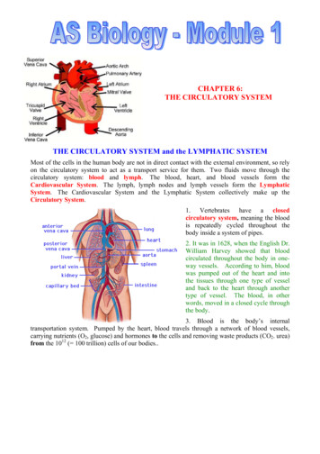
CHAPTER 6:THE CIRCULATORY SYSTEMTHE CIRCULATORY SYSTEM and the LYMPHATIC SYSTEMMost of the cells in the human body are not in direct contact with the external environment, so relyon the circulatory system to act as a transport service for them. Two fluids move through thecirculatory system: blood and lymph. The blood, heart, and blood vessels form theCardiovascular System. The lymph, lymph nodes and lymph vessels form the LymphaticSystem. The Cardiovascular System and the Lymphatic System collectively make up theCirculatory System.1. Vertebrates have a closedcirculatory system, meaning the bloodis repeatedly cycled throughout thebody inside a system of pipes.2. It was in 1628, when the English Dr.William Harvey showed that bloodcirculated throughout the body in oneway vessels. According to him, bloodwas pumped out of the heart and intothe tissues through one type of vesseland back to the heart through anothertype of vessel. The blood, in otherwords, moved in a closed cycle throughthe body.3. Blood is the body’s internaltransportation system. Pumped by the heart, blood travels through a network of blood vessels,carrying nutrients (O2, glucose) and hormones to the cells and removing waste products (CO2. urea)from the 1012 ( 100 trillion) cells of our bodies.
THE HEART1. The central organ of the cardiovascular system is the heart. This is a hollow, muscular organ thatcontracts at regular intervals, forcing blood through the circulatory system.2. The heart is cone-shaped, about the size of a fist, and islocated in the centre of the thorax, between the lungs,directly behind the sternum (breastbone). The heart istilted so that the base is tilted to the left.3. The walls of the heart are made up of three layers oftissue:a) The outer and inner layers are epithelial tissue.b) The middle layer, comprising the cardiac muscle ofthe heart itself, is called the myocardium.4. For obvious reasons, the cardiac muscle is not under theconscious control of the nervous system, and can generateits own electrical rhythm (myogenic). For the samereasons, cardiac muscle cannot respire anaerobically andso the muscle cannot get tired (or develop cramp!)5. Cardiac muscle has a rich supply of blood, which ensuresthat it gets plenty of oxygen. This is brought to the heartthrough the coronary artery. Since the heart relies onaerobic respiration to supply its energy needs, cardiac muscle cells are richly supplied withmitochondria.6. Our hearts beat about once every second of every day of our lives, or over 2.5 million times in anaverage life span. The only time the heart gets a rest is between beats.HOW THE HEART WORKS1. The heart can be thought of as two pumps sitting side by side – each of which has an upperatrium and a lower ventricle – a total of 4 chambers. It functions as two pumps inside one.2. The right side of the heart pumps ‘deoxygenated blood’ (actually, blood low in oxygen) fromthe body into the lungs, where gas exchange takes place. In that process, carbon dioxide is lost tothe air and oxygen is absorbed. This oxygen is almost all carried by the Red Blood Cells (RBC’s).3. The left side of the heart pumps oxygenated blood from the lungs to the rest of the body.4. The heart is enclosed in a protective membrane-like sac called the pericardium, which surroundsthe heart and secretes a fluid that reduces friction as the heart beats.5. The atria (upper chambers) of the heart receive blood coming into the heart. Then have thinwalls, so allowing them to be filled easily. They pump the blood into the ventricles (lowerchambers), thus filling them.6. The ventricles pump blood out of the heart and the left ventricle has the thickest walls of theheart because it has to do most of the work to pump blood to all parts of the body. This is wherethe blood has the highest pressure.7. Vertically dividing the two sides of the heart is a wall, known as the septum. The septumprevents the mixing of oxygenated (left side) and deoxygenated (right side) blood.8. It also carries electrical signals instructing the ventricles when to contract. These impulses passdown specially-modified muscle cells (Purkinje fibres), collectively known as the Bundle of His.
THE RIGHT SIDE OF THE HEART1. Deoxygenated blood from the body enters the right side of the heart through two large veinscalled the vena cavae. The superior vena cava returns blood from the head and arms; the inferiorvena cava from the rest of the body (except, of course, the lungs!)2. Both empty into the right atrium. This is where the blood pressureis lowest (even negative).When the heart relaxes (between beats),pressure in the circulatory system causes the right atrium to fill withblood.3. When the atria contract, pressure inside it rises, the right atrioventricular (AV) valve opens, and blood is squeezed from the rightatrium into the right ventricle. This valve is also known as the tricuspidvalve. The closing of this valve makes a sound – ‘lub’.4. When the atrium is empty, the pressure inside it falls, and the pressureinside the ventricle begins to rise. This causes the atrio-ventricular valve toAtria contractshut quickly, preventing the back-flow of blood.5. The general purpose of all valves in the circulatory system is to prevent the back-flow of blood,and so ensure that blood flows in only one direction.6. When the right ventricle contracts, blood is forced out through the semi-lunar valve (also knownas the pulmonary valve), into the pulmonary arteries, where it goes to the lungs. These are theonly arteries to carry deoxygenated blood.7. When the right ventricle is empty, the pressure inside falls below that in the pulmonary artery,and this causes the semi-lunar valve to snap shut. The closing of these valves also causes a sound –‘dup’. A normal heart-beat is thus ‘lub dup’.THE LEFT SIDE OF THE HEART1. Oxygenated blood leaves the lungs and returns to the heart through thepulmonary veins. These are the only veins to carry oxygenated blood.2. This blood enters the left atrium, which, when full, forces blood intothe left ventricle, filling it. The valve which opens is called the left atrioventricular (AV) valve, (or bicuspid or mitral valve). As on the rightside of the heart, this valve closes when the atrium is empty and pressurebegins to rise in the ventricle.3. From the left ventricle, blood is forced at very high pressure throughanother semi-lunar valve (the aortic valve), into the aorta, which carriesblood throughout the body (apart from the lungs!).Ventricles contract4. This surge of blood from the ventricles causes the walls of the aorta toexpand and the muscles within to stretch – we can detect this as a pulse.5. When the ventricle is almost empty, the pressure begins to fall below that in the aorta, and thiscauses the semi-lunar valve to snap shut, as the elastic walls of the aorta recoil, thus preventingback-flow of blood into the heart.
THE CARDIAC CYCLE1. The cardiac cycle is the sequence of events in one heartbeat. In its simplest form, the cardiaccycle is the simultaneous contraction of both atria, followed a fraction of a second later by thesimultaneous contraction of both ventricles.2. The heart consists of cardiac muscle cells that connect with each other – they are branched – andso when one contracts, they stimulate their neighbours and they all contract. The heart is an ‘all-ornothing’ muscle, getting its rest between beats. It can only respire aerobically.3. A heartbeat has two phases:A. Phase 1 - Systole is the term for contraction. This occurs when the ventricles contract,closing the A-V valves and opening the Semi-Lunar valves to pump blood into the two majorvessels leaving the heart.B. Phase 2 – Diastole is the term for relaxation. This occurs when the ventricles relax, allowingthe back pressure of the blood to close the semi-lunar valves and opening the A-V valves.4. The cardiac cycle also creates the heart sounds: each heartbeat produces two sounds, often calledlub-dup, that can be heard with a stethoscope. The first sound is caused by the contraction of theventricles (ventricular systole) closing the A-V valves. The second sound is caused by thesnapping shut of the Aortic and Pulmonary Valves (Semi-lunar valves). If any of the valves do notclose properly, an extra sound called a heart murmur may be heard.5. Although the heart is a single muscle, it does not contractall at once. The contraction spreads over the heart like awave, beginning in a small region of specialized cells in theright atrium called the Sino-Atrial Node (SAN). This isthe hearts natural pacemaker, and it initiates each beat6. The impulse spreads from the SAN through the cardiacmuscle of the right and left atrium, causing both atria tocontract almost simultaneously.7. When the impulse reaches another special area of theheart, right in the centre of the septum, known as the AtrioVentricular (or AV) Node, the impulse is delayed forapproximately 0.2 s. This allows time for the ventricles tofill completely.8. The AV Node relays the electrical impulse down the septum, along the Bundle of His, to thebase of the ventricles. The ventricles then contract simultaneously, from the bottom upwards,thus allowing them to empty completely with each beat.9. The heartbeat is initiated by the Sino-Atrial Node and passes through the Atrio-Ventricular Node,remaining at the same rhythm until nerve impulses cause it to speed up or to slow down. Unlikeother muscles, it does not require a new nerve impulse for each contraction.10. The autonomic nervous system controls heart rate. The accelerator nerve of the sympatheticnervous system increases heart rate and the vagus nerve of the parasympathetic nervous systemdecreases heart rate.11. For most people, their resting heart rate is between 60 and 80 b.p.m. During exercise that canincrease to as many as 200 beats per minute for an athlete; for the rest of us, 150 b.p.m. is about allwe can safely manage!
BLOOD VESSELS (ARTERIES, VEINS and CAPILLARIES)1. The Circulatory System is known as a closed system because the blood is contained within eitherthe heart or blood vessels at all times – always flowing in one direction. The path is the same –heart (ventricles) arteries arterioles organ (capillaries) veins heart (atrium)2. Except for the capillaries, all blood vessels havewalls made of 3 layers of tissue. This provides forboth strength and elasticity:A. The inner layer is made of epithelial tissue.B. The middle layer is smooth muscle.C. The outer layer is connective tissue.ARTERIES and ARTERIOLES1. Arteries carry blood from the heart to thecapillaries of the organs in the body.2. The walls of arteries are thicker than those of veins. The smooth muscle and elastic fibres thatmake up their walls enable them to withstand the high pressure of blood as it is pumped from theheart. The force that blood exerts on the walls of blood vessels is known as blood pressure and itcycles with each heart-beat (see below).3. Each artery expands when the pulse of blood passes through and the elastic recoil of the fibrescause it to spring back afterwards, thus helping the blood along. This is known as secondarycirculation, and it reduces the load on the heart.4. Other than the pulmonary arteries, all arteries carry oxygenated blood.5. The aorta carries oxygenated blood from the left ventricle toof the body except the lungs. It has the largest diameter(25mm) and carries blood at the highest pressure.6. As the aorta travels away from the heart, it branches intosmaller arteries so that all parts of the body are supplied. The smallestare called arterioles.allpartsof these7. Arterioles can dilate or constrict to alter their diameter and so alter the flow of blood throughthe organ supplied by that arteriole. Examples include muscles (when running) and skin (when hotor blushing). Since the volume of blood remains the same, if more blood flows through one organ,less must flow through another.8. Two organs which always have the same blood flow are the brain and the kidneys. Popularorgans to have blood flow reduced are the guts (between meals), muscles (when resting) and skin(when cold).CAPILLARIES1. Arterioles branch into networks of very small blood vessels – the capillaries. These have a verylarge surface area and thin walls that are only one (epithelial) cell thick.2. It is in the capillaries that exchanges take place between the blood and the tissues of the body.3. Capillaries are also narrow. This slows the blood down allowing time for diffusion to takeoccur. In most capillaries, blood cells must flow in single file.4. Tissue fluid is formed in the capillaries, for their walls are leaky (see below).
VEINS1. After leaving the capillaries, the blood enters a network of small venules, which feed into veins.These, in turn, carry the blood back to the atria of the heart.2. Like arteries, the walls of veins are lined with epithelium and contain smooth muscle. The wallsof veins are thinner and less elastic than arteries, but they are also more flexible.3. Veins tend to run between the muscle blocks of the body and nearer to the surface than arteries.4. The larger veins contain valves that maintain the direction of blood-flow. This is important whereblood must flow against the force of gravity.5. The flow of blood in veins is helped by contractions of the skeletal muscles, especially those inthe arms and legs. When muscles contract they squeeze against the veins and help to force theblood back towards the heart. Once again, this is known as secondary circulation.PATTERNS OF CIRCULATION1. Blood moves through the body in a continuous fashion:Left ventricle systemic circulation (body) right atrium right ventricle pulmonarycirculation (lungs) left atrium left ventricle.2. Deoxygenated blood is pumped from the right ventricle into the lungs through the pulmonaryarteries – the only arteries to carry deoxygenated blood.3. Blood returns to the heart through the pulmonary veins, the only veins to carry oxygenated blood.4. The systemic circulation starts at the left ventricle and ends at theright atrium. It carries blood to and from the rest of the body.5. The heart itself receives its supply of blood from the two coronaryarteries leading from the aorta. Blood enters into capillaries that leadto veins through which blood returns to the right atrium.6. There are three parts of the systemic circulation that you need toknow:A. coronary circulation - supplying blood to the heart muscle(coronary artery).B. renal circulation – supplying blood to the kidneys (renalartery). Nearly 25% of the blood leaving the heart flows to thekidneys, which are pressure filters for waste.C. hepatic portal circulation- nutrients picked up by capillaries inthe small intestines are transported directly to the liver in the hepaticportal vein, where excess nutrients are stored. This is about 70% of the liver’s blood supply. Theliver also receives oxygenated blood from the hepatic artery, which branches off the aorta, andprovides 30% of its blood. All blood leaves the liver through the hepatic vein.
BLOOD PRESSURE1. Blood moves through our circulation system because it is under pressure, caused by thecontraction of the heart and by the muscles that surround our blood vessels. The measure of thisforce is blood pressure.2. Blood pressure will always be highest in the two main arteries, just outside the heart, but, becausethe pulmonary circulation is inaccessible, blood pressure is measured in the systemic circulationonly, i.e. blood leaving the left ventricle only – normally in the upper arm.3: To measure blood pressure:a) Ensure the patient is relaxed and has not taken anyexercise for at least 10 mins.b) A cuff is inflated around a persons arm - stopping theflow of blood through the artery.c) The pressure in the cuff is slowly released – whilstlistening for the first sounds of blood passing through theartery. This means that the ventricles are pumping withenough force to overcome the pressure exerted by thecuff. This is the systolic pressure.d) Normal systolic pressure is about 120 mm Hg formales; 110mm Hg for females. Average systolic pressurerises with age so 100 your age is a safe maximum.e) The pressure continues to be released – now listeningfor the disappearance of sound - indicating a steady flowof blood. This is the diastolic pressure, when thepressure of the blood is sufficient to keep the arteries openeven when the ventricles relax.f) Normal diastolic pressure is about 80 mm Hg for malesand 70 mm Hg for females.g) Blood pressure readings are given as two numbers –the systolic (higher) figure over the diastolic (lower) figure e.g. 120/80mm Hg.h) Hypertension (high blood pressure) is diagnosed when the diastolic pressure is 10mm Hgabove the norm; the systolic pressure is of less concern.4. Blood pressure is maintained by:a) The kidneys, which regulate blood pressure by removing excess water (and salt) from the body.The higher the blood pressure, the more water is forced out in the nephrons; this reduces the volumeof lymph and lowers the blood pressure. But it makes the blood thicker (thus more likely to clot).b) The nervous system, which regulates heart rate. The level of CO2 in the blood is monitored inthe carotid artery and the aorta and this information is sent to the cardiovascular centre in thebrain. This sends impulses down either the accelerator nerve (of the sympathetic nervoussystem), which speeds up heart rate, or down the vagus nerve (of the parasympathetic nervoussystem), which slows it down. Both nerves lead to the sino-atrial node (SAN).c) Stretch receptors in the walls of the heart. When exercising, more blood is returned to the heart,causing the walls to stretch more than normal. The heart responds to this by beating faster andharder.5) Blood pressure that is too high (risk of thrombosis) or too low (risk of fainting) are undesirable.
TISSUE FLUID and the LYMPHATIC SYSTEM1. As blood passes through the capillaries, about 10%of its fluid leaks into the surrounding tissues. This isknown as tissue fluid.2. This fluid carries chemicals such as glucose andhormones to the cells of the body that are not next tothe capillary, and removes waste products, such asurea and CO2.3. The mechanism behind the formation of this fluidis a common question!a) The high blood pressure (‘hydrostaticpressure’)at the arteriole end of the capillary bed ismuch greater than the solute potential (‘osmoticpressure’) of the surrounding cells. Thus fluid is forced out of the capillary.b) at the venous end of the capillary bed, the blood pressure (‘hydrostatic pressure’) is low,whilst the solute potential (‘osmotic pressure’) of the blood is much stronger, since the blood ismore concentrated. [The proteins in the blood are generally too big to leave the capillaries, whilstthe blood cells (and their proteins) all remain behind]. This causes some water to be returned to theblood in the capillaries by osmosis. (see diagram above)c) The overall effect is to ensure that the tissuefluid is constantly on the move and so every cell in thebody receives a fresh supply of nutrients.4. Not all of the fluid forced out of the capillaries isreturned by osmosis (which anyway only moves water –what about the other chemicals?) and a network of vesselsknown as the lymphatic system collects this excess fluidand returns it to the circulatory system.5. This fluid – lymph – flows through wider and widervessels which contain valves to ensure a one-way flow,before it is returned to the blood in the vena cava, justoutside the right atrium (where blood pressure is lowest).6. The lymphatic system has no pump, so lymph must bemoved through vessels by the squeezing of skeletal muscles.7. These lymph vessels pass through small bean-shaped enlargements (organs) called lymph nodes,which produce one type of white blood cell (lymphocytes) which are an important source ofantibodies and help us to fight infection. Examples of lymph nodes are the tonsils, the appendix,the spleen and the thymus gland (in children only – it disappears from the age of 10 or so).8. If the blood pressure is too high, or if the person is inactive, the lymph can build up in thetissues, particularly around the ankles and feet. This is known as oedema and is common in olderpeople and can also happen on long-distance flights. With the blood now thicker, it is more likelyto clot, forming DVT or deep vein thrombosis. A simple precaution against this is to take oneaspirin tablet, 24 and 12 hours before flying, and also to regularly move your feet and ankles duringthe flight. However, going for a jog is not recommended!
BLOODWe have between 4 and 6 litres of blood, the liquid connective tissue that is the transport mediumof the circulatory system. The two main functions of blood are to transport nutrients and oxygen tothe cells and to carry CO2, urea and other wastes away from the cells. Blood also transfers heat tothe body surface and plays a role in defending the body against disease.1. Blood is composed of 55% liquid - plasma – and 45% cells, almost all of which are Red BloodCells (RBC’s). together, they transport all the materials around our bodies that every cell needs tofunction and the hormones that are an important part of coordination.2. Blood also regulates body temperature, pH, and electrolytes, so it is important in homeostasis.3. Blood helps to protect us from infection and reduces fluid loss when we are injured.BLOOD PLASMA1. Approximately 55% of blood is made up of plasma, the straw-coloured liquid portion of blood; itis 90% water and 10% dissolved molecules (mainly plasma proteins).2. These can be divided into three types:a) Albumins - these help to regulate water potential, by maintaining normal blood volume andpressure. They are the most common plasma protein.b) Immunoglobins (antibodies) – These are very large proteins that target infection and socause infected or foreign cells to be attacked by white blood cells (WBC’s). Together with theWBC’s they form the immune system.c) Fibrinogen – these are tightly coiled proteins that unwind to form a blood clot.BLOOD CELLSThese comprise Red Blood Cells RBC’s (also known as haemocytes or erythrocytes); White BloodCells (WBC’s) of several different types and platelets. Together, they make up 45% of blood.RED BLOOD CELLS (RBC’s) 1. RBC’s are by far the most numerous. One cubic millimetre(one microlitre, or 1µl) contains roughly 5 million RBCs. This figure canrise to over 8 million as an adaptation to living at high altitudes – thereason why endurance athletes train at altitude. The liver destroys excessRBC’s on returning to sea-level, so training must continue untilimmediately before the event, if possible.2. RBC’s are biconcave disks about 8 µ across, thus giving them a largersurface area (Fick’s Law), and allowing them to fold up and pass throughthe smallest capillaries.3. They are produced from stem cells in the bone marrow; are full of haemoglobin; have no nucleusor mitochondria and their function is to transport respiratory gases. A mature RBC becomeslittle more than a membrane sac containing haemoglobin and this gives blood its red colour.4. RBC’s stay in circulation for about 120 days before they are destroyed in the liver and spleen,giving a turnover rate of about 2 million per second!
MonocyteWHITE BLOOD CELLS (WBC)1. These are outnumbered by RBC’sapproximately 500 to 1 and their numbersfluctuate, rising during infection andfalling at other times. Like the RBC’s,they are formed from stem cells in thebone marrow, but may also reproduce inthe lymph nodes, thymus and spleen.They are larger than RBC’s, almostcolourless,anddonotcontainhaemoglobin.Red Blood CellsGranulocyteLymphocyte2. WBC’s have a nucleus and whilst most live for a few days, others can live for many months oryears, thus providing us with life-long immunity from repeat infections (memory cells)3. WBC’s protect us from infection and invasion by foreign cells or substances.Whilstlymphocytes produce antibodies, the other two types of WBC can also engulf bacteria, in aprocess called phagocytosis (a form of active transport!). Granulocytes also produce histamine,which is important in allergies.DescriptionSite ofproductionMode of actionLymphocytes(Cell A)Large roundnucleus. Clearcytoplasm.lymph nodesand spleenMake antibodies orkill infected cells.Granulocytes(Cell B)Lobed nucleus.bone marrowGranularcytoplasm.Destroy bacteriaby ingestion(phagocytosis)Monocytes(Cell C)Oval or kidneyshape nucleus.Lymph nodesClearcytoplasm.Ingest foreignparticlesAppearancePLATELETS AND BLOOD CLOTTING1. Platelets are not true cells; they are tiny fragments of other cells –megakaryocytes - that were formed in the bone marrow; their lifespan is 7-11 days.2. Platelets play an important role in blood clotting, by adhering to thesite of the wound and releasing clotting factors known asprothrombin.5. Clotting factors are part of a cascade reaction which begins withchemicals released by injured cells and ends with a sticky meshwork offibrin stop bleeding by producing a clot.6. A genetic disorder of Factor VII is called haemophilia, suffers (allmale – why?) may bleed extensively from even a small cut or scrape.7. Unwanted clotting of blood within blood vessels can block the flowof blood – a thrombosis. If this happens in the brain, brain cells may die, causing a stroke; in thecoronary artery, it may cause the death of heart cells – a coronary thrombosis.
BLOOD TYPES1. Blood group is determined by the antigenspresent on the surface of RBC’s.2. An antigen is a molecule (in this case acarbohydrate) that acts as a signal, enabling thebody to recognize foreign substances in the body.3. Human blood is classified into 4 main groups:A, B, AB and O. Each can be either Rhesus veor Rhesus –ve, giving 8 groups in all.4. Blood typing is the identification of theantigens in a blood sample. The ABO system is based on the A and B antigens. It classifies bloodby the antigens on the surface of the RBC’s and the antibodies circulating in the plasma.5. An individual's RBC’s may carry an A antigen, a Bantigen, both A and B antigens, or no antigen at all. Theseantigen patterns are called blood types A, B, AB and O,respectively.6. Type AB is known as a universal recipient, meaning thatthey can receive any type blood, whilst O is the universaldonor, meaning they can donate blood to anyone.Rhesus system1. An antigen that is sometimes on the surface of RBC is the Rh factor named after the Rhesusmonkey in which it was first discovered. Of the UK population, 85% are Rh ve, meaning that Rhantigens are present. The other 15% are Rh-ve.2. If an Rh- person receives a transfusion of blood that has Rh antigens, anti-Rh antibodies willbe formed and will react with the Rh antigen and agglutination (clumping) will occur.3. The most serious problem with Rh incompatibility occurs during pregnancy. If the mother is Rhand the father is Rh , the child may inherit the dominant Rh allele (gene) from the father. Thebaby’s Rh blood will then get into the mother’s blood during delivery, causing her to developantibodies to the Rh factor.4. If a second Rh child is later conceived, the mother's antibodies will cross the placenta andattack the blood of the foetus, causing a condition known as rhesus baby syndrome. Thesymptoms include damaged liver and so fewer RBC’s, brain (due to lack of oxygen) and skin.5. To prevent this, any Rhmother will automatically begiven an injection of anti-Rh antibodies(known,confusingly, as anti-D) atchildbirth. These antibodiesattack and destroy all Rh antigens in the mother’s blood,thus preventing her frombecoming sensitised to the Rh antigen. This tricks her bodyinto believing she has not had aRh ve child, and so the nextpregnancy will be protectedfrom attack, since she will haveno antibodies to Rh ve blood. IHW October 2005
THE CARDIAC CYCLE 1. The cardiac cycle is the sequence of events in one heartbeat. In its simplest form, the cardiac cycle is the simultaneous contraction of both atria, followed a fraction of a second later by the simultaneous contraction of both ventricles. 2. The heart consists of cardiac muscle cells that connect with each other - they are branched - and