Transcription
BRIEF HISTORY OF ANATOMY(with emphasis on cardiovascular anatomy)Professor Carlos Alberto Mandarim-de-Lacerda, M.D., Ph.D.The University of the State of Rio de Janeiro(www.lmmc.uerj.br)Rio de Janeiro, BrazilNovember, 2010
-1-The development of anatomy as science extends from the first examinations of victimsof sacrifices to the sophisticated analyzes currently done by modern scientists. It ischaracterized, over time, by the continuous development of the understanding of thestructure of the body and the function of the organs. Human Anatomy has a prestigioushistory and is considered the most prominent of the biological sciences until the 19thcentury and early 20th century. Methods of study improved dramatically, allowing studyfrom examination through dissection of bodies to the use of technologically complex.Anatomy is one of the foundations of medical education and is taught since at least theend of middle age. The format and amount of information prepared to young doctors haveevolved and changed in association with the demands of the medical profession. What istrained today differs significantly from the past, but the methods used to teach have notchanged much. For example, the famous public dissections that occurred at the end ofmiddle age and early renaissance can now be considered the 'anatomical demonstrations'used in practical classes.Ancient anatomyCharak born in 300 BCE1and was a significant contributor to the ancient Indian art andscience of Ayurveda2. Charak studied the anatomy of the human body. He described thenumber of bones as being 360, including the teeth and mistakenly believed that the hearthad only one cavity connected to the rest of the body through thirteen channels. Fromthese channels, there would be numerous others of varying sizes that would supply thevarious tissues. He also said that obstruction in one of these channels would lead todisease and deformity of the body.EgyptThere are reports of the anatomical study of the human body since the year 1600 BCE,date of the surgical papyrus of Edwin Smith. This treatise shows that the ancient Egyptiansrecognized the heart and its vessels, the liver, the spleen, the kidneys, the hypothalamus,the uterus, and the bladder. They also knew that the blood vessels were from the heart.Other vessels were described, some containing air, others containing mucus. Two vesselsCommon Era is the period that measures the time from the first year in the Gregorian calendar. It is an alternate termfor Anno Domini, Latin for "in the year of (Our) Lord", also translated as the Christian Era. All these names of eras arechronologically equivalent, that is, the number of any given year is the same regardless of which of these names ofages is used. When using the term 'Common Era', background years are described as 'Before the Common Era' (BCE).When using the terms anno Domini or Christian Era, antecedent years are described as before Christ or before theChristian Era. None of the designations uses a zero year and the two designations are numerically equivalent; then'2012 EC' corresponds to 'AD 2012' and '399 BCE' corresponds to '399 BC'.1Ayurveda is the name given to medical knowledge developed in India about 7,000 years ago, which makes it one ofhumanity's oldest medical systems. Ayurveda means, in Sanskrit, Science (veda) of life (ayur).2
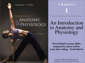
-2-connected to the right atrium were said to hold 'the breath of life,' while two vesselsconnected to the left atrium were said to contain 'the breath of death.' Egyptianiconography illustrates the story of the 'weighing of the heart' (Fig. 1), as a necessary testbefore the dead being raised by Osiris.The Ebers papyrus (1550 BCE) deals with the 'heart' theme and regards the organ as thecenter of the blood supply, with vessels attached to each limb of the body. The Egyptiansknew little of the function of the kidneys and made the heart the meeting point ofnumerous vessels that carry the body fluids - blood, tears, urine, and sperm.Fig. 1 Weighing the heart at the resurrection ceremonyGreeceThe earliest scientific medical work that has survived to this day is due to Hippocrates(460-377 BCE), who had a basic knowledge of skeletal and muscular structure, and thebeginning of the understanding of the function of some organs, such as the kidneys.Hippocrates was the first to recognize the tricuspid heart valve, which he documented inthe treatise on the heart (in the Hippocratic Body).Diogenes (of Apollonia) was a pre-Socratic philosopher who lived in the 5th century BCEand made the first systematic description of the architecture of blood vessels in man. Inthe 4th century BCE, Aristotle (Fig. 2) and several of his contemporaries produced asystem of knowledge based on the dissection of animals.Praxagoras is recognized as being the first to identify the differences between arteriesand veins. The first use of cadavers for anatomical research occurred in the 4th centuryBCE with Herophilus and Erasistratus. They could dissect still living individuals(vivisections), in the case criminals of Alexandria under the auspices of the Ptolemydynasty. Herophilus in particular synthesized anatomical knowledge more closely to
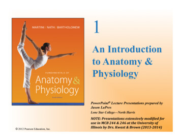
-3-current expertise than any other until that time. The Greek Theophrastus (? - 287 BCE),a disciple of Aristotle, also carried out dissections in humans. He coined the term'anatomy' (Greek, 'anna temnein'), which became generalized, encompassing the wholefield of biology that studies the form and structure of living beings, existing or extinct.Fig. 2 - Aristotle (Louvre Museum)Fig. 3 - GalenGalenAelius Galenus or Claudius Galenus (Pergamon, 129, now Bergama in Turkey - probablySicily, 200) was a prominent Greek physician (educated in Alexandria), surgeon andphilosopher. Galen contributed much to the understanding of numerous scientificdisciplines, including anatomy, physiology, pathology, pharmacology, neurology, andphilosophy. Galen's anatomy and medicine were influenced by the 'humorism' theorypracticed by many Greek physicians, including Hippocrates.Galen's theories dominated and influenced Western medical science for almost 'twomillennia.' His anatomical analyzes, based on the dissection of animals (apes, pigs, anddogs), remained incontestable until 1543 when detailed descriptions and drawings ofdissections of the human body were published in De Humani Corporis Fabrica (byAndreas Vesalius). Galen's theory for the functioning of the circulatory system still lasteduntil 1628, when William Harvey published the Anatomical Exercitatio of Motu Cordis etSanguinis in Animalibus and established that the blood circulated, and the heart propelledhim like a bomb.Galen elucidated the anatomy of the trachea and was the first to demonstrate that thelarynx generates the voice. Galen probably understood the importance of artificialventilation because one of his experiments used to inflate the lungs of a dead animal. Oneof his most significant contributions to medicine was work on the circulatory system. Hewas the first to recognize that there are differences between venous (dark) and arterial(bright) blood. With his anatomical experiments on animals, he better understood thecirculatory, nervous, respiratory and other structures. However, his work still containedmany inaccuracies. Galen believed that the circulatory system consisted of twounidirectional blood distribution systems, rather than a single unified circulation system.
-4-His understanding was that venous blood would be generated in the liver (from where itwould be distributed to the body and then consumed). Galen also postulated that thearterial blood would be produced in the heart (from where it would be distributed to thebody and then consumed). The liver and heart, then, would be responsible forregenerating the blood, completing the cycle. Galen believed in the existence of a groupof blood vessels called the rete mirabile, near the dorsal part of the brain.Unfortunately, Galen's original work was lost in time. We are aware of a fraction of hiswork thanks to the compilations made by Arab medicine, which were recovered in theRenaissance in Europe.Middle AgesAfter the decline of the RomanEmpire, the study of anatomy stagnated in ChristianEurope as it flourished in the Islamic world. The physician Persian Avicenna (980-1037)absorbed the anatomical teachings of Galen by expanding them in his "Principle ofMedicine" (1020)3.The physician Ibn Zuhr (1091-1161) was the first Arab to perform dissections in man, aswell as necropsies to study the cause of death. He recognized that scabies was causedby a parasite, a finding that was contrary to the 'mood theory' that came from the Greeks.Removal of the parasite from the patient's body produced healing and did not involve anypurging of humor, bleeds, or any other traditional treatment associated with the fourhumors. In the 12th century, the private physician of the great politician and conquerorSaladin, Ibn Jumay, also performed dissections in the human body and urged his peersto do the same to understand medicine better. Another Arab physician, Abd-el-Latif,during the famine in Egypt in 1200 observed and examined many bodies, which led himto disagree with the teachings of Galen on the formation of bones, especially the jaw andthe sacrum.Ibn al-NafisThe Arab physician Ibn al-Nafis (1213-1288) was prominent in dissections of humanbodies and performing necropsy. In 1242 he described, for the first time, the pulmonarycirculation and the coronary circulation, and was therefore considered the 'father of thecirculation theory.' Ibn al-Nafis also issued the first concept of metabolism and developednew systems of anatomy that replaced the doctrine of the four humors of Avicenna andGalen. He described the pulse, bones, muscles, intestines, sensory organs, bile ducts,esophagus, stomach, and anatomy of almost every part of the human body.This was the most important treatise on anatomy in the Islamic world until the appearance of Ibn al-Nafis in the 13thcentury, whose book dominated medical education in medieval Europe until the 16th century.3
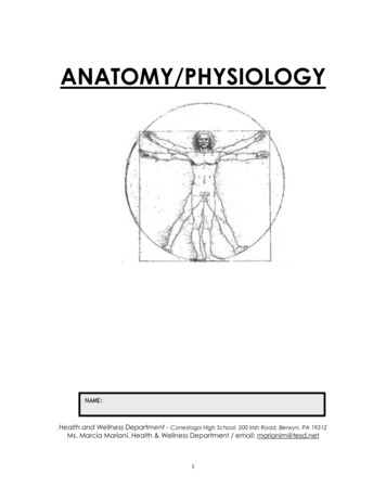
-5-Early modern anatomyThe 'Principle of Medicine' (from Avicenna, which incorporated the teachings of Galen)was translated into Latin. With this, it remained the most important text of anatomy inmedical education in Europe until the 16th century. The first significant development inanatomical knowledge in Christian Europe since the fall of Rome occurred in Bolognabetween the 14th and 16th centuries. There several anatomists dissected corpses andcontributed with more precise descriptions of organs, identifying their functions.The first significant challenge of Galen's doctrine in Europe occurred in the 16th century.Thanks to the advent of the Gutenberg press4 there was in Europe a collective effort tocirculate the works of Galen and Avicenna. Andreas Vesalius (1514-1564) published atreatise on anatomy in 1543, De humani corporis fabrica libri septem5, which was achallenge to Galen. Vesalius went from Leuven to Padua, where he can dissect bodies ofcriminals condemned to death (hangings) without fear of being persecuted. His drawingsare detailed descriptions of the human anatomy that evidence the differences about thereports made by Galen (in animals). Many other anatomists who came after Vesalius alsochallenged 'galenic knowledge,' but it still reigned for another century.Fig. 4 – Illustration of the work of Vesalius(1543) with realistic drawing of a dissection.Johannes Gensfleisch zur Laden zum Gutenberg (Mainz, 1398-1468) German inventor and graphic designer whointroduced the modern form of book printing. His invention of the mobile mechanical type for printing began theRevolution of the Press and is widely considered the most important event of the modern period. He played a key rolein the development of the Renaissance, the Reformation and the Scientific Revolution, and laid the material foundationfor the modern knowledge-based economy and the spread of mass learning.4This book, along with Nicolaus Copernicus's "On the Revolutions of the Celestial Bodies" and Isaac Newton's "PrincipiaMatematica" compose the list of the three books that most revolutionized human knowledge.5
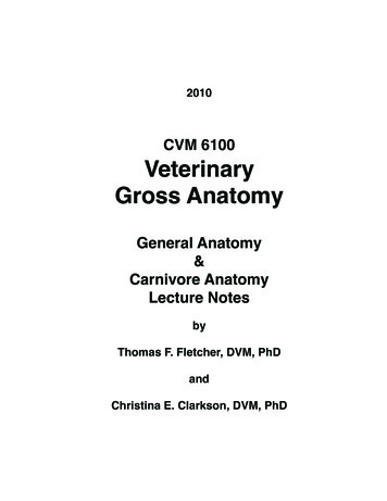
-6-The foundation of the School of Medicine of the University of Bologna was a longprocess that began in 1063, with the first Professorial Professors appearing around theyear 1170. The statute of the city of Bologna for the teaching of medicine dates from 1378.Many prominent personalities have contributed their activities through the centuries tothe celebration of the University of Bologna. Between the end of the 13th century and thebeginning of the 14th century, Mondino Dei Liuzzi (1270-1326) reestablished theAlexandrian school tradition of vivisection practice and published his observations in ananatomy book that was used until the late 16th century (in fact, can be considered thefirst book of experimental research in anatomy). In Bologna, Alessandro Achillini (14631512) studied the bile duct and the gallbladder; Berengarario da Carpi (1466-1530) wasa famous surgeon for his description of the vermiform appendix, the thymus, the functionof the heart valves (he also talked about fracture treatment and already used mercury inthe treatment of syphilis); Giulio Cesare Aranzio (1530-1589) became interested inembryology ('the duct of Arancio') and blood vessels (bodies of Arancio); CostanzoVarolio (1543-1575) studied the encephalon ('bridge of Vario').Miguel Servet (1511-1553) was a Spanish theologian, physician, cartographer, andhumanist, the first European to describe the function of pulmonary circulation. Heparticipated in the Protestant Reformation and, finally, created 'non-TrinitarianChristology' (which ignored the Holy Trinity), which was condemned by both Catholicsand Protestants. He was arrested in Geneva and burned on a stake as a heretic by orderof the Protestant governor of Geneva under the direction of John Calvin 6. Servet studiedmedicine in Paris, graduating degree in 1536. Among his professors were Sylvius, Fernel,and Guinter (who spoke of him to Vesalius as 'his most skilled assistant in dissections').Despite Servet's contribution to the knowledge of pulmonary circulation, his work was notrecognized in his time mainly because his descriptions were made in a treatise ontheology, Christianismi Restitutio, and not in a medical book. Many copies of this treatisewere quickly burned after publication in 1553 because of persecution by religiousauthorities. Only three copies were spared, but they remained hidden for many years.The most prominent 'son' of Bologna was Marcello Malpighi (1628-1694), 'father ofmicroscopic anatomy' and a defender of the use of experimental methods. Malpighigraduated as a Doctor of Medicine at the University of Bologna in 1653. He marriedFrancesca Massari, the younger sister of his anatomy professor, in 1654. Malpighi usedthe microscope to study the capillaries of the pulmonary alveoli (Fig 5), corpuscles of thekidney, the spleen corpuscles, and the epidermis follicles. His pupil, Antonio MariaValsalva, investigated the vagus nerve and created the 'Valsalva maneuver.'A succession of anatomists refined anatomical knowledge and lent their names tonumerous anatomical structures. The 16th and 17th centuries witnessed remarkableadvances in the understanding of the circulatory system, the function of the heart valvesand venous valves. Blood flow was described, and the hepatic veins and lymphatic vesselswere identified as separate portions of the circulatory system.John Calvin (1509-1564), French Christian theologian (Calvinism), was never ordained a priest. After his departure from theCatholic Church, he was the voice of the Protestant movement. He was persecuted in France and fled to Geneva in 1536, wherehe died.6
-7-17th and 18th CenturiesThe study of anatomy flourished in the 17th and 18th centuries with the advent of thepress, which facilitated the dissemination and exchange of ideas. Because the study ofanatomy was heavily based on observation and drawings (the anatomists' popularity wasrelated to their ability and talent in the drawing).William Harvey (1578-1657) was an English physician who studied at the University ofPadova, a disciple of Fabrizio d'Acquapendente (1533-1619) (who built the AnatomicalTheater in Padua, where he taught for 50 years; the exact description of the venousvalves). Harvey received his medical degree in 1602 and maintained a long friendshipwith D'Acquapendente for a lifetime. It was Harvey's interest in D'Acquapendente's work,'De Venarum Ostiolis,' which led him to study the circulation of blood.Fig. 5 – Illustration from the Malpighi book describing the 'pulmonary capillaries' (left) and theplace where Malpighi is buried in Bologna (right).Harvey was not the first anatomist to postulate that blood circulation was through thearteries and veins, but he was the first to demonstrate this fact convincingly. He also didexperiments on the function of the heart by pumping blood. Harvey's mathematicalreasoning led him to calculate the volume of blood in the body, which counteracted theGalenian theory that the blood was made in the liver.In De Motu Cordis (Fig. 6A) Harvey adapted the diagram used by his masterD'Acquapendente in De Venarum Ostiolis. There are distended veins in the forearm andthe position of the venous valves. When the vein is pressed centrally (milked), and itsextremity is closed (compressed), the vein only fills with blood when pressure is relaxed.Blood cannot be forced into the 'wrong' direction (Fig. 6B).
-8-Many famous artists studied anatomy, dissected bodies and published their drawingsfor money, from Michelangelo and Da Vinci to Rembrandt. Leonardo Da Vinci (1452-1519)learned anatomy with Andrea del Verrocchio (1435-1488). As an artist, he quickly becamemaster of topographical anatomy, designed many studies of muscles, tendons and otheranatomical parts, always seeking perfection. Being a successful artist, he was allowed todissect human bodies at the Hospital of Santa Maria Nuova in Florence and then inhospitals in Milan and Rome.ABFig. 6 – Illustration from Harvey's book on blood circulation. A - Frontispiece of the book(Thesis), B - direction of blood flow in the veins of the forearm.Leonardo Da Vinci (1452-1519) did many studies on the skeleton and muscles. We cansay that these studies are a harbinger of the modern science of biomechanics. He alsodesigned the heart and vascular system, sexual organs, and other internal organs andmade the first scientific drawing of copulation (Fig. 7) and the development of the fetus inthe uterus. He also designed the effects of aging and emotion on the human face, as wellas the 'three-dimensional anatomy' of body segments (Fig. 8). From 1510 to 1511 Da Vincicollaborated with Marcantonio della Torre (1481-1511), and together they prepared ananatomy work where Da Vinci drew more than 200 boards (which was only published in1680, 161 years after Da Vinci's death, under the title of Treaty of Painting).
-9-Many European cities such as Amsterdam, London, Copenhagen, Padua, and Parishad Anatomical Academies held by the local government. Thus, Nicolaes Tulp7, mayor ofAmsterdam, can perform numerous dissections and public demonstrations (Fig. 9). Atthat time, students traveled through Europe where bodies were available (hanged, forexample) to perform dissections (no way to keep bodies in study conditions long afterdeath and their dissection should be done quickly).Fig. 7 – Drawing by Leonardo Da Vinciillustrating copulation.Fig. 8 – Leonardo Da Vinci drew different levels ofdissection of a body (possibly that of a 100-year-old elderwhom he met still alive, dead on the same day).Europeans interested in anatomy traveled to Italy, which was the high center of theanatomy of the time. Only in Italy was it possible to dissect a woman's body, for example.Renaldus Columbus (1516-1559) and Gabriele Falloppio (1523-1562) were disciples ofVesalius. Columbus was his successor in Padua and later a professor in Rome. Columbusaccurately described the bones, the shape of the heart cavities, the pulmonary artery, theaorta, and their leaflets, and traced the course of blood from the right side to the left sideof the heart. Also, it made a good report of the encephalon and its vessels and theventricle of the larynx. The name of Columbus is also associated with the description ofthe clitoris (which he considered Amor Veneris, Vel Dulcedo Appelletur, that is, "must becalled love or sweetness of Venus"). Columbus was not the first to designate the clitorisDr. Tulp's "Anatomy Lesson" (Fig. 9) is an oil painting on canvas by Rembrandt, painted in 1632. It is one of his mostfamous and revolutionary works. The work portrays an anatomy class of Dr. Nicolaes Tulp. The body that appears inthe picture is of a marginal who had been condemned to death by robbery the day before the lesson. Anatomy lessonsreally existed and took place in amphitheaters, given by anatomist doctors.7
-10-but was one of the first to propose its role in female sexual pleasure. Fallopio describedthe uterine tubes ('Fallopian tubes').Fig. 9 – Anatomy Lesson of Dr Tulp (Rembrandt).19th centuryDuring the 19th century, the anatomists finalized and systematized the human descriptiveanatomy that they inherited from the anatomists of the preceding centuries. The increasein anatomy research has increased the demand for corpses, which led to the mistrust thatsome used obscure means to obtain them (including committing a crime8). Discipline hasalso progressed and has increasingly established connections with the histology andbiology of development, not only in man but also in other animals. The SociétéAnatomique de Paris is one of the oldest medical societies still in operation, founded byDupuytren and Lænnec in 1803, under the aegis of Emperor Napoleon Bonaparte(currently works at UFR Biomédicale des Saints-Pères (Fig. 10).Why does the heart pulse? The issue is known as myogenic theory versus neurogenictheory dominated cardiac research in the 19th century. Marie Francois Xavier-Bichat andNysten reported experiments with beheaded individuals (in Paris, 1800-1802) in whomthey caused the heart to resume pulsating using electric shock (during the FrenchRevolution it should not have been a problem to obtain bodies of beheaded9).Two Irishmen William Hare and William Burke of Edinburgh in the mid-19th century committed a series of murders inorder to sell the victims' bodies for dissection in anatomy classes.8Individual rights were suspended and daily, with popular applause, carried out publicly and en masse. The Jacobinleader Robespierre, sanctioning the summary executions, had announced that France needed no judges, but moreguillotines. The result was the death sentence of 35,000 to 40,000 people.9
-11-Fig. 10 – UFR Biomédicale des SaintsPères (Université Rene Descartes, ParisV), where the Société Anatomique de Parisstill operates today.Jan Evangelista Purkinje (1787-1869) discovered in 1839 fibers in the subendocardiumof the ventricles ('Purkinje fibers' of the conduction system of the heart). Walter Gaskell in1886 described specialized muscle fibers connecting the atria and ventricles, which whensectioned cause blockage. It also identified the area of onset of cardiac excitation in theregion derived from the venous sinus. Wilhelm His, Jr (1863-1934) examined a series ofhistological sections of the heart of human embryos and showed that there is connectivetissue surrounding a beam from the right atrium to the ventricles, the bundle of His. SunaoTawara (1873-1952) followed the 'bundle of His' (atrioventricular, AV) to its connectionwith the ‘compact’ portion of the AV node at the base of the interatrial septum. Tawaraconcluded that the 'AV connection system' originates from the AV node, penetrates theseptum (like the His bundle) and divides into right and left branches ending in the 'Purkinjefibers.' Arthur Keith (1866-1955) and Martin Flack (1882-1931) found, in 1907, a peculiarstructure at the sinoatrial junction that recalled the structure of the AV node; theyconsidered that the heart rhythm started there and called it the sinoatrial node region. In2006 and 2007 was celebrated the 100th anniversary of the anatomical discoveries of thecardiac conduction system.A unique opportunity for in vivo experiments occurred in 1882. Catharina Serafin(Prussia), a 46-year-old woman, had a chest tumor that was excised along with the leftwall of the left hemithorax, exposing the heart, which could be seen covered by a thinlayer of skin. Hugo Von Ziemssen stimulated the heart of Mrs. Serafin using electriccurrent, which altered the frequency of heartbeats. Years later, Einthoven'selectrocardiogram showed a better understanding of the electrical phenomenon thatoccurs in the contraction of the heart.Modern AnatomyTheanatomical research in the last 100 years has benefited from technologicalinnovations and the growing understanding of related sciences such as 'evolution' and'molecular biology.' Endocrinology explained the purpose of the glands that the
-12-anatomists could not tell. Sophisticated medical devices allow us to study anatomy inliving people. Today, progress in anatomy is mainly in the study of ontogenetic andphylogenetic development and the study of the function of specific structures, usingtechniques such as immunohistochemistry / confocal microscopy by laser scanning,neuronal tracers or others.Increased knowledge of cardiac anatomy and congenital heart disease led to the firstsurgery to treat congenital heart disease in November 1944 at Johns Hopkins Hospital (in1938 the persistence of ductus arteriosus had been corrected, but now for the first time,there was a specific procedure to correct a congenital heart defect). The operation wascalled the Blalock-Taussig shunt and opened the door for new methods to be attemptedin this area.Anatomy is already thoroughly documented in man, but the nonhuman anatomy is still fullof possibilities. Modern anatomists are still interested in performing experiments onanimals since they allow us to understand the primary organization of organs and theprinciples of functioning of structures with the use of advanced techniques of microscopy,physiology, cellular and molecular biology.There are still mysteries in the human body that need clarification. The current challengeof cardiovascular morphology is to characterize the exact role of stem cells10. Stem cellresearch has been a field of increasing activity since the works of Ernest McCulloch andJames Till in the 1960s at the University of Toronto. There are two different types of stemcells in mammals: embryonic stem cells, isolated from the inner cell mass of the blastocystand stem cells found in adult tissues. Possible mechanisms of stem cell activation in heartcell therapy include the generation of cardiomyocytes, stimulation and growth of newblood vessels (angiogenesis), secretion of growth factors, and possibly some othermechanism still unknown.Fig. 11 – Diagram illustrating the innumerablepossible ways of stimulating the cardiomyocyte thatlead to cardiac hypertrophy (Opie & Gersh, 2008).10Stem cells are cells found in every multicellular organism. They are characterized by the ability to renew by mitoticdivision and differentiate into a wide range of specialized cell types.
-13-ReferencesBasalla G. William Harvey and the heart as a pump. Bull Hist Med 36:467-470, 1962.Castiglioni A. Andreas Vesalius: Professor at the Medical School of Padua. Bull N YAcad Med 19:766-777, 1943.DiDio L.J. Marcello Malpighi: the father of microscopic anatomy. Ital J Anat Embryol 100Suppl 1:3-9, 1995.Dunn P.M. Galen (AD 129-200) of Pergamun: anatomist and experimental physiologist.Arch Dis Child Fetal Neonatal Ed 88:F441-443, 2003.Fogazzi G.B. The description of the renal glomeruli by Marcello Malpighi. Nephrol DialTransplant 12:2191-2192, 1997.Harvey E.D. Anatomies of Rapture: Clitoral Politics/Medical Blazons. Signs: Journal ofWomen in Culture and Society 27:315-346, 2002 (doi: 10.1086/495689)Joutsivuo T. Vesalius and De humani corporis fabrica: Galen's errors and the change ofanatomy in the sixteenth century. Hippokrates (Helsinki):98-112, 1997.MacDonald H. Procuring corpses: the English anatomy inspectorate, 1842 to 1858. MedHist 53:379-396, 2009.Mellick S. The anatomy lesson of Dr. Nicolaes Tulp painted by Rembrandt in 1632. AnzJ Surg 79:496; 496-497, 2009.Meyer A.W. Malpighi as Anatomist. Science 72:234-238, 1930.Opie LH, Gersh BJ. Drugs for the heart. Saunders-Elsevier, Philadelphia, 2008).Pranghofer S. "It could be seen more clearly in unreasonable animals than in humans":the representation of the rete mirabile in early modern anatomy. Med Hist 53:561586, 2009.Rengachary S.S., Colen C., Dass K., Guthikonda M. Development of anatomic science inthe late middle ages: the roles played by Mondino de Liuzzi and Guido da Vigevano.Neurosurgery 65:787-793; 793-784, 2009.Shoja M.M., Tubbs R.S. The history of anatomy in Persia. J Anat 210:359-378, 2007.Wiltse L.L., Pait T.G. Herophilus of Alexandria (325-255 B. C.). The father of anatomy.Spine 23:1904-1914, 1998.
treatise on anatomy in 1543, De humani corporis fabrica libri septem. 5, which was a challenge to . Galen. Vesalius. went from Leuven to Padua, where he can dissect bodies of criminals condemned to death (hangings) without fear of being persecuted. His drawings are detailed descriptions of the human anatomy that evidence the differences about the