Transcription
12/19/2018 11:00 AMFOUNDATIONAL LEARNING SYSTEMv Johnson & Johnson Services Inc. 2018 All rights reserved.092892-1812191SAY: Welcome to Module 1: Anatomy & Physiology of the Brain. This module willstrengthen your understanding of basic neuroanatomy, neurovasculature, and functionalroles of specific brain regions.1
12/19/2018 11:00 AMLesson 1: Introduction to the BrainThe brain is a dense organ with variousfunctional units. Understanding the anatomy ofthe brain can be aided by looking at it fromdifferent organizational layers.In this lesson, we’ll discuss the principle brainregions, layers of the brain, and lobes of thebrain, as well as common terms used to orientneuroanatomical discussions.2SAY: The brain is a dense organ with various functional units. Understanding theanatomy of the brain can be aided by looking at it from different organizational layers.(Purves 2012/p717/para1)In this lesson, we’ll explore these organizational layers by discussing the principle brainregions, layers of the brain, and lobes of the brain. We’ll also discuss the terms used byscientists and healthcare providers to orient neuroanatomical discussions.2
12/19/2018 11:00 AMLesson 1: Learning Objectives Define terms used to specify neuroanatomical locations Recall the 4 principle regions of the brain Identify the 3 layers of the brain and their relative location Match each of the 4 lobes of the brain with their respective functions3SAY: Please take a moment to review the learning objectives for this lesson.3
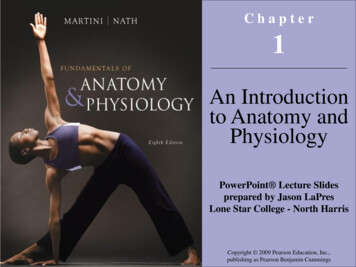
12/19/2018 11:00 AMDirectional TermsUsed in Anatomy4SAY: Specific directional terms are used when specifying the location of a structure orarea of the brain.Note that the terms anterior, posterior, superior, and inferior refer to the long axis of thebody (which is straight), and doesn’t change. (Purves 2012/p718/Figure A1/caption)However, the terms dorsal, ventral, rostral, and caudal are relative to the nervoussystem axis they are describing,which bends at the brainstem. (Purves 2012/p718/Figure A1/caption)Image Reference: (Purves 2012/p718/Figure A1)4
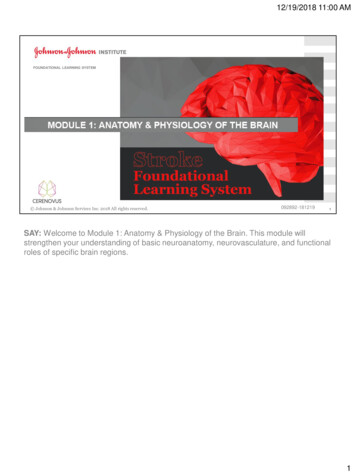
12/19/2018 11:00 AMSectional Planes Used in Anatomy Coronal: taken in the plane of the face Sagittal: taken in the plane dividing the 2hemispheres, perpendicular to the coronal plane Horizontal (axial transverse): taken parallel tothe rostral-caudal axis of the brain; so if anindividual is standing upright, these sections areparallel to the groundThis is not the case for the brainstem; remember that in humans, therostral-caudal axis of the forebrain is tilted. Therefore, a transversesection of the brainstem is perpendicular to its long axis and theposterior-anterior axis indicate the same directions5SAY: When studying the internal anatomy of the brain, you may notice different sectionalplanes. (Purves 2012/p718/c2/para1)The terms used to describe these planes help provide a common frame of reference whendiscussing the locations of brain structures. (Purves 2012/p718/c2/para1)Image Reference: (Purves 2012/p718/Figure A1B; Tortora 2009/pg 56/Figure14.4)There are 3 key planes to note, the coronal plane, the sagittal plane, and the horizontal, or axialtransverse plane. Coronal (frontal): taken in the plane of the face (Purves 2012/p718/c1/para2) Sagittal: taken in the plane dividing the 2 hemispheres, perpendicular to the coronal plane(Purves 2012/p718/c1/para2) Horizontal (also called axial sections): taken parallel to the rostral-caudal axis of the brain; soif an individual is standing upright, these sections are parallel to the ground (Purves2012/p718/c1/para2)This is not the case for the brainstem; remember that in humans, the rostral-caudal axis of theforebrain is tilted (Purves 2012/p718/c1/para2).Therefore, a transverse section of the brainstem is perpendicular to its long axis and theposterior-anterior axis indicate the same directions (Purves 2012/p718/c2/para1).5
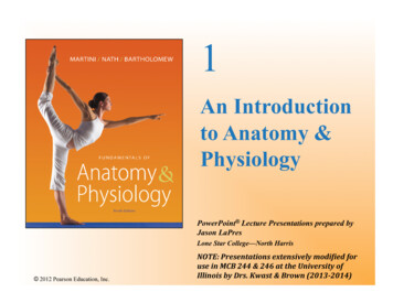
12/19/2018 11:00 AMPrincipal Regions of the BrainThere are 4 principal regions of theadult brain: Cerebrum Cerebellum Diencephalon Brainstemv6SAY: There are 4 principal regions of the adult brain, which are shown here. Let’s beginreviewing these regions starting with the cerebrum.Image Reference: (Marieb 2016/p432/Figure 12.2)6
12/19/2018 11:00 AMPrincipal Regions of the Brain: Cerebrum Largest portion of the brain– 83% of brain mass Responsible for complex aspects ofconsciousness– Memory– Personality– Intelligence7SAY: The cerebrum is the largest portion of our brain, accounting for 83% of the totalbrain mass. (Marieb 2016/p434/c2/para1)It is responsible for complex aspects of our consciousness, such as memory,personality, and intelligence. (Tortora 2009/p508/Table 14.2/c2/last para)Image Reference: (Marieb 2016/p454/Table 12.1/row 1)7
12/19/2018 11:00 AMPrincipal Regions of the Brain: CerebrumDivided into 2 cerebral hemispheres: Outer Cerebral Cortex Inner regions of grey matter andwhite matter8SAY: The cerebrum is divided into 2 cerebral hemispheres (Marieb 2016/cerebrum)each of which consists of an outer cerebral cortex, and inner regions of grey matter andwhite matter. (Tortora 2009/p514/c1/para1)Image Reference: Marieb 2016/p44/Figure 12.10b)8
12/19/2018 11:00 AMCerebrum: Cerebral Cortex 2-4 mm thick Billions of neurons Folds triple surface area– Ridges (gyri)– Grooves (sulci)9SAY: The cerebral cortex is 2 to 4 millimeters thick, contains billions of neurons, and hasfolds that nearly triple its surface area (Marieb 2016/p435/c2/para3). The folds on thesurfaces of the cerebral hemispheres are comprised of ridges of tissue, called gyri,separated by shallow grooves, called sulci (Marieb 2016/p435/c1/para 3)Image Reference: (Marieb 2016/p434/Figure 12.5)9
12/19/2018 11:00 AMCerebrum: Cerebral Cortex Areas:1. Sensory areas that deal with the perception ofsensory information2. Motor areas that control voluntary movementexecution3. Association areas that integrate complex functions Each hemisphere predominantly controlsthe opposite side of the body No functional area acts alone Conscious behavior requires entire cortex10SAY: The cerebral cortex contains (Tortora 2009/p508/Table 14.2/c2/last para) Sensory areas that deal with the perception of sensory information, Motor areas that control voluntary movement execution, and Association areas that integrate complex functions.Each hemisphere predominately controls the opposite side of the body; however, no onefunctional area of the cortex acts entirely alone—conscious behavior requires the entirecortex to play a role. (Marieb 2016/p437/c1/para 1)Image Reference: (Marieb 2016/Figure 12.7a)10
12/19/2018 11:00 AMPrinciple Regions of the Brain: Cerebellum “Little Brain” Second largest part of brain 11% of brain mass Folds increase surface area and allow for greaternumbers of neurons Functions1. Coordinates skeletal muscle contractions2. Regulates balance and posture3. May have a role in language processing and recognition11SAY: The cerebellum sits in the inferior and posterior part of the cranial cavity (Tortora2009/p507/c2/para4). It is also known as the “little brain” and is the second largest partof the brain, accounting for 11% of total brain mass (Tortora 2009/p496/c2/para1)(Marieb 2016/p434/c2/para1’450/c1/para8).Similar to the cerebrum, the cerebellum has folds that increase surface area and allowfor a greater number of neurons. (Tortora 2009/p507/c2/para4). The cerebellumcoordinates skeletal muscle contractions, regulates balance and posture, and may havea role in language processing and cognition. (Tortora 2009/p508/Table 14.2/c1/lastpara)Image Reference: (Marieb 2016/p455/Table 12.1/last row)11
12/19/2018 11:00 AMPrinciple Regions of the Brain: Diencephalon Partners with the cerebrum and cerebellum to:1.2.3.4.5.Coordinate motor functionsPlay a role in consciousnessControl and integrate the autonomic nervous systemRegulate eating and thirstcontrol body temperature and circadian rhythm12SAY: The diencephalon is surrounded by the cerebral hemispheres. (Marieb2016/p443/c2/para2) It contains structures that partner with the cerebrum and thecerebellum to coordinate motor functions, play a role in consciousness, control andintegrate the autonomic nervous system, regulate eating and thirst, and control bodytemperature and circadian rhythm. (Tortora 2009/p508/Table 14.2/c2/para1)Image Reference: (Marieb 2016/p454/Table 12.1/row2)12
12/19/2018 11:00 AMPrinciple Regions of the Brain: BrainstemLocated between the diencephalon and the spinal cord, it is composed of 3 parts:1. Midbrain2. Pons3. Medulla Oblongata13SAY: The brainstem is located between the diencephalon and the spinal cord. It iscomposed of 3 parts. (Tortora 2009/p504/c1/para1)The MidBrain, the Pons, and the Medulla OblongataImage Reference: (Purves 2012/p730/FigureA12) (Marieb 2016/p446/Figure 12.13)13
12/19/2018 11:00 AMBrainstem: MidbrainThe Midbrain contains tracks for nerveimpulses between motor areas of the cerebralcortex to the spinal cord. Some of the otherfunctions that relay through the midbrain includereflexes for the head, eyes and trunk responseto visual stimuli, and impulses for auditorystimuli (eg, the startle reflex)14SAY: The midbrain contains tracks for nerve impulses between motor areas of thecerebral cortex to the spinal cord. (Tortora 2009/p507/c1/para1) Some of the otherfunctions that relay through the midbrain include reflexes for the head, eyes, and trunkin response to visual stimuli, and impulses for auditory stimuli (e.g., the startle reflex).(Tortora 2009/p503/c2/para2)14
12/19/2018 11:00 AMBrainstem: PonsThe pons serves as a bridge to connect areas ofthe brain to one another. The pons contains areasthat relay signals for voluntary movements,equilibrium information from the inner ear, andareas that (together with the medulla oblongata)help control breathing15SAY: The pons serves as a bridge to connect areas of the brain to one another. Thepons contains areas that relay signals for voluntary movements, equilibrium informationfrom the inner ear, and areas that (together with the medulla oblongata) help controlbreathing. (Tortora 2009/p505/c2/para2)15
12/19/2018 11:00 AMBrainstem: Medulla OblongataThe medulla oblongata (also calledmedulla) contains areas that control keyvital body functions: the cardiovascular center, whichregulates the rate and force of theheartbeat and the diameter of bloodvessels he respiratory center, which adjuststhe basic rhythm of breathing. reflexes for swallowing, sneezing,vomiting, hiccupping, and coughing16SAY: The medulla oblongata (also called medulla) contains areas that control key vitalbody functions. For example, the medulla contains the cardiovascular center, whichregulates the rate and force of the heartbeat and the diameter of blood vessels. Themedulla also contains the respiratory center, which adjusts the basic rhythm ofbreathing. (Tortora 2009/p503/c2/para2) Other sections of the medulla control reflexesfor swallowing, sneezing, vomiting, hiccupping, and coughing. (Tortora2009/p503/c2/para3)16
12/19/2018 11:00 AMLayers of the BrainMeninges: Connective tissue 3 layers:– Dura Mater– Arachnoid Mater– Pia Mater Cover and protect the brain and bloodvessels, contain cerebrospinal fluid,and create partitions in the skull17SAY: Now that we have discussed the principle parts of the brain, let’s look at the layersof the brain from the outside moving in. The brain is surrounded by 3 layers ofconnective tissue membrane called meninges. These layers cover and protect the brainand blood vessels, contain cerebrospinal fluid, and create partitions in the skull. (Marieb2016/p460/c2/para1)17
12/19/2018 11:00 AMLayers of the Brain: MeningesDura Mater1 Outermost layer Strongest of the 3 meninges 2-layered sheet1. Marieb 2016/p460/c2/para118SAY: The outermost layer of the meninges, the dura mater, is the strongest of the 3meninges. The dura mater is a 2-layered sheet, the most superficial of which is attachedto the inner surface of the skull. (Marieb 2017/p460/c2/para3) In some places, the foldsof the dura mater extend into the cerebral hemispheres to limit excessive movement ofthe brain within the cranium. (Marieb 2017/p460/c2/para3)18
12/19/2018 11:00 AMLayers of the Brain: MeningesArachnoid Mater Middle layer Forms a loose brain covering,separated from the dura mater by anarrow cavity Beneath the arachnoid membrane isthe subarachnoid space, whichcontains web-like extensions thatsecure the arachnoid mater to the piamater below it Term come from the word “arachnida”,meaning spider19SAY: The middle layer of the meninges is the arachnoid mater. This layer forms a loosebrain covering that is separated from the dura mater by a narrow cavity. Beneath thearachnoid membrane is the subarachnoid space, which contains web-like extensionsthat secure the arachnoid mater to the pia mater below it. (Marieb 2017/p461/c1/para4)The term arachnoid comes from the word arachnida, meaning spider. The arachnoidmater is named for its web-like extensions. (Marieb 2017/p461/c1/para4)19
12/19/2018 11:00 AMLayers of the Brain: MeningesPia Mater Innermost layer Composed of delicate connective tissueand small blood vessels20SAY: The pia mater is the innermost layer of the meninges and closely adheres to theshape of the brain. (Marieb 2017/p461/c1/para5-c2/para1) The pia mater is composedof delicate connective tissue and many small blood vessels. (Marieb2017/p461/c1/para4) As blood vessels pass along the surface of the brain and turninward, they are covered by a loose-fitting sleeve of pia mater. (Tortora2009/p496/c2/para2)20
12/19/2018 11:00 AMLobes of the Brainv21SAY: Let’s shift our attention to another form of anatomical subdivision, the lobes of thebrain.Image Reference: (Purves 2012/p720/Figure A3a)21
12/19/2018 11:00 AMLobes of the BrainFill in the blank: The Brain has lobes.22SAY: How many lobes does the brain have?22
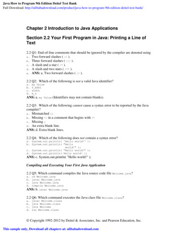
12/19/2018 11:00 AMLobes of the BrainFill in the blank: The Brain has lobes.There are 4 Lobes: Frontal Parietal Occipital Temporal23SAY: There are 4 lobes of the brain. Specific gyri and sulci on the surface of thecerebrum serve as anatomical landmarks to divide the brain into 4 lobes: frontal,parietal, occipital, and temporal. (Purves 2012/p720/c1/para3)Landmarks that divide the brain into lobes include the central sulcus that divides thefrontal lobe from the parietal lobe. Additionally note the lateral fissure, also known as theSylvian fissure, dividing the temporal lobe from the frontal and parietal lobes. (Purves2012/p720/c1/para1)Looking at the brain from the hemisected view allows us to note the parieto-occipitalsulcus separates the parietal and occipital lobes.Image Reference: (Purves 2012/p720/Figure A3a)23
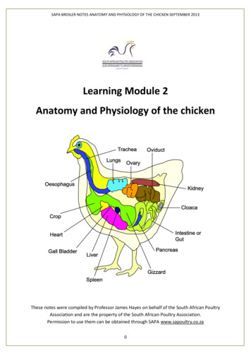
12/19/2018 11:00 AMFunctional Areas of the Brain: Frontal LobeAssociated with executive functions, motor performance, and production of language Prefrontal Cortex:– Executive functions (e.g., personality & recognizingconsequence) Primary Motor Cortex:– Motor Performance (e.g., initiation of voluntarymovement as well as the premotor andsupplementary motor areas, which coordinate theplanning and initiation of voluntary movement) Broca’s area:– Production of language– Damage causes Broca’s aphasia, a condition inwhich the patient understands many written andspoken words, but has difficulty speaking them24SAY: The frontal lobe is associated with executive functions, motor performance, andproduction of language. (Vanderah 2016/p61/c2/last para-p62/c2/para1)The first of these functions occupies a large portion of the frontal lobe and is called theprefrontal cortex. This area is involved with executive functions, which includepersonality, and recognizing future consequences of current actions. (Vanderah2016/p62/c1/para2)Areas of the frontal lobe associated with motor performance include the primary motorcortex, which is involved in the initiation of voluntary movement, as well as the premotorand supplementary motor areas, which coordinate the planning and initiation ofvoluntary movement.The motor aspects of language (ie, the production of language) are controlled by anarea in 1 hemisphere (typically the left) of the frontal lobe called Broca’s area.(Vanderah 2016/p62/c1/para3) Damage to this area causes Broca’s aphasia, acondition in which the patient understands many written and spoken words but hasdifficulty speaking them. (Dorland’s/broca’s asphasi)Image Reference: (Patton 2014/p255/Figure 10-13)24
12/19/2018 11:00 AMParietal LobeAssociated with integrating sensory information, contains the spatial orientationsystem, and is involved in the comprehension of language Primary somatosensory cortex:– Concerned with processing of proprioceptive andtactile stimuli Parietal Cortex:– Controls aspects of spatial orientation and directingattention Wernicke’s area:– Located partially in the parietal lobe– Involved in recalling, recognizing, and interpretingwords and other sounds in the process of usinglanguage25SAY: The parietal lobe is associated with integrating sensory information, contains thespatial orientation system, and is involved in the comprehension of language.(Vanderah 2016/p64/c1/para2-c2/para3)An area of the parietal lobe, called the primary somatosensory cortex, is concernedwith the processing of proprioceptive and tactile stimuli. (Vanderah 2016/p64/c1/lastpara-c2/para1)The parietal cortex controls complex aspects of spatial orientation and directingattention. (Vanderah 2016/p64/c2/para3)A large portion of the parietal lobe of one of the hemispheres (typically the left) workswith portions of the temporal lobe to comprehend language.Wernicke’s area is located partially in the parietal lobe and is involved in recalling,recognizing, and interpreting words and other sounds in the process of using language.(Dorland’s/Wernicke aphasia) (Taber’s/Wernicke).25
12/19/2018 11:00 AMOccipital LobeChiefly responsible for visual functions Contains the primary visual cortex andthe majority of the visual associationarea, involved in higher order processingof visual information26SAY: The occipital lobe is chiefly responsible for visual functions. This lobe contains theprimary visual cortex and the majority of the visual association area, involved in higherorder processing of visual information. (Vanderah 2016/p65/c2/para2)26
12/19/2018 11:00 AMTemporal Lobe Contains the primary auditory cortexand part of Wernicke’s area Medial parts of the temporal lobe areinvolved in aspects of memory andlearning27SAY: The temporal lobe contains the primary auditory cortex (involved with auditoryperception (Taber’s/auditory area) and part of Wernicke’s area. (Vanderah2016/p65/c1/para2) Recall that Wernicke’s area is involved in the comprehension oflanguage; a patient with damage to Wernicke’s area (Wernicke aphasia) would beunable to understand written, spoken, or tactile speech symbols. (Dorland’s/Wernickeaphasia) Medial parts of the temporal lobe are involved in aspects of memory andlearning. (Vanderah 2016/p65/c1/para3)27
12/19/2018 11:00 AMLobes of the BrainWhich lobe’s primary function is integrating sensory information?a)Occipital lobeb)Parietal lobec)Frontal lobed)Temporal lobe28SAY: Let’s briefly review. Which of the lobe’s primary function is integrating sensoryinformation?28
12/19/2018 11:00 AMLobes of the BrainWhich lobe’s primary function is integrating sensory information?a)Occipital lobeb) Parietal lobec)Frontal lobed)Temporal lobe29SAY: That’s correct – the parietal lobe’s primary function is integrating sensoryinformationORSAY: That’s incorrect – the parietal lobe’s primary function is integrating sensoryinformation29
12/19/2018 11:00 AMLobes of the BrainWhich lobe is associated with executive functions, motor performance, and productionof language?a)Occipital lobeb)Parietal lobec)Frontal lobed)Temporal lobe30SAY: Let’s do another review. Which lobe is associated with executive functions, motorperformance, and production of language?30
12/19/2018 11:00 AMLobes of the BrainWhich lobe is associated with executive functions, motor performance, and productionof language?a)Occipital lobeb)Parietal lobec)Frontal lobed)Temporal lobe31SAY: That’s correct – the frontal lobe is associated with executive functions, motorperformance, and production of languageORSAY: That’s incorrect - the frontal lobe is associated with executive functions, motorperformance, and production of language31
12/19/2018 11:00 AMLesson 2:Blood Supply to the BrainIn this lesson, we’ll discuss the arterial system and the branches of the aorta thatsupply our brains with this critical substance.32SAY: Let’s move on to the next topic: blood supply to the brain. In this lesson, we’lldiscuss the arterial system and the branches of the aorta that supply our brains with thiscritical substance.32
12/19/2018 11:00 AMLesson 2: Learning Objectives Define the types of arteries in the arterial system, and the flow of bloodthrough these arteries List the 3 portions of the aorta Label the aortic arch, the brachiocephalic artery, and their respectivebranches Name common types and variations in the aortic arch Recall the 2 sources for the brain’s arterial blood supply33SAY: Please take a moment to review the learning objectives for this lesson.33
12/19/2018 11:00 AMBlood Supply to the BrainHow quickly do we lose consciousness after losing blood supply to the brain?34SAY: How quickly do we lose consciousness after losing blood supply to the brain?34
12/19/2018 11:00 AMBlood Supply to the BrainHow quickly do we lose consciousness after losing blood supply to the brain?After just 10 seconds35SAY: To function correctly, our brains require a continuous supply of well-oxygenatedblood. After just 10 seconds without it, we lose consciousness. After a few minutes,irreversible damage usually begins to manifest. To account for this dependency, neuraltissues are supplied by a dense meshwork of blood vessels. (Vanderah2016/p126/c1/para1).35
12/19/2018 11:00 AMThe Arterial SystemThe arterial system is divided into 2 major circulation systems1. Systemic arteries:– Carry blood from the heart to the whole body– Direct or indirect branches from the aorta, the main andlargest artery of the body2. Pulmonary arteries:–Carry deoxygenated blood from the heart to the lungs.36SAY: The arterial system is divided into 2 major circulation systems. (SEER2018/p1/para1)The first are the systemic arteries that carry blood from the heart to the whole body.(SEER 2018/p2/para1)All systemic arteries are either direct or indirect branches from the aorta, (SEER2018/p2/para3), the main and largest artery of the body. (Patton 2014/p705/aorta) Thesecond are the pulmonary arteries that carry deoxygenated blood from the heart to thelungs. (Patton 2014/p399/c1/para2-c2/parra1) (Marieb 2016/p729/c1/para3)36
12/19/2018 11:00 AMSystemic and Pulmonary CirculationFlow of oxygenated and nutrient rich blood in systemiccirculation: Left ventricle of the heart Systemic arteries Capillaries in body tissues Returns deoxygenated bloodback to the heart throughsystemic veins37SAY: Systemic circulation carries oxygenated and nutrient-rich blood from the leftventricle of the heart, through systemic arteries to capillaries in the body’s tissues, andthen returns deoxygenated blood back to the heart through systemic veins. (Patton2014/p399/c1/para2-c2/para1) (Marieb 2016/p729/c1/para3)Image Reference: (Marieb 2016/p664/Figure 18.1)37
12/19/2018 11:00 AMSystemic and Pulmonary CirculationFlow of blood in pulmonary circulation: Right ventricle of the heart Left and right pulmonaryarteries Lungs for gas exchange38SAY: Pulmonary circulation brings blood from the right ventricle of the heart, through theleft and right pulmonary arteries, to the lungs for gas exchange. (Marieb2016/p728/c1/para1-2)38
12/19/2018 11:00 AMThe AortaThe aorta can be divided into 3 main portions1. Ascending aorta2. Aortic arch3. Descending aorta– Thoracic part– Abdominal part39SAY: The aorta can be divided into 3 main portions. (SEER 2018/p2/para3):The first portion is the ascending aorta, which begins where the aorta arises from theleft ventricle of the heart. The second part begins as it then bends into the aortic arch.Finally, the third portion arises as it proceeds downward as the descending aortathrough the upper thoracic part and the lower abdominal part. At that point, it divides intothe 2 common iliac arteries, the internal and external. (Dorlands/aorta)(Taber’s/artery/p7(external); p10(internal))Image Reference: (Dorland’s/aorta)39
12/19/2018 11:00 AMAortic Arch Brachiocephalic trunk/artery– Brachiocephalic artery or innominateartery– First and largest branch– Ascends beside the trachea Common Carotid artery Subclavian artery– Vertebral artery– Internal thoracic artery40SAY: The brachiocephalic trunk, also called the brachiocephalic artery or the innominateartery, is the first and largest branch that arises from the aortic arch. (Osborn1999/p12/c2/para2)The brachiocephalic artery ascends beside the trachea, and branches into the rightcommon carotid artery and the right subclavian artery (Osborn 1999/p12/c2/paara2)Arteries that further branch from the right subclavian artery are the right vertebral artery,the internal thoracic artery, and the thyrocervical artery. (Osborn 1999/p13/Figure 1-7)Image Reference: (Osborn 1999/p12/Figure1-6;p13/Figure 1-7)40
12/19/2018 11:00 AMAortic Arch Types Aortic arch differences– Important for designing and therapeuticinterventions Types– Type I: Distance from origin to top of thearch is 1 diameter of the left commoncarotid artery– Type II: Distance from origin to top of thearch is between 1 and 2 diameters of theleft common carotid artery– Type III: Distance from origin to top of thearch is 2 diameters of the left commoncarotid art41SAY: Knowledge of the differences in each individual’s aortic arch is important forsurgeons and interventionalists (Demertzis 2010/p588/c1/para1) in designing andoptimizing therapeutic intervention. (Demertizis 2010/p588/c2/para1).One way the aortic arch can be described is by aortic arch type, which measures thevertical distance from the origin of the brachiocephalic trunk to the top of the arch.(Demertzis 2010/p589/c2/para1)The 3 types are shown here First is the type I arch, where the distance from origin to top of the arch is 1 diameterof the left common carotid artery. Next is the Type II arch, where the distance from origin to top of the arch is between 1and 2 diameters of the left common carotid artery. Finally, there is the Type III arch, where the distance from origin to top of the arch is 2diameters of the left common carotid artery. (Demertzis 2010/p589/c2/para1)Image Reference: (Demertzis 2010/p590/Figure3)41
12/19/2018 11:00 AMBovine Aortic Arch Bovine arch refers to anatomicalvariations in the configuration of the 3vessels originating from the aortic arch Important for therapeutic interventions(e.g., stenting) Less usual variation (Figure A) More usual variation (Figure B) No resemblance to branching patternfound in cattle42SAY: The bovine arch refers to anatomical variations in the configuration of the 3 vesselsoriginating from the aortic arch. (Spacek 2012/p166/para1)Knowledge of the aortic arch branching variations is important for therapeutic interventions,especially during stenting of the left internal carotid artery, which is performed by passagethrough the bovine arch. (Spacek 2012/p166/para3)In the less usual variation, or about 7% of the population, the brachiocephalic artery trifurcatesinto the left common carotid artery, the right common carotid artery and the right subclavianartery, shown in Figure A here. (Spacek 2012/p166/para1)In the more usual variation, or about 20% of the population, the brachiocephalic artery and theleft common carotid artery share a common origin, shown in Figure B here. (Spacek2012/p166/para1)Note that the name of the bovine arch is a common misnomer in medical literature. Theanatomical configuration it describes in humans does not have any resemblance with the aorticarch branching pattern found in cattle. (Layton 2006/p1541/c1/para1).Instead, it is believed to have been named for the resemblance the pattern has to a bovine’shead and horns. (Spacek 2012/p166/para2; p167/Figure2).Image Reference: (Spacek 2012/p167/Figure 1)42
12/19/2018 11:00 AMBlood Supply to the LimbsBrachial Artery– Major artery of upper armFemoral Artery– Major artery of thigh43SAY: The brachial artery is the major arterial supply of the upper arm. (Patton2014/p395/Table 15-1) It is the continuation of the axillary artery. At the point of theelbow joint, it divides into the ulnar and radial arteries. (Stedmans/brachial artery)(Marieb 2016/p731/Figure 19.4)The femoral artery is the major arterial supply of the thigh. (Patton 2014/p395/Table 151)(Marieb 2016/p740/c2/para4) Near the knee, the femoral artery passes posteriorlyand through a gap in the muscle, where its name changes to the popliteal artery.(Figure 2.1, Table 2.1). (Marieb 2016/p740/c2/para4)Image Reference: (Marieb 2016/p731/Figure 19.4)43
12/19/2018 11:00 AMMajor Arterial Supply of Upper and Lower Extremities44SAY: These are the major arteries that supply the upper and lower extremities.Image Reference: (Patton 2015/p395/Table 15-1)44
12/19/2018 11:00 AMBlood Supply to the Brain45SAY: The brain receives its arterial blood supply from 2 sources: The internal carotid arteries, known as anterior circulation The vertebral arteries, known as posterior circulation (Brain in Morton 2011/p5/para3)Anterior and posterior circulation will be covered in greater detail in Lesson 3 andLesson 4. Here, we will begin with an overview of the arteries that make up thesecircuits.Image Reference: (Purves 2012/p736/Figure A15A)45
12/19/2018 11:00 AMBlood Supply to the Brain46SAY: The le
5 Sectional Planes Used in Anatomy Coronal: taken in the plane of the face Sagittal: taken in the plane dividing the 2 hemispheres, perpendicular to the coronal plane Horizontal (axial transverse): taken parallel to the rostral-caudal axis of the brain; so if an individual is standing upright, these sections are