Transcription
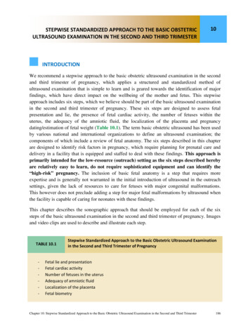
STEPWISE STANDARDIZED APPROACH TO THE BASIC OBSTETRICChapter 1010ULTRASOUND EXAMINATION IN THE SECOND AND THIRD TRIMESTERINTRODUCTIONWe recommend a stepwise approach to the basic obstetric ultrasound examination in the secondand third trimester of pregnancy, which applies a structured and standardized method ofultrasound examination that is simple to learn and is geared towards the identification of majorfindings, which have direct impact on the wellbeing of the mother and fetus. This stepwiseapproach includes six steps, which we believe should be part of the basic ultrasound examinationin the second and third trimester of pregnancy. These six steps are designed to assess fetalpresentation and lie, the presence of fetal cardiac activity, the number of fetuses within theuterus, the adequacy of the amniotic fluid, the localization of the placenta and pregnancydating/estimation of fetal weight (Table 10.1). The term basic obstetric ultrasound has been usedby various national and international organizations to define an ultrasound examination; thecomponents of which include a review of fetal anatomy. The six steps described in this chapterare designed to identify risk factors in pregnancy, which require planning for prenatal care anddelivery in a facility that is equipped and staffed to deal with these findings. This approach isprimarily intended for the low-resource (outreach) setting as the six steps described herebyare relatively easy to learn, do not require sophisticated equipment and can identify the“high-risk” pregnancy. The inclusion of basic fetal anatomy is a step that requires moreexpertise and is generally not warranted in the initial introduction of ultrasound in the outreachsettings, given the lack of resources to care for fetuses with major congenital malformations.This however does not preclude adding a step for major fetal malformations by ultrasound whenthe facility is capable of caring for neonates with these findings.This chapter describes the sonographic approach that should be employed for each of the sixsteps of the basic ultrasound examination in the second and third trimester of pregnancy. Imagesand video clips are used to describe and illustrate each step.TABLE 10.1-Stepwise Standardized Approach to the Basic Obstetric Ultrasound Examinationin the Second and Third Trimester of PregnancyFetal lie and presentationFetal cardiac activityNumber of fetuses in the uterusAdequacy of amniotic fluidLocalization of the placentaFetal biometryChapter 10: Stepwise Standardized Approach to the Basic Obstetric Ultrasound Examination in the Second and Third Trimester186
STEP ONE: FETAL LIE AND PRESENTATION IN THE UTERUSThe lie of the fetus in the uterus is defined by the orientation of the fetal spine to the maternalspine. A longitudinal lie is defined when the fetal spine is in parallel orientation to the maternalspine. A transverse lie is defined when the fetal spine is in a transverse orientation to thematernal spine, and an oblique fetal lie is defined when the fetal spine is in an oblique orientationto the maternal spine. Determining the lie of the fetus by ultrasound therefore requires obtaininga mid-sagittal plane of the fetal spine (Figure 10.1), which is a technically difficult plane toacquire for the novice ultrasound examiner. We therefore recommend that the fetal lie beinferred from determining the fetal presentation. If the fetal presentation is cephalic or breech, atechnically easy step to determine by ultrasound, then a longitudinal fetal lie can be inferred. Ifneither a cephalic nor a breech presentation is noted in the lower uterine segment on ultrasound,an oblique or a transverse fetal lie should then be suspected and an attempt for a confirmation ofsuch should be done by obtaining a mid-sagittal plane of the fetal spine (Figure 10.1) andassessing the orientation of the fetal spine to the maternal spine.Figure 10.1: Mid-Sagittal view of the fetal spine (labeled) by ultrasound in the late secondtrimester of pregnancy. This plane is used to determine fetal lie in the uterus. The location ofthe fetal head is noted for orientation purposes. See text for details.Step One-Technical Aspect of Determining Fetal Presentation in the UterusPlace the transducer transversely in the lower abdomen just above the symphysis pubis as shownin Figures 10.2 and 10.3, and angle inferiorly towards the cervix as shown in Clip 10.1. TheChapter 10: Stepwise Standardized Approach to the Basic Obstetric Ultrasound Examination in the Second and Third Trimester187
presence of a fetal head on the ultrasound monitor confirms a cephalic presentation (Figure10.4) and the presence of fetal buttocks confirms a breech presentation (Figure 10.5). Note thatthe presence of either a cephalic or a breech presentation implies a longitudinal lie of the fetus. Ifneither cephalic nor breech fetal parts are seen in the lower uterine segment on step one (Figure10.6), further evaluation is needed to assess for an abnormal fetal lie. Note that the presence of aplacenta previa is commonly associated with abnormal fetal presentation and lie.Figure 10.2: Initial transducer placement for determining fetal presentation (step 1). Note theplacement transversely in the lower abdomen just above the symphysis pubis. Uterine fundusis labeled. This picture is taken from the patient’s left side.Figure 10.3: Initial transducer placement for determining fetal presentation (step 1). Note theplacement transversely in the lower abdomen just above the symphysis pubis. This representsthe same transducer placement as in Figure 10.2, imaged from a different angle. Uterinefundus is labeledChapter 10: Stepwise Standardized Approach to the Basic Obstetric Ultrasound Examination in the Second and Third Trimester188
Figure 10.4: Step 1: determining fetal presentation. Note the transverse orientation of thetransducer. This figure shows a cephalic presentation. See text for details.Chapter 10: Stepwise Standardized Approach to the Basic Obstetric Ultrasound Examination in the Second and Third Trimester189
Figure 10.5: Step 1: determining fetal presentation. Note the transverse orientation of thetransducer. This figure shows a breech presentation. See text for details.Chapter 10: Stepwise Standardized Approach to the Basic Obstetric Ultrasound Examination in the Second and Third Trimester190
Figure 10.6: Step 1: determining fetal presentation. Note the transverse orientation of thetransducer. This figure infers the presence of a transverse or oblique fetal lie given that nofetal presenting parts (asterisk) are noted. See text for details.Chapter 10: Stepwise Standardized Approach to the Basic Obstetric Ultrasound Examination in the Second and Third Trimester191
STEP TWO: FETAL CARDIAC ACTIVITYConfirming fetal viability by noting the presence of fetal cardiac activity should be an essentialcomponent of the obstetric ultrasound examination and performed in the early steps of theexamination. In the second and third trimester of pregnancy, this is easily accomplished by thevisualization of the movements of the heart on ultrasound. Color Doppler, if available on theultrasound equipment, can help in identifying the moving heart but is not an essential part of thisstep, as the heart motion can be easily imaged on real-time grey scale ultrasound. Documentationof fetal cardiac activity can be performed by saving a movie (cine-loop) clip of the moving hearton the hard drive of the ultrasound equipment or by using M-Mode. M-Mode, which stands forMotion mode, is an application that is available on most ultrasound equipment. When M-Mode isactivated, a line appears on the ultrasound screen, which detects any motion along its path andcan be moved by the track ball. By placing the M-Mode line across the cardiac chambers, motionof the cardiac chambers can thus be documented and a still image reflecting cardiac activity canbe printed (Figure 10.7) and stored for documentation. See chapters 1 and 2 for more details.Figure 10.7: M-Mode documenting fetal cardiac activity in the second trimester ofpregnancy. Note the M-Mode line (labeled) intersecting the cardiac chambers and notethe presence of cardiac chamber contractions (labeled) in the tracing section. See textfor details. Chapters 1 and 2 provide more information on M-Mode.Chapter 10: Stepwise Standardized Approach to the Basic Obstetric Ultrasound Examination in the Second and Third Trimester192
STEP TWO - TECHNICAL ASPECT OF DETERMINING FETAL CARDIACACTIVITYPlace the transducer transversely in the lower abdomen just above the symphysis pubis as shownin Figures 10.1 and 10.2, and slide superiorly in the mid-abdomen towards the umbilicus whilemaintaining the transverse orientation of the ultrasound transducer as shown in Clip 10-2. Fetalcardiac activity can be seen along the path of the transducer in the majority of ultrasoundexaminations. If fetal cardiac activity is not seen following the steps outlined here, slide theultrasound transducer from the mid-abdomen to the right and/or the left lateral abdomen whilemaintaining the transverse orientation as shown in Clip 10.3. These steps show cardiac activitywhen present, in almost all fetal presentations.STEP THREE-NUMBER OF FETUSES IN THE UTERUSOne of the most important benefits of ultrasound in obstetrics is in its ability to identify thepresence of twins or higher order multiple pregnancy. Twin pregnancy is associated with anincreased risk of preterm delivery, preeclampsia, abnormal labor and growth restriction (seechapter 7). By identifying twin pregnancy prenatally, pregnancy surveillance can be initiated andplanning for delivery can be optimized which may significantly minimize the risk for pregnancycomplications.The diagnosis of a twin pregnancy in the second and third trimester is commonly first suspectedwhen 2 fetal heads are seen in the uterine cavity during the ultrasound examination. Confirmingthe presence of twins is thus dependent on the identification of 2 separate fetal bodies within oneuterus. A dividing membrane is seen when twin pregnancy is of the dichorionic-diamniotic ordichorionic-monoamniotic twin type. When two fetal heads are seen on ultrasound within theuterine cavity, the presence of a dividing membrane confirms the presence of a multiplepregnancy (Figure 10.8).Chapter 10: Stepwise Standardized Approach to the Basic Obstetric Ultrasound Examination in the Second and Third Trimester193
Figure 10.8: Transabdominal ultrasound of a twin pregnancy showing a thick dividingmembrane (arrow) confirming the presence of twins. A and B denote the locations ofgestational sacs for twin A and twin B respectively.Step Three-Technical Aspect of Identifying the Number of Fetuses in the UterusThe technical aspect of identifying the number of fetuses within a uterus is dependent uponmapping the entire uterine cavity by ultrasound in a systematic and standardized way, lookingfor the number of fetal heads (crania) within the uterus. If more than one fetal head is identified,confirmation of the presence of twins should then be performed. Mapping the uterus byultrasound involves scanning the uterus in its entirety both in a longitudinal and transverseapproach.Technique for mapping the uterine cavity by ultrasound, searching for 2 fetal heads, involvesimaging the uterine cavity from a transverse (part 1) and sagittal (part 2) orientations as follows:start by placing the transducer in a transverse orientation in the right lower abdomen as shown inFigure 10.9 and slide the transducer superiorly towards the upper right abdomen whilemaintaining the transverse orientation (Figure 10.10 and Clip 10.4). Repeat these steps in themid and left abdomen in similar fashion as the right abdomen (Figure 10.10 and Clip 10.4).Chapter 10: Stepwise Standardized Approach to the Basic Obstetric Ultrasound Examination in the Second and Third Trimester194
Place the transducer in a sagittal orientation in the right upper abdomen as shown in Figure10.11 and slide the transducer towards the left upper abdomen while maintaining the sagittalorientation as shown in Figure 10.12 and Clip 10.5. Repeat these steps in the lower abdomen insimilar fashion to the upper abdomen as shown in Figure 10.12 and Clip 10.5. Look for thepresence of more than one fetal head, which indicates the presence of a multiple pregnancy.When a false diagnosis of twins is made by ultrasound, a common source of error involvesimaging a single fetal head from multiple angles. This error occurs when the ultrasoundtransducer is oblique and not maintained in a perpendicular orientation to the abdomen (floor) asshown in Figures 10.9 to 10.12. It is therefore important to maintain the ultrasound transducer ina perpendicular orientation to the floor while performing this technique. When the presence of asecond fetus is suspected, provide confirmation of twin pregnancy by the identification of twoseparate bodies and a dividing membrane when present. Imaging both fetal heads or bodies in asingle image when feasible, is proof of a twin pregnancy.Figure 10.9: Initial transverse transducer placement for determining number of fetuses inuterine cavity (step 3-part 1). Note the transverse placement in the right lower abdomen.Uterine fundus is labeled.Chapter 10: Stepwise Standardized Approach to the Basic Obstetric Ultrasound Examination in the Second and Third Trimester195
Figure 10.10: Transverse transducer movement for determining number of fetuses in uterinecavity (step 3- part 1). Note that the uterine cavity is scanned inferiorly to superiorly alongtracks 1, 2 and 3, while maintaining the perpendicular orientation of the transducer to thefloor. Uterine fundus is labeled.Figure 10.11: Initial sagittal transducer placement for determining number of fetuses inuterine cavity (step 3-part 2). Note the sagittal placement in the right upper abdomen and theperpendicular orientation of the transducer to the floor. Uterine fundus is labeled.Chapter 10: Stepwise Standardized Approach to the Basic Obstetric Ultrasound Examination in the Second and Third Trimester196
Figure 10.12: Sagittal transducer movement for determining number of fetuses in uterinecavity (step 3-part 2). Note that the uterine cavity is scanned from right to left along tracks 1and 2 while maintaining the transducer perpendicular to the floor. Uterine fundus is labeled.Step Four: Placental Localization in the UterusThe presence of abnormal placental implantation such as placenta previa increases the risk ofmaternal hemorrhage before, during and after delivery. Ultrasound is the most optimal imagingmodality for the diagnosis of placental abnormalities and the diagnosis of placenta previa byultrasound is one of the most important benefits of incorporating ultrasound in prenatal care.Detailed description of placenta previa and its associated pregnancy complications are outlinedin chapter 8. This section deals with the technical aspect of placental localization by ultrasound.Step Four-Technical Aspect of Placental Localization in the UterusPlace the transducer in the sagittal orientation in the right upper abdomen, just above the uterinefundus and scan longitudinally towards the lower right abdomen as shown in (Figure 10.13).Repeat the same steps in the mid and left abdomen as shown in (Figure 10.13 and Clip 10.6). Itis important to start at the fundus of the uterus and ensure that you see the fundal contour of theuterus at the beginning of this step in order not to miss a fundal placenta. Look for the placentaon ultrasound and determine its location on the uterine wall. The placenta can be located in thefundal, anterior, posterior, right lateral, or left lateral uterine walls (Figures 10.14 – 10.18respectively). When the placenta is on the posterior uterine wall, shadowing may occur from theChapter 10: Stepwise Standardized Approach to the Basic Obstetric Ultrasound Examination in the Second and Third Trimester197
fetus especially in the third trimester, which makes placental imaging suboptimal. This can beovercome by placing the transducer on the lateral aspect of the abdomen as shown in Figures10.19 and 10.20. The lower placental edge should be assessed and its relationship to the loweruterine segment and the cervix should be evaluated and documented. If the lower placental edgeis noted to be in the lower uterine segment (Figure 10.21) and suspected to be close to orcovering the cervix, a transvaginal ultrasound is recommended in order to confirm the presenceor absence of placenta previa. The diagnosis of a placenta previa is best performed by thetransvaginal ultrasound approach.Figure 10.13: Sagittal transducer movement for determining placental localization (step 4).Note that the uterine cavity is scanned from superior (fundal region) to inferior along tracks 1,2 and 3 while maintaining the transducer perpendicular to the floor. Uterine fundus islabeled.Chapter 10: Stepwise Standardized Approach to the Basic Obstetric Ultrasound Examination in the Second and Third Trimester198
Figure 10.14: Fundal placenta (labeled) shown on ultrasound obtained from a sagittal view ofthe uterus. The uterine fundus is labeled. See text for details.Figure 10.15: Anterior placenta (labeled) shown on ultrasound obtained from a sagittal viewof the uterus. The uterine fundus is labeled. See text for details.Chapter 10: Stepwise Standardized Approach to the Basic Obstetric Ultrasound Examination in the Second and Third Trimester199
Figure 10.16: Posterior placenta (labeled) shown on ultrasound obtained from a sagittal viewof the uterus. The uterine fundus is labeled. See text for details.Figure 10.17: Right lateral placenta (labeled) shown on ultrasound obtained from a sagittalview of the uterus. Right lateral uterine wall is labeled. See text for details.Chapter 10: Stepwise Standardized Approach to the Basic Obstetric Ultrasound Examination in the Second and Third Trimester200
Figure 10.18: Left lateral placenta (labeled) shown on ultrasound obtained from a sagittalview of the uterus. Left lateral uterine wall is labeled. See text for details.Figure 10.19: Ultrasound imaging of the uterus from the lateral aspect of the abdomen forplacental localization in the third trimester when fetal shadowing obstructs view and theplacenta is on the posterior uterine wall. The uterine fundus is labeled.Chapter 10: Stepwise Standardized Approach to the Basic Obstetric Ultrasound Examination in the Second and Third Trimester201
Figure 10.20: Ultrasound imaging of the uterus from the lateral aspect of the abdomen forplacental localization in the third trimester when fetal shadowing obstructs view and theplacenta is on the posterior uterine wall. Note the orientation of the transducer, almostlateral to the floor. This represents the same transducer placement as in figure 10.19, imagedfrom a different angle. The uterine fundus is labeled.Figure 10.21: Placenta (labeled) shown on transabdominal sagittal ultrasound to be reachingthe lower uterine segment, in close proximity to the cervical internal os (asterisk - labeled). Atransvaginal ultrasound is indicated for accurate localization of placental edge. See text fordetails.Chapter 10: Stepwise Standardized Approach to the Basic Obstetric Ultrasound Examination in the Second and Third Trimester202
STEP FIVE: AMNIOTIC FLUID ESTIMATIONEstimation of amniotic fluid is an important part of the ultrasound examination. Severaltechniques for estimation of amniotic fluid have been proposed during the ultrasoundexamination including a subjective assessment, single deepest maximal vertical pocket (MVP)and amniotic fluid index (AFI). We recommend the use of the MVP technique as it is easy tolearn and has been shown to have a lower false positive diagnosis for oligohydramnios inrandomized studies (1). The term oligohydramnios (decreased amniotic fluid), which is definedby a MVP of less than 2 cm (Figure 10.22), is associated with genitourinary abnormalities in thefetus, premature rupture of the membranes, uteroplacental insufficiency and posterm pregnancy.Oligohydramnios has been linked to increased rates of perinatal morbidity and mortality (2). Theterm polyhydramnios or hydramnios (increased amniotic fluid), which is defined by a MVP ofequal to or greater than 8 cm (Figure 10.23), is often idiopathic but can be associated withgestational diabetes, isoimmunization, fetal structural or chromosomal abnormalities orcomplicated multiple pregnancy. More discussion on ultrasound and amniotic fluid assessment ispresent in chapter 9.Figure 10.22: Oligohydramnios noted on ultrasound with a maximal vertical pocket (MVP) of1.5 cm.Chapter 10: Stepwise Standardized Approach to the Basic Obstetric Ultrasound Examination in the Second and Third Trimester203
Figure 10.23: Polyhydramnios noted on ultrasound with a maximal vertical pocket (MVP) of20.2 cm. Note the presence of fetal hydrops.Step Five-Technical Aspect for Amniotic Fluid EstimationThe estimation of amniotic fluid using the MVP involves finding the single deepest pocket ofamniotic fluid in the amniotic cavity on ultrasound examination, free of cord and fetal parts, andthen measuring the greatest vertical dimension with the ultrasound transducer in sagittalorientation and perpendicular to the floor. In order to be a measurable pocket on ultrasound, thewidth of the pocket must be at least 1 cm.This step requires mapping of the uterine cavity initially in order to identify the location of theMVP. Mapping of the uterus is performed by scanning the entire amniotic cavity with thetransducer in sagittal orientation and perpendicular to the floor (Figure 10.24 and 10.25 and Clip10.7). When the deepest pocket is identified, measurement is performed by placing the calipersin a straight vertical line avoiding any cord or fetal parts in the image as shown in Figure 10.22and 10.23.Chapter 10: Stepwise Standardized Approach to the Basic Obstetric Ultrasound Examination in the Second and Third Trimester204
Figure 10.24: Accurate transducer orientation for amniotic fluid measurement for theAmniotic Fluid Index (AFI) or the Maximal Vertical Pocket (MVP) methods. Note that theultrasound transducer is in sagittal orientation and is perpendicular to the floor.Figure 10.25: Sagittal transducer movement for amniotic fluid assessment (step 5). Notethat the uterine cavity is scanned from right lateral to left lateral along tracks 1 and 2,while maintaining the transducer in sagittal orientation and perpendicular to the floor.Uterine fundus is labeled.Chapter 10: Stepwise Standardized Approach to the Basic Obstetric Ultrasound Examination in the Second and Third Trimester205
STEP SIX- FETAL BIOMETRYThe final step (step 6) in the basic obstetric ultrasound evaluation in the second and thirdtrimester includes fetal biometric measurements. Fetal biometric measurements of the biparietaldiameter, head circumference, abdominal circumference and femur length have been discussedin details in chapter 5 and 6, including estimation of fetal weight and the technical aspect of eachmeasurement. The reader should review these chapters for more detailed information on thissubject.Chapter 10: Stepwise Standardized Approach to the Basic Obstetric Ultrasound Examination in the Second and Third Trimester206
CLIP 10.1CLIP 10.2Chapter 10: Stepwise Standardized Approach to the Basic Obstetric Ultrasound Examination in the Second and Third Trimester207
CLIP 10.3CLIP 10.4Chapter 10: Stepwise Standardized Approach to the Basic Obstetric Ultrasound Examination in the Second and Third Trimester208
CLIP 10.5CLIP 10.6Chapter 10: Stepwise Standardized Approach to the Basic Obstetric Ultrasound Examination in the Second and Third Trimester209
CLIP 10.7Chapter 10: Stepwise Standardized Approach to the Basic Obstetric Ultrasound Examination in the Second and Third Trimester210
References:1) Chauhan S, Doherty D, Magann E, Cahanding F, et al. Amniotic fluid index vs. singledeepest pocket technique during modified biophysical profile: A randomized clinicaltrial. Am J Obstet Gynecol 2004; 191:661-8.2) The Cochrane Collaboration. Amniotic fluid index versus single deepest vertical pocketas a screening test for preventing adverse pregnancy outcome. 2009; Issue 3, pp (1 – 31).Chapter 10: Stepwise Standardized Approach to the Basic Obstetric Ultrasound Examination in the Second and Third Trimester211
STEP ONE: FETAL LIE AND PRESENTATION IN THE UTERUS The lie of the fetus in the uterus is defined by the orientation of the fetal spine to the maternal . This picture is taken from the patient's left side. Figure 10.3: Initial transducer placement for determining fetal presentation (step 1). Note the