Transcription
THE KOREAN JOURNAL ofORTHODONTICSOriginal ArticlepISSN 2234-7518 eISSN 329Prevalence of incidental maxillary sinus findings inItalian orthodontic patients: a retrospective conebeam computed tomography studyAntonio GraccoaSerena Incerti ParentibChristian IoelecGiulio Alessandri BonettibEdoardo StellinidaDepartment of Orthodontics, School ofDentistry, University of Padua, Padua,ItalybDepartment of Orthodontics, Schoolof Dentistry, University of Bologna,Bologna, ItalycDepartment of Orthodontics, Schoolof Dentistry, University of Ferrara,Ferrara, ItalyObjectives: To determine the prevalence of incidental maxillary sinus findingsin a large sample of orthodontic patients by cone-beam computed tomography(CBCT) with a wide field of view and assess the relationships of such abnor malities with age and gender. Methods: Five hundred thirteen CBCT scansobtained for orthodontic diagnosis and treatment planning in a Northern Italianpopulation (N 513; 292 female and 221 male subjects; 1,026 maxillary sinuses)were studied. The frequencies of pseudocysts and mucosal thickening of themaxillary sinus were recorded. Logistic regression analysis was used to determinethe influence of age and gender on these abnormalities. Results: Pseudocysts weredetected in 52 patients (10.1%) and 59 sinuses (5.75%). Mucosal thickening wasobserved in 206 patients (40.1%) and 258 sinuses (25.1%). Gender and age weresignificantly associated with pseudocysts (p 0.027) and mucosal thickening (p 0.001), respectively. Conclusions: Half of the orthodontic patients had incidentalmaxillary sinus findings. Men were more likely to show pseudocysts, and olderpatients (aged 41 - 60 years) were more likely to show mucosal thickening.[Korean J Orthod 2012;42(6):329-334]Key words: Computed tomography, Three dimensional diagnosis and treatmentplanningdDepartment of Pedodontics, School ofDentistry, University of Padua, Padua,ItalyReceived June 2, 2012; Revised September 8, 2012; Accepted September 10, 2012.Corresponding author: Serena Incerti Parenti.Visiting Professor, Department of Orthodontics, School of Dentistry, University of Bologna,Via San Vitale 59, 40125, Bologna, Italy.Tel 39-0512088133 e-mail serena.incerti2@unibo.itThe authors report no commercial, proprietary, or financial interest in the products or companiesdescribed in this article. 2012 The Korean Association of Orthodontists.This is an Open Access article distributed under the terms of the Creative Commons Attribution Non-Commercial 3.0) which permits unrestricted non-commercial use, distribution, andreproduction in any medium, provided the original work is properly cited.329
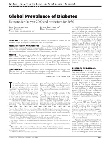
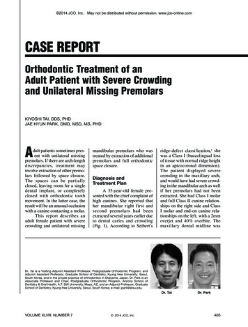
Gracco et al Maxillary sinus findings in orthodontic patientsINTRODUCTIONMATERIAL AND METHODSThe advent of cone-beam computed tomography (CBCT)represents a great improvement in the field of craniofacialimaging. 1 Unlike traditional two-dimensional (2D)radio g raphy, CBCT avoids structural superimpositionand image enlargement and distortion, thus allowingprecise three-dimensional (3D) visualization of dentaland maxillofacial structures at a lower radiation dosethan multislice computed tomography (CT). 1 Suchimaging frequently presents the dentist or orthodontistwith incidental findings and may raise concerns aboutthe need for further diagnostic tests or referral to otherspecialists.2,3Incidental findings are common in the maxillary sinus.4Although earlier studies using CBCT for dental diagnosticpurposes have already addressed this issue,2,4 only onestudy using CBCT focused on incidental maxillary sinusfindings in orthodontic patients.5 However, the studywas limited by the small sample size and imaging with anarrow field of view (FOV), which does not allow accu rate assessment of sinus pathology.The aims of this retrospective study were to determinethe prevalence of incidental maxillary sinus findings in alarge sample of orthodontic patients by CBCT with a wideFOV and assess the relationships of such abnormalitieswith age and gender.SampleThis study included 680 CBCT images obtained fororthodontic diagnosis and treatment planning at theDepart ment of Orthodontics, University of Ferrara, Italy,between January 2005 and December 2010. The protocolwas approved by the local Institutional Review Board.Images of patients with fixed orthodontic appliances,implants, or metallic prosthesis in the maxillary posteriorregion that could generate scattering phenomena, cranio facial syndromes, or cleft palate were excluded. Finally,513 CBCT scans of Northern Italian patients (N 513;292 female and 221 male subjects; 1,026 maxillarysinuses) were selected. For statistical analysis, the subjectswere divided into the following age groups4: 12 - 18,19 - 25, 26 - 40, and 41 - 60 years. The age and genderdistributions are shown in Table 1.Table 1. Age and gender distributions of the sampleCharacteristicAge (year)Data12 - 1819 - 2526 - 4041 - lCBCT scanningCBCT was performed by using a NewTom 3G volumescanner (QR srl, Verona, Italy) under the followingconditions: 12-inch FOV, 110 kV (anterior posteriorlatero lateral), 2.00 mA (anterior posterior), 1.00 mA(latero lateral), 5.4-s exposure, and 0.50-mm slice thick ness.The resulting images were elaborated by using NNTNewTom 3G software to obtain the axial sections inwhich the maxillary sinus could best be distinguished.Then, a line was traced through the greatest mesiodistaldimension of the most suitable sections and various0.2-mm-spaced coronal sections of the maxilla perpen dicular to this line were obtained.The CBCT scans were reviewed independently by 2orthodontists trained by a radiologist with experience ofover 1,000 NewTom scans at the time of the study. Thediagnostic criteria were developed on the basis of thepublished literature, and in the case of a disagreement,a consensus was reached after a discussion between theobservers. Each observer separately performed all themeasurements by using the same computer (50-inchFigure 1. CBCT scans withincidental findings of pseu docysts associated with mu cosal thickening in the rightmaxillary 329www.e-kjo.org
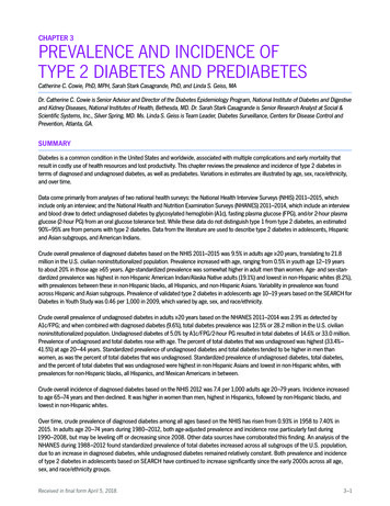
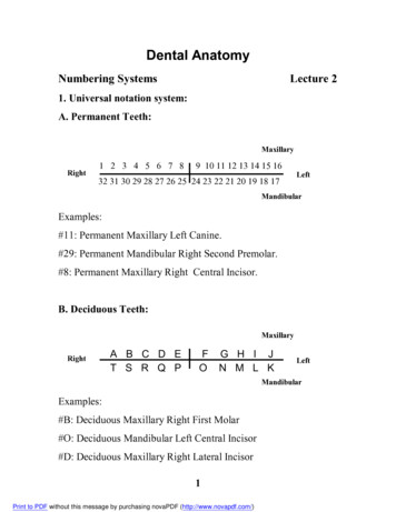
Gracco et al Maxillary sinus findings in orthodontic patientsAssessment of mucosal thickeningMucosal thickening was considered present when thethickness of the sinus mucosa was 1 mm as measuredfrom the floor of the sinus to the highest border of themucosa (Figure 2).8 For statistical analysis, this patho logywas classified according to the degree of sinus opaci fication (limited, 1/3; moderate, 1/3 - 2/3; severe, 2/3)and location (right, left, or bilateral).liquid crystal display monitors, ASUS VW246H; ASUS,Taipei, Taiwan) and screen resolution (bright ness, 300 cd/m2; resolution, 1,920 1,080).Assessment of pseudocystsPseudocysts were diagnosed as homogeneous, domeshaped, noncorticated soft tissue opacities with a smoothand well-defined outline in the maxillary sinus (Figure1).6,7 For statistical analysis, the pseudocysts were cate gorized according to their greatest diameter (small, 1cm; medium, 1 - 2 cm; large, 2 cm) and location (right,left, or bilateral).Calibration and reliabilityThe 2 observers were calibrated for evaluation by toge ther reviewing and discussing the findings of 20 sam ple CBCT images (10 per pathology) that had beenFigure 2. CBCT scans with in cidental findings of mucosalthickening in the right maxil lary sinus.Table 2. Unilateral incidental findings in the maxillary sinusesPseudocystsSmallMucosal thickeningLimitedModerateSevereLeft sinus20Medium7Large2892116Right sinus18102873015Total381741765131Table 3. Data of the unilateral and bilateral incidental findings in the maxillary sinusesIncidental findingPseudocysts95% CI of the total percentageTotalpercentageUpper limit Lower dium132152.94.81.62130.61.70.1LargeMucosal thickening evere215265.17.33.3CI, Confidence od.2012.42.6.329331
Gracco et al Maxillary sinus findings in orthodontic patientsdiagnosed as either pseudocysts or mucosal thickening byan experienced radiologist. Each examiner measured andevaluated all the CBCT images twice, on 2 days a weekapart. The intra-observer and interobserver agreementswere excellent for the linear measurements (intraclasscorrelation coefficients [ICCs], 0.92) and very good forthe degree of sinus opacification (kappa, 0.80).Statistical analysisThe descriptive statistics are presented as frequencies,and the corresponding 95% confidence intervals (CI)were also calculated. Logistic regression analysis wasperformed to determine the effects of gender and age onthe pathologies. The results are presented in terms of oddsratio increments with respect to a baseline profile, whichwas set as that pertaining to a 14-year-old female patient.Statistical analyses were performed by using SPSS forWindows software (version 16.0; SPSS Inc., Chicago, IL,USA). The level of significance was set at 0.05.RESULTSTables 2 and 3 show the results of the incidental maxil lary sinus findings. In total, 258 patients (50.3%) and 317sinuses (30.9%) had pathologies, with 38.8% and 11.5%of the subjects showing bilateral and unilateral incidentalfindings, respectively. Pseudocysts were detec ted in 52patients (10.1%) and 59 sinuses (5.75%), and 38 of the59 pseudocysts were small. Further, mucosal thickeningwas observed in 206 patients (40.1%) and 258 sinuses(25.1%), and 176 of these sinuses presented limitedopacification. Either one or both sinuses of 3 (0.6%) and26 (5.1%) patients showed large pseudocysts and severeopacification, respectively.Gender was a significant predictor of pseudocysts (p 0.027), with male subjects showing a 196.3% higherrelative risk of this pathology. Age was a significantpredictor of mucosal thickening (p 0.001), with subjectsaged 41 - 60 years showing a 401% higher odds ratio thanthose aged 12 - 18 years.DISCUSSIONIn this study, 513 CBCT images obtained for orthodonticdiagnostic purposes in a Northern Italian population wereretrospectively evaluated. Overall, half of the pa t ientshad incidental findings in the maxillary sinus. A lowerprevalence (19%) in 5,021 Finnish dental patients hasbeen reported by using panoramic radiography,9 whichis not as reliable as 3D imaging techniques for diag no sing maxillary sinus pathologies.9,10 The present re sult issomewhat in accordance with the frequencies (40.0 - 42.5%)reported by using CT and magnetic resonance imagingin the ear-nose-throat field11,12 and is confirmed by the332reported prevalence of 56.3% in 1,029 German patients byusing CBCT for dental purposes.4By focusing on CBCT scans for orthodontic purposes,Pazera et al.5 reported a prevalence of 46.8% in 134 Swisspatients (mean age, 17.5 years), but Cha et al.2 reportedairway-related findings in only 14.3% of 252 Californianpatients (mean age, 18.6 10.1 years). However, directcomparison with the current study is inappropriatebecause of the different age distribution and geographicorigin of the sample. The existing data vary among thestudies as a function of different imaging modalities anddifferent age and patient groups. Further, the definitionsof pathologic changes vary among the published articlesand are sometimes not fully described. In the presentwork, the diagnostic criteria were predefined and theobservers were calibrated to strengthen the reproducibilityof the results. Moreover, the ICCs for interobserver andintra-observer reliability were very good, confirming thereliability of the obtained data.Although pseudocysts, sometimes called mucosalcysts or polypoid mucosal thickening,4,5,8 should be dif ferentiated from mucous retention cysts, which are epi the lium-lined true cystic structures usually detected byhistologic examination,6,9 many authors use the term“retention cyst” as “pseudocyst.” 2 Their incidence onpanoramic radiographs has been reported to be between5.8% in an orthodontic population13 and 7.0 - 9.7% in ageneral dental population. 6,9 In the present study, theywere diagnosed in 10.1% of the subjects and male patientsshowed a greater risk, in accordance with previous find ings.9,13 The reported incidences on CBCT scans varybetween 6.5% and 16.4% in a dental population4,8 andfrom 3.5% to 19.4% in an orthodontic sample. However,comparisons with the published data should be madecautiously because of the lack of adequate diagnosticcriteria.2,4,5Because the normal thickness of the maxillary sinusmucosa is 0.8 - 1.0 mm, 8 we considered a thicknessof 1 mm as evidence of mucosal thickening. Theincidence of this pathology on panoramic radiographshas been reported to be between 3% in an orthodonticpopulation and 12% a general dental population. 9,10In the present study, we noted mucosal thickening in40.1% of the subjects, in accordance with the incidences(38.1% and 42.0%, res p ec t ively) reported by Ritter etal.4 and Phothikhun et al.8 among dental patients. Thisresult is higher than the prevalences (23.7% and 7.5%,respectively) reported by Pazera et al.5 and Cha et al.2 inorthodontic patients. Again, the different age and patientgroups as well as the different definitions and diagnosticcriteria for the pathologic changes may account for thesediscrepancies. Patients aged 41 - 60 years showed thehighest risk of mucosal thickening in the maxillary sinus,supporting previous reports of a higher prevalence e-kjo.org
Gracco et al Maxillary sinus findings in orthodontic patientsmucosal thicke ning in older individuals.4,8So far, no studies have directly and rigorously addressedthe prevalence of pseudocysts and mucosal thickeningby using CBCT for orthodontic purposes in an Italianpopulation. These sinus abnormalities are benign andself-limiting conditions, generally seen in asymptomaticpatients.8 No treatment is required unless they are asso ciated with symptoms or considerable radiographic ex pan sion.8 The high prevalence of incidental maxillarysinus findings in the present study is relevant to ortho dontics because of the increasing use of miniscrewsas temporary anchorage devices.14,15 As postoperativemaxillary sinusitis is one of the most concerning sequelaeafter sinus perforation,16,17 CBCT evaluation for signs ofinflammation in the maxillary sinus before miniscrewplacement is very important from both the clinical andthe medicolegal points of view. Further research is neededto determine whether these findings can be generalized toa nationally representative sample.CONCLUSIONHalf of the orthodontic patients showed incidental find ings in the maxillary sinus, with 64.4% of the pseudocystsbeing smaller than 1 cm and 68.2% of the sinusespresenting less than one-third opacification. CBCT scansobtained for orthodontic purposes should be carefullyexamined in all 3 views (axial, coronal, and sagittal) toexclude any pathologic changes in the maxillary sinusand refer the patient to a specialist if necessary. Detectionof signs of inflammation in the maxillary sinus is alsorelevant when the use of orthodontic miniscrews isplanned.REFERENCES1. Horner K, Islam M, Flygare L, Tsiklakis K, Whaites E.Basic principles for use of dental cone beam computedtomography: consensus guidelines of the EuropeanAcademy of Dental and Maxillofacial Radiology.Dentomaxillofac Radiol 2009;38:187-95.2. Cha JY, Mah J, Sinclair P. Incidental findings in themaxillofacial area with 3-dimensional cone-beamimaging. Am J Orthod Dentofacial Orthop 2007;132:7-14.3. Rogers SA, Drage N, Durning P. Incidental findingsarising with cone beam computed tomography ima ging of the orthodontic patient. Angle Orthod 2011;81:350-5.4. Ritter L, Lutz J, Neugebauer J, Scheer M, DreiseidlerT, Zinser MJ, et al. Prevalence of pathologic findingsin the maxillary sinus in cone-beam /kjod.2012.42.6.329tomography. Oral Surg Oral Med Oral Pathol OralRadiol Endod 2011;111:634-40.5. Pazera P, Bornstein MM, Pazera A, Sendi P, KatsarosC. Incidental maxillary sinus findings in orthodonticpatients: a radiographic analysis using cone-beamcomputed tomography (CBCT). Orthod CraniofacRes 2011;14:17-24.6. Carter LC, Calamel A, Haller A, Aguirre A. Seasonalvariation in maxillary antral pseudocysts in a generalclinic population. Dentomaxillofac Radiol 1998;27:224.7. Carter L, Farman AG, Geist J, Scarfe WC, Angelo poulos C, Nair MK, et al; American Academy of Oraland Maxillofacial Radiology. American Academy ofOral and Maxillofacial Radiology executive opinionstate ment on performing and interpreting diagnosticcone beam computed tomography. Oral Surg OralMed Oral Pathol Oral Radiol Endod 2008;106:561-2.8. Phothikhun S, Suphanantachat S, ChuenchompoonutV, Nisapakultorn K. Cone-beam computed tomo graphic evidence of the association between perio dontal bone loss and mucosal thickening of the maxil lary sinus. J Periodontol 2012;83:557-64.9. Vallo J, Suominen-Taipale L, Huumonen S, SoikkonenK, Norblad A. Prevalence of mucosal abnormalitiesof the maxillary sinus and their relationship to den tal disease in panoramic radiography: results fromthe Health 2000 Health Examination Survey. OralSurg Oral Med Oral Pathol Oral Radiol Endod 2010;109:e80-7.10. Bondemark L, Jeppsson M, Lindh-Ingildsen L, RangneK. Incidental findings of pathology and abnormalityin pretreatment orthodontic panoramic radiographs.Angle Orthod 2006;76:98-102.11. Gordts F, Clement PA, Buisseret T. Prevalence of sinu sitis signs in a non-ENT population. ORL J Otorhino laryngol Relat Spec 1996;58:315-9.12. Havas TE, Motbey JA, Gullane PJ. Prevalence ofincidental abnormalities on computed tomographicscans of the paranasal sinuses. Arch Otolaryngol HeadNeck Surg 1988;114:856-9.13. Bóio JA, Tanaka O, Rovigatti E, de Gruner SK. Theincidence of maxillary sinus retention cysts in ortho dontic patients. World J Orthod 2009;10:e7-8.14. Kravitz ND, Kusnoto B. Risks and complications ofor t ho dontic miniscrews. Am J Orthod DentofacialOr thop 2007;131(4 Suppl):S43-51.15. Papadopoulos MA, Papageorgiou SN, Zogakis IP. Cli ni cal effectiveness of orthodontic miniscrew im plants:333
Gracco et al Maxillary sinus findings in orthodontic patientsa meta-analysis. J Dent Res 2011;90:969-76.16. Gracco A, Tracey S, Baciliero U. Miniscrew insertionand the maxillary sinus: an endoscopic evaluation. JClin Orthod 2010;44:439-43.33417. Graham JW, Cope JB. Potential complications withOrthoTADs: classification, prevention, and treatment.In: Cope JB, ed. OrthoTADs: The clinical guide andatlas. Dallas: Under Dog Media LP; 2007. p. 9www.e-kjo.org
liquid crystal display monitors, ASUS VW246H; ASUS, Taipei, Taiwan) and screen resolution (brightness, 300 cd/ m2 Assessment of pseudocysts Pseudocysts were diagnosed as homogeneous, dome-shaped, noncorticated soft tissue opacities with a smoot

![Clinical significance of incidental [18 F]FDG uptake in the .](/img/32/s12876-016-0545-x.jpg)