Transcription
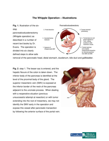
The Whipple Operation – IllustrationsFig. 1. Illustration of the sixsteppancreaticoduodenectomy(Whipple operation) asdescribed in a number ofrecent text books by Dr.Evans. The operation isdivided into six clearlydefined steps to allow saferemoval of the pancreatic head, distal stomach, duodenum, bile duct and gallbladder.Fig. 2. step 1. The lesser sac is entered, and thehepatic flexure of the colon is taken down. Theinferior body of the pancreas is identified at thelevel of the proximal body of the gland. Thesuperior mesenteric vein (SMV) is exposed atthe inferior border of the neck of the pancreasadjacent to the uncinate process. When dealingwith a reoperative situation (previousunsuccessful attempt at resection) or with tumorextending into the root of mesentery, we may notidentify the SMV early in the operation andexpose this vessel after pancreatic transectionby following the anterior surface of the portal vein.
Fig. 3. Illustration of step 2. A Kochermaneuver has been performed by firstidentifying the inferior vena cava (IVC) atthe level of the proximal portion of thetransverse segment of the duodenum(D3). One can then mobilize theduodenum and pancreatic head off ofthe IVC in a cephalad direction therebyremoving all soft tissue anterior to theIVC. Note that the Kocher maneuver iscontinued to the left lateral border of theaorta (AO).Fig. 4. Illustration of step 3.Dissection of the portahepatis begins withidentification of the commonhepatic artery (CHA), byremoval of the large lymphnode which commonly sitsanterior to this vessel. TheCHA is then followed distallyto allow identification anddivision of the right gastricartery (not shown) and the gastroduodenal artery (GDA). This allows the CHA-properhepatic artery to be mobilized off of the underlying anterior surface of the portal vein(PV). The PV is always identified prior to division of the common hepatic duct (CHD).
Fig. 5. Illustration of step 4. Theantrum of the stomach is resectedwith the main specimen by dividingthe stomach at the level of the third orfourth transverse vein on the lessercurvature. CHA, common hepaticartery; CHD, common hepatic duct;SMA, superior mesenteric artery;SMV, superior mesenteric vein.Sometimes the entire stomach ispreserved; when this is done, theoperation is called a pyloruspreserving Whipple (or pancreaticoduodenectomy).Fig. 6. Illustration of step 5.Transection of the jejunum is followedby ligation and division of its mesentery.The loose attachments of the ligamentof Treitz are taken down, and the fourthand third portions of the duodenum aremobilized by dividing their shortmesenteric vessels. The duodenumand jejunum are then reflectedunderneath the mesenteric vessels inpreparation for the final and mostimportant part ofpancreaticoduodenectomy.
Fig. 7. Illustration of step 6.The pancreatic head anduncinate process are separatedfrom the superior mesentericportal vein confluence. Thepancreas has been transectedat the level of the portal veinand the pancreatic head isreflected laterally, allowingidentification of small venoustributaries from the portal veinand superior mesenteric vein(SMV). These tributaries are ligated and divided. CHA, common hepatic artery.Fig. 8. Illustration of the continuation ofstep 6, and the final step in resection ofthe tumor. Medial retraction of thesuperior mesenteric-portal veinconfluence facilitates dissection of thesoft tissues adjacent to the lateral wallof the proximal superior mesentericartery (SMA). This is the mostimportant step in the operation from anoncologic perspective.
Fig. 9. Illustration of the importantsurgical anatomy of the superiormesenteric vein (SMV). The SMVusually bifurcates into two mainbranches, one to the ileum and oneto the jejunum. Adequate venousreturn from the small bowel requiresthat one or the other of these twomain SMV-tributaries is intact. Thejejunal branch of the SMV (oftenreferred to as the first jejunal branch)drains the proximal jejunum, travelsposterior to the superior mesenteric artery (SMA), and enters the SMV along itsposterolateral wall. Very rarely the jejunal branch will travel anterior to the SMA.
Figs. 10-11. Illustration of the final step in pancreaticoduodenectomy when segmentalvenous resection is required and the splenic vein is preserved. The intact splenic veintethers the portal vein, making a primary anastomosis impossible in most cases. Aninterposition graft is used to repair the segment of vein which is removed; the leftinternal jugular vein from the neck is used for the graft in most case2.
Fig. 12. Illustration of the different types of venousreconstruction used at the time ofpancreaticoduodenectomy. When a patch is neededwe use the saphenous vein from the leg and when aninterposition graft is needed we use the left internaljugular (IJ) vein. PV, portal vein; SMV, superiormesenteric vein; Spl V, splenic vein.
Fig. 13-14. Illustration of pancreaticojejunostomy. A two-layer, end-to-side, duct-tomucosa retrocolic pancreaticojejunostomy is performed with (when the pancreatic ductis not dilated) or without a small stent. When used, the stent (4-5 cm long) is sewn tothe pancreatic duct with a single absorbable monofilament suture.Fig. 15. Illustration ofhepaticojejunostomy. A onelayer, end-to-sidehepaticojejunostomy isperformed with 4-0 or 5-0absorbable monofilamentsutures distal to thepancreaticojejunostomy. Astent is rarely placed in thisanastomosis.
Fig. 16. Illustration of thecompleted reconstructionafterpancreaticoduodenectomy.The falciform ligament,mobilized upon opening theabdomen, is placed over thehepatic artery to cover thestump of the gastroduodenalartery, thereby separating thehepatic artery from theafferent jejunal limb. CHA, common hepatic arteryFig. 17. Our care team of nurses who work on thespecialty floor at Froedtert Hospital where patientsrecover from their pancreatic surgery. Such highly trainednurse specialists understand all aspects of this type ofcancer surgery.
The Whipple Operation – Illustrations . Fig. 1. Illustration of the six-step pancreaticoduo