Transcription
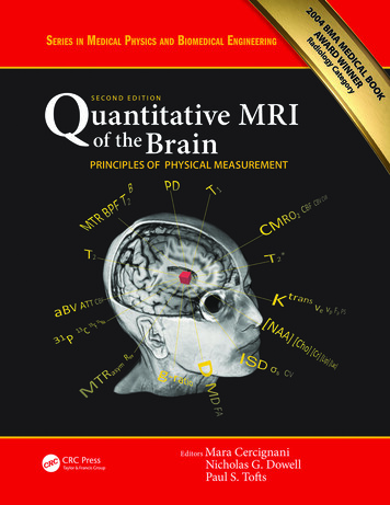
Quantitative MRIof theBrainPRINCIPLES OF PHYSICAL MEASUREMENT
Series in Medical Physics and Biomedical EngineeringSeries Editors: John G. Webster, E. Russell Ritenour, Slavik Tabakovand Kwan-Hoong NgRecent books in the series:Quantitative MRI of the Brain: Principles of Physical Measurement, Second editionMara Cercignani, Nicholas G. Dowell and Paul S. Tofts (Eds)A Brief Survey of Quantitative EEGKaushik MajumdarHandbook of X-ray Imaging: Physics and TechnologyPaolo Russo (Ed)Xun Jia and Steve B. Jiang (Eds)Todd A. Kuiken, Aimee E. Schultz Feuser and Ann K. Barlow (Eds)Emerging Technologies in BrachytherapyWilliam Y. Song, Kari Tanderup, and Bradley Pieters (Eds)Environmental Radioactivity and Emergency PreparednessMats Isaksson and Christopher L. RääfMichael G. StabinRadiation Protection in Medical Imaging and Radiation OncologyRichard J. Vetter and Magdalena S. Stoeva (Eds)Statistical Computing in Nuclear ImagingArkadiusz SitekJohn G. Webster (Ed)Radiosensitizers and Radiochemotherapy in the Treatment of CancerShirley LehnertDiagnostic EndoscopyHaishan Zeng (Ed)Medical Equipment ManagementKeith Willson, Keith Ison, and Slavik TabakovQuantifying Morphology and Physiology of the Human Body Using MRIL. Tugan Muftuler (Ed)
Quantitative MRIof theBrainPRINCIPLES OF PHYSICAL MEASUREMENTEdited byMara CercignaniNicholas G. DowellPaul S. Tofts
CRC PressTaylor & Francis Group6000 Broken Sound Parkway NW, Suite 300Boca Raton, FL 33487-2742 2018 by Taylor & Francis Group, LLCCRC Press is an imprint of Taylor & Francis Group, an Informa businessNo claim to original U.S. Government worksThe first edition of this book, entitled Quantitative MRI of the Brain: Measuring Changes Caused by Disease, was published by Wiley in 2003Printed on acid-free paperInternational Standard Book Number-13: 978-1-138-03285-9 (Hardback)This book contains information obtained from authentic and highly regarded sources. Reasonable efforts have been made to publish reliable data andinformation, but the author and publisher cannot assume responsibility for the validity of all materials or the consequences of their use. The authorsand publishers have attempted to trace the copyright holders of all material reproduced in this publication and apologize to copyright holders if permission to publish in this form has not been obtained. If any copyright material has not been acknowledged please write and let us know so we mayrectify in any future reprint.Except as permitted under U.S. Copyright Law, no part of this book may be reprinted, reproduced, transmitted, or utilized in any form by any electronic, mechanical, or other means, now known or hereafter invented, including photocopying, microfilming, and recording, or in any informationstorage or retrieval system, without written permission from the publishers.For permission to photocopy or use material electronically from this work, please access www.copyright.com (http://www.copyright.com/) or contact the Copyright Clearance Center, Inc. (CCC), 222 Rosewood Drive, Danvers, MA 01923, 978-750-8400. CCC is a not-for-profit organization thatprovides licenses and registration for a variety of users. For organizations that have been granted a photocopy license by the CCC, a separate systemof payment has been arranged.Trademark Notice: Product or corporate names may be trademarks or registered trademarks, and are used only for identification and explanationwithout intent to infringe.Library of Congress Cataloging‑in‑Publication DataNames: Cercignani, Mara, editor. Dowell, Nicholas G., editor. Tofts, Paul S., editor.Title: Quantitative MRI of the brain : principles of physical measurement /editors, Mara Cercignani, Nicholas G. Dowell, Paul S. Tofts.Other titles: Series in medical physics and biomedical engineering.Description: Second edition. Boca Raton, FL : CRC Press, Taylor & FrancisGroup, [2017] Series: Series in medical physics and biomedicalengineering Includes bibliographical references and index.Identifiers: LCCN 2017034655 ISBN 9781138032859 (hardback ; alk. paper) ISBN 1138032859 (hardback ; alk. paper) ISBN 9781315363578 (e-book) ISBN 1315363577 (e-book) ISBN 9781315363554 (e-book) ISBN 1315363550(e-book) ISBN 9781315363561 (e-book) ISBN 1315363569 (e-book) ISBN9781315363547 (e-book) ISBN 1315363542 (e-book)Subjects: LCSH: Brain--Magnetic resonance imaging.Classification: LCC RC386.6.M34 Q365 2017 DDC 616.8/04754--dc23LC record available at https://lccn.loc.gov/2017034655Visit the Taylor & Francis Web site athttp://www.taylorandfrancis.comand the CRC Press Web site athttp://www.crcpress.com
ContentsForeword. viiForeword to the First Edition.ixEditors. .xiContributors. xiiiIntroduction. xvIntroduction to the First Edition.xvii1Concepts: Measurement in MRI. 12Measurement Process: MR Data Collection and Image Analysis. . 133Quality Assurance: Accuracy, Precision, Controls and Phantoms. . 334PD: Proton Density of Tissue Water. . . 555T1 : Longitudinal Relaxation Time. . . 736T2 : Transverse Relaxation Time. 837T 2* : Susceptibility Weighted Imaging and Quantitative Susceptibility Mapping. . 978D: The Diffusion of Water (DTI). . . 1119Advanced Diffusion Models. 13910Paul S. ToftsPaul S. ToftsPaul S. ToftsShir Filo and Aviv A MezerRalf Deichmann and René-Maxime GracienNicholas G. Dowell and Tobias C. WoodSagar Buch, Saifeng Liu, Yongsheng Chen, Kiarash Ghassaban and E. Mark HaackeFrancesco Grussu and Claudia A.M. Gandini Wheeler-KingshottAurobrata Ghosh, Andrada Ianus and Daniel C. AlexanderMT: Magnetisation Transfer. 161Marco Battiston and Mara Cercignani11CEST: Chemical Exchange Saturation Transfer. . .18512MRS: 1 H Spectroscopy. . . 203Mina Kim, Moritz Zaiss, Stefanie Thust and Xavier GolayYan Li and Sarah J. Nelsonv
viContents13Multinuclear MR Imaging and Spectroscopy. 23514T1-Weighted DCE MRI. 25115Functional and Metabolic MRI. 26916ASL: Blood Perfusion Measurement Using Arterial Spin Labelling. 28317Image Analysis. 30318Future of Quantitative MRI. 325Wieland A. Worthoff, Aliaksandra Shymanskaya, Chang-Hoon Choi, Jörg Felder, Ana-Maria Oros-Peusquensand N. Jon ShahLeonidas Georgiou and David L. BuckleyClaudine J. Gauthier and Audrey P. FanLisa A. van der Kleij and Esben Thade PetersenSiawoosh Mohammadi and Martina F. CallaghanMara CercignaniAppendix A: Greek Alphabet for Scientific Use. 329Appendix B: MRI Abbreviations and Acronyms . 331Index. . . 333
ForewordIt is remarkable how much progress has occurred since the pub lication of Paul S. Tofts’ first edition in 2003. Remember thatradiology began as a highly qualitative field where shapes andpatterns were correlated with diseases. Initially, magnetic res onance imaging thrived because it could produce undistortedviews in three planes. It has evolved considerably from beautifulimages to being able to truly predict in vivo pathology. The pre vious edition of this book was ahead of its time in articulatingthe vision of quantitative MRI. How far the field will go in thefuture depends on the creativity of investigators and their abilityto understand and solve technical problems.To reliably predict biology on combinations of techniquescould change approaches to disease. There are indeed manypossibilities for the future including accuracy in differentiatingaspects of disease such as tumor versus edema, necrosis ver sus tumor, benign versus malignant, and the grail – biologicalaggressiveness of lesions. The opportunities are abundant.I will cite two examples. Think about being able to predictaggressiveness and tumor volume of prostate cancer – totallychanging how surveillance is carried out. MR may also be ableto accurately separate therapeutic tissue changes such as theeffects of radiation or immunotherapy from recurrent tumor.This is essential in detecting infiltrating neoplasms such asglioma.For those engaged in imaging, understanding how physicalprinciples can be applied to produce quantitative results hasthe prospect of transforming communications, monitoring,and reporting. It is the path where MR and medicine are head ing. Quantitation provides objective measures of activity and disease – vital to assessing the extent of pathology and deter mining the success of particular treatment protocols. The powerof quantitative MR is underscored today by its incorporation inclinical trials to demonstrate efficacy.Quantitative MRI of the Brain begins with basic discussion ofdata collection and image generation. Chapter 3 discusses qual ity assurance, precision, and accuracy. These concepts are criti cal to consistency, an essential element of quantitative imaging.Once grounded in the essentials of reproducibility, the text leadsthe reader through relaxation times and biophysical principlesof susceptibility, diffusion, and magnetisation transfer. The lastchapters focus on techniques including functional MRI, spec troscopy, and perfusion.Paul S. Tofts and his co-editors, Mara Cercignani andNicholas G. Dowell, have done a magnificent job assembling contributors with appropriate expertise and have been success ful in compiling the requisites necessary to achieve a high levelof understanding in quantitative MR. This is not a small feat andis necessary for advancing the field. Congratulations on a sig nificant accomplishment!Robert I. GrossmanNYU Langone Healthvii
Foreword to the First EditionPaul S. Tofts has succeeded brilliantly in capturing the essence ofwhat needs to become the future of radiology in particular, andmedicine in general – quantitative measurements of disease. Thisis a critical notion. The discipline of radiology started with the ability to discern the shadows that were abnormal. On chest x-rays,one could see the ‘white in the right’ and that was correlated tothe clinical diagnosis of pneumonia. It is truly amazing how longsuch descriptions were adequate and indeed, the state of the art.This transcended the modern era of cross-sectional imaging. CTand then MR heralded the ability to not only observe pathologicalstates but to specifically define and locate such conditions. Basedupon absorption of biophysical parameters combined with position, one could suggest that a particular abnormality was a strokerather than a tumour or an infection. Make no mistake about it, thiswas an incredible scientific leap and has totally changed the calculusof medicine. In the twenty-first century radiologists have becomeboth the diagnostician and the arbiter of therapeutic efficacy. Inclinical neuroscience the function of the neurologic exam has beendiminished by the unbiased, reliable nature of imaging. This hasbeen mirrored throughout the body. The preeminent role of imaging now requires a new level of metric-quantitative measurements.Why is quantitative methodology so vital? First and foremost,it is relatively unbiased compared with qualitative descriptions.Second, it lends itself easily to statistical modelling. Lastly, if performed correctly, the data can be pooled over multiple centres toprovide power regarding a clinical trial or longitudinal study.Thus, the natural history of a disease such as multiple sclerosis may be ascertained by a time-dependent study. This was firstmade apparent when the FDA in the United States approvedthe use of interferon beta-1b in 1993 based upon MRI data thatrevealed a decrease in disease activity and lesion burden. Theeffect of the drug could not be ascertained from the clinicalmeasure of disability, the acknowledged gold standard for multiple sclerosis. Approval of interferon beta changed the courseof clinical treatment trials. What has ensued is a discussion ofsurrogate markers in imaging, sensitivity, specificity, reproducibility, etc. The bottom line is the emergence of the mandatoryneed to incorporate quantitative imaging techniques into treatment trails.This book addresses the measurement process, the measures, what the measures mean biologically, and image analysismethodology. Any physician/scientist participating in a scientificstudy or clinical trial must be familiar with the concepts elucidatedin this book. Although the text is focused on the brain, the concepts pertain to any imaging study. How to ensure that your resultswill stand the test of the critical review is the underlying theme ofthe first section on the concept of measurement in MR. Thoroughknowledge of the principles of accuracy, precision, and qualityassurance are essential to the writing of any imaging proposal andthe subsequent performance of the study.The second section is focused on the metrics themselves. Here,there are lucid discussions of MR parameters that are the windowson the pathological processes we wish to study. This is complete andcovers the intrinsic MR parameters (T1, T2, PD), diffusion, magnetisation, transfer, spectroscopy, dynamic contrast, perfusion, andfMRI. To appreciate the strengths and limitations of these measuresenables the reader to identify optimal parameters for particularstudies. It also assists in the interpretation of the current literature.The section offers a complete survey of all of the metrics used inclinical MR today.The chapter on the biological significance of the MR parametersin multiple sclerosis translates the imaging parameters to theirbiological correlates. This is important, for if the measures are justabstract it is hard to argue for their implementation.The last major section of the book deals with the topics of registration of images and other measures, including atrophy,texture, and volumetric analysis. Image registration is fundamental when performing any longitudinal analysis. Just thinkabout it. When a radiologist is asked if a lesion has changedon two different studies, one must be careful that the apparentchange is not the result of technical differences (slice alignment,slice thickness, etc.).I was honoured to be asked by Professor Tofts to write thisforeword. In my opinion this text is a beautifully executed work,capturing what is essential for radiologists and scientists to understand about quantitative MR measures. There is no more qualified individual up to this task than Dr. Tofts. He is a lucid andmost thoughtful scientist. I wish to extend my congratulations tohim and the other authors on this effort. This book will becomea classic and the first of many on this significant topic.Robert I. GrossmanNew York University School of Medicineix
EditorsMara Cercignani is professor of Medical Physics at the Brightonand Sussex Medical School (BSMS). She has worked in the field ofMRI since 1998, and received her Doctorate from University CollegeLondon in 2007. Before moving to BSMS in 2011, she worked at SanRaffaele Hospital in Milan (1998–2002), the Institute of Neurologyin London (2002–2007) and Santa Lucia Foundation in Rome (2007–2011). Her main research interests lie with the field of quantitativeMRI, spanning from diffusion MRI to quantitative magnetisationtransfer imaging.Nicholas G. Dowell is a lecturer in Imaging Physics at the Brightonand Sussex Medical School (BSMS). He received his Doctorate insolid-state nuclear magnetic resonance from the University of Exeterin 2004 before moving to the Institute of Neurology, UniversityCollege London, to work on quantitative magnetic resonance imaging. He moved to the newly opened Clinical Imaging Sciences Centreat BSMS in 2007. His research interests lie principally with quantitative magnetisation transfer, diffusion-weighted imaging, data modelling and analysis techniques in the brain.Paul S. Tofts is emeritus professor at the Brighton and SussexMedical School (BSMS). After obtaining a BA in Physics fromOxford University in 1970, the new University of Sussex provided anideal contrasting environment where his D Phil was in experimentalNMR studies of helium at low temperature. When biomedical NMRhardly existed, from 1975 he researched radioisotope and CT imaging at the Royal Postgraduate Medical School, London. At UniversityCollege London, in 1978, he developed a prototype 31P NMR machinefor newborn babies and started a career in quantitative MR with thefirst measurement of absolute metabolite concentrations in vivo.An early MRI machine in 1985 devoted to multiple sclerosisstudies at the Institute of Neurology, Queen Square (now partof University College London), enabled Paul to develop a wholerange of quantitative imaging techniques and to edit the first book on quantitative MRI. Analysisof dynamic Gd-enhanced image data enabled the quantifying of blood–brain barrier leakage, andhis mathematical model is now used extensively.The new imaging centre at BSMS attracted Paul to return in 2006 to Sussex as the foundationchair of Imaging Physics until 2009. He has 215 publications, 15,000 citations and an h-factor of 62.xi
ContributorsDaniel C. AlexanderCentre for Medical Image Computing(CMIC)Department of Computer ScienceUniversity College London (UCL)London, UKMarco BattistonDepartment of NeuroinflammationUCL Institute of NeurologyUniversity College London (UCL)London, UKSagar BuchThe MRI Institute for Biomedical ResearchWaterloo, ON, CanadaDavid L. BuckleyDivision of Biomedical ImagingUniversity of LeedsLeeds, UKMartina F. CallaghanWellcome Trust Centre for NeuroimagingUCL Institute of NeurologyUniversity College London (UCL)London, UKMara CercignaniDepartment of NeuroscienceBrighton and Sussex Medical SchoolBrighton, UKYongsheng ChenDepartment of RadiologyWayne State UniversityDetroit, MI, USAChang-Hoon ChoiInstitute of Neuroscience and Medicine(INM-4)Forschungszentrum JülichJülich, GermanyRalf DeichmannBrain Imaging CenterGoethe-UniversityFrankfurt/Main, GermanyNicholas G. DowellDepartment of NeuroscienceBrighton and Sussex Medical SchoolBrighton, UKAudrey P. FanRichard M. Lucas Center for ImagingStanford UniversityStanford, CA, USAJörg FelderInstitute of Neuroscience and Medicine(INM-4)Forschungszentrum JülichJülich, GermanyShir FiloEdmond and Lily Safra Center for BrainSciences (ELSC)The Hebrew University of JerusalemJerusalem, IsraelClaudia A.M. GandiniWheeler-KingshottDepartment of NeuroinflammationUCL Institute of NeurologyUniversity College London (UCL)London, UKandBrain MRI 3T Mondino Research CentreC. Mondino National Neurological InstitutePavia, ItalyandDepartment of Brain and BehaviouralSciencesUniversity of PaviaPavia, ItalyClaudine J. GauthierDepartment of Physics/PERFORMCentreConcordia UniversityMontreal, QC, CanadaLeonidas GeorgiouDivision of Biomedical ImagingUniversity of LeedsLeeds, UKKiarash GhassabanMagnetic Resonance Innovations Inc.Detroit, MI, USAAurobrata GhoshCentre for Medical Image Computing(CMIC)Department of Computer ScienceUniversity College London (UCL)London, UKXavier GolayUCL Institute of NeurologyUniversity College London (UCL)London, UKRené-Maxime GracienDepartment of NeurologyUniversity HospitalGoethe-UniversityFrankfurt/Main, GermanyFrancesco GrussuDepartment of NeuroinflammationUCL Institute of NeurologyUniversity College London (UCL)London, UKxiii
xivEwart Mark HaackeThe MRI Institute for Biomedical ResearchWaterloo, ON, CanadaandDepartment of RadiologyWayne State UniversityDetroit, MI, USAandMagnetic Resonance Innovations Inc.Detroit, MI, USAAndrada IanusCentre for Medical Image Computing(CMIC)Department of Computer ScienceUniversity College London (UCL)London, UKMina KimUCL Institute of NeurologyUniversity College London (UCL)London, UKYan LiDepartment of Radiology andBiomedical ImagingUniversity of California San FranciscoSan Francisco, CA, USASaifeng LiuThe MRI Institute for Biomedical ResearchWaterloo, ON, CanadaAviv A. MezerEdmond and Lily Safra Center for BrainSciences (ELSC)The Hebrew University of JerusalemJerusalem, IsraelContributorsSiawoosh MohammadiMedical Center Hamburg-EppendorfDepartment of Systems NeuroscienceHamburg, GermanyStefanie ThustUCL Institute of NeurologyUniversity College London (UCL)London, UKSarah J. NelsonDepartment of Radiology andBiomedical ImagingUniversity of California SanFranciscoSan Francisco, CA, USAPaul S. ToftsBrighton and Sussex Medical SchoolBrighton, UKAna-Maria Oros-PeusquensInstitute of Neuroscience and Medicine(INM-4)Forschungszentrum JülichJülich, GermanyEsben Thade PetersenDanish Research Centre for MagneticResonanceCopenhagen University HospitalHvidovre,DenmarkN. Jon ShahInstitute of Neuroscience and Medicine(INM-4)Forschungszentrum JülichJülich, GermanyAliaksandra ShymanskayaInstitute of Neuroscience and Medicine(INM-4)Forschungszentrum JülichJülich, GermanyLisa A. van der KleijUniversity Medical Center UtrechtUtrecht, The NetherlandsTobias C. WoodDepartment of NeuroimagingInstitute of Psychiatry, Psychology andNeuroscienceKing’s College London (KCL)London, UKWieland A. WorthoffInstitute of Neuroscience and Medicine(INM-4)Forschungszentrum JülichJülich, GermanyMoritz ZaissDepartment of Medical Physics inRadiologyDeutsches Krebsforschungszentrum(DKFZ)Heidelberg, Germany
Recent books in the series: Quantitative MRI of the Brain: Principles of Physical Measurement, Second edition Mara Cercignani, Nicholas G. Dowell and Paul S. Tofts (Eds) A Brief Survey of Quantitative EEG Kaushik Majumdar Handbook of X-ray Imaging: Physics and Tec
