Transcription
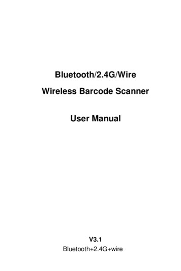
PET/CT Scanner Designs andCharacteristicsMaking a diagnosis from imagingFDG brain scanHave a seat Kermit. What I’m about to tell you mightcome as a big shock .David W Townsend PhDDepartments of Medicine and RadiologyUniversity of Tennessee, Knoxville, TNNormal CTAbnormalPETNormal PETPET/CTFDG-PETDiagnosis from anatomy .Dual-modality prototypes: 1995 - 1998MRCTDiagnosis from function.The Geneva PRT Camera Project 1990 - 1992PETPRT-1CTPETSPECTPET/MRCherry,y, Marsden et alPET/CTPET/CTBeyer, Nutt et alPET detectors (BGO)SPECT/CTHasegawa, Lang et alCTSPECTPETFirst FDG brain study on PRT-1, May 1991MouseCT detectors (Xe)PET/CT: artist’s impression
Early PET/CT recognitionPET/CT prototype design, 1998CT: 160 mAs; 130 KVp; pitch 1.6; 5 mm slicesPET: 7 mCi FDG; 2 x 15 min; 3.4 mm slicesTransverseSagittalPET/CT scannerUniversity of Pittsburgh Medical CenterSomatomSoa o AR.SPSSNM Image of the Year 1999ECATC ARTCTPETPET/CTTIME magazine, December, 2000CT consolePET consolePET/CT imaging, 1998-2001PET/CT Project, 1995 - 1998As always .the skepticsPET/CTprototypeThomas BeyerPaul KinahanClaude ComtatDavid BrasseHugo EmbertJNM Outstanding Basic Science paper 2000Ron NuttRaymond RoddyTony BrunLarry ByarsJohn YoungJohn IsraelKen BakerFinancially supported by the National Cancer Institute
skepticism: an attitude of doubt or a disposition to incredulity either ingeneral or toward a particular object (e.g. PET/CT)1999Fusing images2008 PET/CT will be too expensive PET will restrict access to CT CT: 5 min; but PET: 45 min significant artifacts with CT-AC makes poor use of CT tech time gives unnecessary CT radiation requires CT and NM physicians results in two separate reports NM and radiology conflictsSpecific biomarkers: now costs less than PET-only majority perform PET/CT scans PET/CT now takes 5-10 min total CT-AC now used routinely now dual certified PET/CT techs standard low dose CT protocols more dual-boarded physicians many places generate one report progress resolving most issues .68Ga-DOTATOCCTxyFused image accuratelylocalizes uptake into alymph node and thusdemonstrates spread ofdisease. Fused imagescanPETimprove staging ofhead and neck cancerθφz νSoftwarePET/CT image different aspects of disease localize functional abnormalities give added value to CT and PET identify non-specific tracer uptakeHardwareAnatomy of a PET/CT scannerPETBrainGantry dimensions:228 cm x 200 cm x 168 cmLungs168 cm80 cmCT rotation: 0.4 s; 16 sliceHeartLiver/spleenPET/CT200 cmKidneysSpineBladderFDG: non-specific biomarkerfunctional anatomyCT58 year-old male. History of removal of carcinoidtumor. He then presented with a bone lesion readas degenerative bone disease from a bone scan.CT and MR were negative. Findings from thePET/CT changed patient management.Michael Hofmann, MDPET156 cmDual-modality imaging range
PET/CT patient support designsPET/CT design choicesCT parametersPET parametersdetectors:ceramic; 1 – 24scintillator:BGO; GSO; LSO; LYSOslices:4, 6, 8, 16, 40, 64detector size:4 x 4 mm; 6 x 6 mmtrans. FOV:45 – 50 cmtrans. FOV:55 – 60 cmrotationt ti speed:d 0.30 3 – 2.020sresolution:l i 4 – 6 mmtube current:80 – 280 mAaxial extent:15 – 22 cmheat capacity:3.5 – 6.5 MHUsepta:2D/3D; 3D onlytopogram:128 – 2000 cmattenuation:CT-basedtime /100 cm:13 – 90 spatient port:70 cmslice width:0.5 – 12 mmpeak NECR:63 @ 12 kBq/mlpatient port:70 cm(3D)CTPETFixed cantilever point; floorfloor-mountedmounted railsCT12Variable cantilever point; dual positionsPET– 160 @ 31 kBq/mlCTVariable cantilever point; support in tunnelHigh performance PET/CT scanner designPET/CT scanner status in 2008Advances in CT:Straton x-ray tubeDiscoveryST, STE, RXBiograph6, 40, 64GeminiGXL TFGXL,BGO, LYSOLSOGSO, LYSO4.7 x 6.3 x 30 mm34.2 x 6.2 x 30 mm32D/3D (septa)8, 16, 64 slice CT70 cm port15.7 cm axial FOV11.7 ns coincidencedual-position bed6.4 x 6.4 x 25 mm34 x 4 x 20 mm33D only (no septa)6, 40, 64 slice CT70 cm port21.8 cm axial FOV4.5 ns coincidencebed on rails4 x 4 x 30 mm34 x 4 x 22 mm33D only (no septa)6, 10, 16, 64 CT71.7 cm port18 cm axial FOV6 ns coincidencebed supported in tunnelSceptreP3LSO6.4 x 6.4 x 25 mm33D only; rotating4 slice CT increased number of axial slices faster gantry rotation times incorporation of dual Straton x-ray tubes very fast scan times for cardiac applications improved use of the radiation dose (TCM, AEC)Advances in PET:AquiduoLSO4 x 4 x 20 mm316 slice CTgantry on railsPETStationary bed; gantries travel on rails new faster scintillators (LSO, LYSO) higher spatial resolution detectors increased sensitivity from extended AFOV overall improved count rate performance iterative reconstruction, accurate system model improved SNR from Time-of-Flight (TOF)
Light outputAdvances in scintillatorsBphototubesCDγ (511 keV)10LSOBGO GSO5scintillatorLSOLYSO7.136.77.47.1Effective Z746166.465.4Decay 258080Timing (ps)Light (ph/MeV)% NaIGEMINI TFImproving signal-to-noise: time-of-flight (TOF)Detector BΔt [(d d1) – (d-d1)]/c ;Patient outlineNaItBdCourtesy Matthias Egger PhD, Philipse d1 c Δt/2SNRTOF (D/Δd) · SNRnon-TOFd1100200300 400Time (ns) after interactionGSODensity (g/ml)69%35%20%4.3%00BGOLSO:NaI:GSO:BGO:e-tAΔt: tB - tADetector Aδt (ps)δx (cm)SNR*1001.55.23004.53.05007.52.3120018.01.5* SNR gain for 40 cm phantom SNRTOF / SNRnon-TOFGemini TFCourtesy Joel Karp PhD, University of PennsylvaniaLYSO4 x 4 x 22 mm3 (LYSO)3D only (no septa)Brilliance 16 CT70 cm port18 cm axial FOV585 ps timingPET detector design Detector design:Crystals:Coincidence time window:Lower level discriminator:Acquisition mode:PIXELAR with continuous lightguide28,336; LYSO: 4 mm x 4 mm x 22 mm3.8 ns440 keVSustained high-rate listmodeTOF PET performance Timing resolution:Sampling rate:Effective sensitivity gain:Effective system sensitivity:Peak effective NEC (1R):650 pslocalization accuracy of 9.75 cm25 ps2 – 4 x depending on patient size 14,400 cps/MBq @ 10 cm 210 kcps* @ 16 kBq/ml*assuming TOF SNR gain (non-TOF: 105 kcps)Non-TOFTOF60 s scan durationRectal carcinoma,with metastaseslocated in themesentery andbilateral iliac chainsmore clearly seenwith TOF.114 kg; BMI 38.112 mCi; 2 hr pi3min/bed positionNon-TOFTOF
Discovery STECourtesy Osama Malawi PhD, MD Anderson Cancer CenterCrystal dimensions, mm4.7 x 6.3 x 30Crystal detectors/block6x8Number of blocks280Number of block rings4Detector blocks/ring70Number of crystals/ring560Transverse resolution@ 1 cm (mm)5.1 (2D), 5.2 (3D)Transverse resolution@ 10 cm (mm)5.6 (2D), 5.6 (3D)Axial resolution@ 1 cm (mm)4.7 (2D), 5.4 (3D)6.0 (2D), 6.0 (3D)Number of detector rings24Axial resolution@ 10 cm (mm)Ring diameter88.6 cmSystem sensitivity – 3D8.47 cps/kBqSystem sensitivity – 2D2.2 cps/kBqPeak NECR – 2D (kcps)87.9 kcps@ 44.9 kBq/ccPeak NECR – 3D (kcps)75.1 kcps@ 12.8 kBq/ccTotal number of crystals13,440Number of slicesPET: 47; CT: 8,16Plane spacing3.27 mmNumber of PMTs280 quad PMTsPhysical axial FOV15.7 cmTransverse FOV70 cmEffective AFOV (cm)12.5 (3D), 13.7 (2D)Scatter fraction – 3D34%9.6 nsec375-650 (2D)425-650 (3D)Scatter fraction – 2DDetector materialBGO511 keV Stopping power95%Energy window setting,keVHygroscopicNoLow BMIScan duration: 18 min6 beds; 3 min/bed17.7 mCi; 60 min pi; 2D71 kg (156 lb) patientHigh BMIScan duration: 18 min6 beds; 3 min/bed19.2 mCi; 60 min pi; 2D113 kg (249 lb) patientBiograph PET CT with TrueVImproved sensitivity with Biograph TrueV cylindrical scanner geometryphototubes thicker LSO crystals 4 rings of 13 x 13 LSO block detectorsscintillator 4 mm x 4 mm x 20 mm pixelsB30 mm LSO volume increase: 50% sensitivity increase:Courtesy Osama Malawi PhD, MD Anderson Cancer Center21%Coincidence window20 mmDiscovery STECD 32,448 individual pixelsγ (511 keV) 109 transaxial image planes 21.8 cm axial field-of-view40%planar sensitivity 192 PMTs extended axial FOV16.2 cm patient port: 70 cm21.8 cm timing: LSO volume increase: 33% sensitivity increase:78%3D (no septa)volume sensitivity4.5 ns resolution: 4.2 mm NECmax:160 kcps LLD:425 keVTotal PET scan duration: 3 min6 beds; 0.5 min/bed; HD recon10.5 mCi; 105 min post-injection
Spatial resolution (Γ)NEMA NU-2 2001 performance measurementsScatter (%)(425 keV – 650 keV)Scatter fractionBiograph TPBiograph TP34Peak NECR (kcps)3.38.6 mCi; 60 min uptake6.4 mm x 6.4 mm8x8Recovery CoefficientsBiograph96 @ 34 kBq/mlBiograph1.3D detector ring diameter (mm)b block decoding factor ( 1 mm)Peak NECR161 @ 31 kBq/mlBiograph TP3.9d detector size (6.4 mm or 4.0 mm)r ‘effective’ positron range (mm)Activity (kBq/ml)3.74.0 mm x 4.0 mmRecovery (%)34NECR (kcps)Biograph2.0Γ 1.25 {(d/2)2 (0.0022D)2 r2 b2}Noise Equivalent Count Rate1008.722 281750374.113 x 138.66.4 mm x 6.4 mm13011.2 mCi; 90 min uptake104 mm x 4 mmSphere diameter (mm)Distortions from a circular geometryImproving the imaging system modelAttenuation-weighted OSEMi kj 1 i kj Lines-of-response become more closely spaced asdistance from center increases{(P - R)·N – S} si pi , j k 1pi i ijj, i pi, j a j iPoint spread function for the central LORs is symmetrical1Point spread function for lines-of-response at large radiusare asymmetrical and broader due to tilt of the crystal anddepth of interactionattenuationtttiOrdinary Poisson OSEMi kj 1 1 p i kj i, j 1 i p i i, j n a ii ns si pi , j i kj ai (ri ni ci ) j randomsAW-OSEM linearly interpolates betweensinogram bins ( )Prompts (P)scatterPSF interpolates between sinogram binswith measured spatially-variant pointspread functions ()-12-10-8-6-4-2024681012The inclusion of the point spread function allows the simulation to bettermatch the data with the effect of improving resolution and lowering noise
HD PET: modeling the system PSFAdvances in reconstruction techniquesUT Molecular ImagingFORE 2D OSEM3DRP7 mm smoothingthi2D OSEM2i/14s; no smoothing2i/14thi3D OSEM2i/14s; no smoothingHD2i/14s; no smoothing3D OSEM88 kg (194 lbs), BMI 253D-OSEM(2mm filter)HD PET(0mm filter)Scan duration: 18 min6 beds; 3 min/bed10.1 mCi; 88 min post-injection168 x 168 matrix; CT-ACHD PETBrainWhole bodyReconstruction algorithms and SUVSUV @ 60 minSUV @ 90 min2D AWOSEM 4i/8s 168 (5 mm)4.596.942D AWOSEM 4i/16s 168 (5 mm)4.557.043D AWOSEM 4i/8s 168 (5 mm)4.467.183D AWOSEM 4i/8s 336 (5 mm)5.197.183D AWOSEM 4i/8s 336 (none)5.478.00Reconstruction method#1#3FBP2D: 1i / 8s2D: 3i / 8s2D: 5i / 8s2D: 3i / 16s3D: 3i / 8sSUVmax / SUVmeanTumor#19.7 / 6.68.7 / 6.19.5 / 6.49.8 / 6.69.9 / 6.69.6 / 6.5#25.9 / 4.23.8 / 2.85.2 / 3.85.9 / 4.25.6 / 4.15.7 / 3.9#310.0 / 6.48.2 / 5.09.0 / 5.710.1 / 6.29.9 / 6.610.0 / 5.9 29% SUV changehforf differentdifft reconstructionst ti 40% SUV change at two different time pointsContrast Mean ROI hot spotContrast#2Mean ROI backgroundIterations
TOF prototype: SNR and detectability6050PSF40PSF TOF2.430PSF20PSF TOF100246802Iteration numberDetectability46Iteration number8SNR0.6Body2.200.4( S - B ) / σB0.2SNR (PSF TOF) / SNR(PSF)SNRSNR543210‐1‐2‐3‐4SNR gain as a function of BMI (SNRTOF / 0246Interation number0.8DetectabilityPSF TOF-0.4815202530354045Body Mass Index (BMI)( S - B ) / (σS2 σB2)Patient studiesReconstruction algorithms with TOF105 kg88 kg0.57BMI: 41PSFFBP2D: 3i / 8s3D: PSFPSF TOFPSF0.24PSF TOF3D: TOF PSFFORE OSEMPSFPSF TOFBMI: 30
Imaging large patientsThe 2D vs 3D debate .1990NECRN(cps)at1010NECR(cps) atmCi mCi1.E 0610619982006BiographNEMA phantomBiograph TP1.E 05105y 124843e-0.015xy 79465e404060608080100100120120140160140160Weight (kg)Decrease in NECR as a function ofpatient weight. There are 15 patients59 year-old male with history for the Biograph and 9 patients for theof lymphoma. PET/CT study Biograph TP. The NECR is for a 10dose.At 7070.5kg, the204 kg;BMIshows hilar and mediastinal mCi injectedfoci that are compatible with Biograph TP NEC1R is 50% higher.metastases.260 lbs (118 kg) male 10%10%–14%15%–19%20%–24%25%–29% 30%AttenuationBe x0I I0 e-μxStrobel K, Rudy M, Treyer V, et al. Objective and subjective comparison of standard2-D and fully 3D reconstructed data on a PET/CT system. Nucl. Med. Comm. 2007;28(7):555-559.Kemp BJ, Lowe VJ, Nathan MA, et al. Clinical evaluation of sequentially acquired 2Dand 3D whole-body PET/CT scans. J Nucl. Med. 2007; 48(2):433P (abstract).x0Patient outline PA exp - μ(x) dx511 keV 511 keVDiscovery RX (LYSO)CT-based attenuation correctionx2Detector Discovery STE (BGO) and RX (LYSO) are 2D and 3DWeight (kg)3 min/bed, 6 bedsBMI 303D2D-0.0142x3 Rings (1080)4 Rings (1094)2020 Biograph PET/CT scanners are fully 3D Aquiduo and SceptreP3 are fully 3D1.E 041041031.E 0310.7 mCi, 93 min pi GEMINI PET/CT scanners are fully 3De-511 keVx1x2PET:µ(E511) ρeσc(E511)x0CT:µ(E70) ρe{σc(E70) σpe(E70, Zeff)} APB exp - μ(x) dxx1Detectorx2I I0 (PA·PB) I0 exp - μ(x) dxx2I0 I exp μ(x) dxx1x1 I· ACFρe electron density; σc(E) Compton; σpe(E) photoelectricZeff effective atomic number
CT-based attenuation correctionRespiration and CT-ACAlgorithmRespiration strategies: bi-linear scaling bone threshold: 80 HU resample CT images scale individual pixels re-project scaled image continuous shallow breathing limited breath hold (over diaphragm) breath hold CT at partial inspiration slow the rotational speedpof CTCoronalScaling functionintravenous contrastrespiration cine CT scan (4DCT)µ(Eγ) (cm-1) respiratory gated CT scan breath hold CT; gated PET scanµPET gating both CT and PET scansµCT breath hold CT; “breath hold” PETµ(Ex) (HU)oral contrastSagittalMR respiration movies courtesy of Jim Hamill PhDmetal artifactsPET/CT scan protocolPET/CT market impacttopogram 120.0 100.0 45spiral CT arms up (except neck)CT scan with breath hold acquiredMixing model: CT-based attenuation correction partial inspiration bi-linear scaling model 10 – 15 s scan timeFusion intravenous water-air /- oral contrast threshold at 60 HUmix kVp-dependentscaling 120 kVp, 140 - 160mAswater-boneCT PET0.180.16 80.0 800.14 490.12 51 60.0 41 42 40.00.1 74 37 39 39Q3Q4020202 89 101 84 94 100 99 99oral contrast some error from iv contrast10 mCi of FDG use CTattenuationor non-correctedPET CT-basedcorrection take period model-basedHounsfield units 15%2 - 3 min per bed position 168 xACF168errorreconstructionmatrix validated in human tissue4 - 11 bed positions0.08 87 83 69attenuationcorrection correctionformix0.060.04 39 13Q2 76 110 Fourier rebinningReconstruction 10 - 30 min scan duration 2D-OSEM,4i/8s; fully 3D-OSEMfused image respiratory gating 5 mm axial smoothingCT PETPET image0.020-1000 8 4 6 3 1 1 1 1 0 0 0 0 0 0 0 0.0FY 105 92 74 48 20.0 0404040505050506060606070707PET/CTPETNEMA - US Shipments ( M)-500050010001500
Mandibular cancerbiograph 16Molecular Imaging andTranslational ResearchPET/CT:- for stagingg g disease- for therapy planning- for monitoring response83 year-old female withmandibular cancer. PET/CTscan acquired pre-surgeryidentified 3 left-side positivenodes 5-12 mm in size withincreased FDG uptake. Postsurgery, pathology identified35 nodes positive for cancer.Primary (1.5 x 3.8 cm)Nodes (12 mm, 7 mm, 5 mm)5 mm lytic spine lesionLung cancerBone lesions, 6-7 mm in diameterRestaging breast cancerBiograph 6 TrueVBiograph 6Molecular ImagingProgramFeb 2007CT10.8 mCi, 92 min pi2 min/bed, 5 beds3i / 8s; 5 mm130 kVp; 50 mAsJuly 2007PET/CTPET/CTScan duration: 10 min44 year-old male (6’, 118 lbs) with recent diagnosis of lung cancer. Smoker for 26 years. Lossof voice and hoarseness. Shortness of breath and 25 lb weight loss in one month. Recent chestpain. Uptake in lymph node obstructing breathing and left pulmonary artery.Patient received radiation therapy to T11and T12 and right and left lateral chin andneck. Sparing of T9-L1 and cervical spine10.2 mCi; 96 min; 6 beds @ 3 minTotal scan time: 18 min71 year-old female (BMI: 25.8) with ahistory of breast cancer and Merkelcell cancer of the chin. Extensive bonemarrow disease of axial and proximalappendicular skeleton. Deceased 7/07.
Staging melanomaBiograph 6Has PET/CT made a real difference?PET/CT compared to PET and CT: average over all cancers is 10-15% improvement Head and neck Esophageal cancerAccuracy: 95% vs 83% PET; 73% CTAccuracy: 92% vs 86% PET Thyroid Colorectal cancerAccuracy: 93% vs 78% CTAccuracy: 89% vs 78% PET Solitary lung nodules LymphomaAccuracy: 96% vs 81% CTAccuracy: 93% vs 78% CT Lung cancer MelanomaAccuracy: 98% vs 80% PET (T stage)Total scan time: 22 minJune 2007Accuracy: 97% vs 93% PET Breast cancer27 year-old female (123 lbs) with ahistory of metastatic melanoma onright lower leg. Evidence of diseaseprogression between two scans Unknown primaryAccuracy: 90% vs 79%No difference; 20-40% detectedCzernin, Allen-Auerbach, Schelbert. J Nucl Med 48 (1, Supplement) 2007: 78S – 88SCan we predict response with FDG-PET?Biological target volume (BTVFDG)0.4000.200GTVCTWeek 1Slope(SUV/week)Slope(SUV / week)0.000Week 73.5-0.200Lung Liver 5LiverTum or10.501BTVFDGGTV defined from the CTBTVFDG defined as FDG-avid volume from PET23456WeekResponderPatient alive 20months after endof chemotherapy71.00.90.80.70.60.50.40.30.20.10.0Week 7Week 1-0.80032.5Probability of survivalMay 2006Metabolic Responders8765432101234567WeekNon-responderPatient survived 2months after endof chemotherapyMetabolic Non-responders0102030 40 50 60Survival (weeks)708090
Combining PET and MR: First studiesChallenges for MR/PETPatient study9 to develop MR-compatible PET detectorsPET PET attenuation correction factors from MR imagesMR/PETMR Six 12 x 12 arrays of 2.5 x 2.5 x 20 mm LSO blocks read out by 3 x 3 APD array Total of 192 LSO APD block detectors FOV: 35.5 cm x 19.25 cm axial Siemens 3T TRIO MR scanner establish a role for MR/PET in research applications for simultaneous MR and PET establish a clinical role for MR/PET develop a whole-body MR/PET systemMR/PETClient-owned dogThe FutureInto the future .PET biomarkersPET/CT16 cm7 x 3 min 21 min22 cm5 x 2 min 10 min92 cm1 x 1 min 1 minTodayPET/MR glucose transport/utilization tumor hypoxia tumor blood flowS. Cherry (UCD) angiogenesisMR magnetPET detectors amino acid synthesis cell proliferationPET detectorsMR magnet apoptosis tumor receptors
UTGSM Molecular Imaging and Tracer Development ProgramAcknowledgements:Jon Wall, PhDKarl Hubner, MDChris Guglielmo, MDGeorge Kabalka, PhDSteve Kennel, PhDWenben Zeng, PhDW i i MiWeiminMiao, PhDBjoern Jakoby, MSCristina Lois, PhDJosh Schaefferkoetter, BSMisty Long, R.T. (R) (N)Chris Carr, R.T. (R) CNMTAlan Stuckey, CNMTPam TrenthamNational Cancer Institute and now on your iPhone
2D/3D (septa) 8, 16, 64 slice CT 70 cm port 15.7 cm axial FOV 11.7 ns coincidence dual-position bed 6, 40, 64 LSO 6.4 x 6.4 x 25 mm 4 x 4 x 20 mm 3D only (no septa) 6, 40, 64 slice CT 70 cm port 21.8 cm axial FOV 4.5 ns coincidence bed on rails GXL, TF GSO, LYSO 4 x 4 x 30 mm3 4 x 4 x 22 mm3 3D