Transcription
Point-of-Care UltrasoundJames H. Moak, MD, RDMS
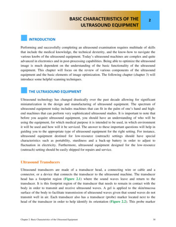
History of UltrasoundEarly use of ultrasound focused on therapyTreatment of gastric ulcers (left) and arthritis (right) in the 1940s.
History of Ultrasound Karl Dussik, 1946– University of Vienna inAustria– Cerebral ventricles
History of Ultrasound Diagnostic ultrasound– George Ludwig, 1940s– Classified experimentswith the Navy– Gall stones
A-mode A-mode ultrasound A amplitude
History of Ultrasound Howry’s somascope 2-D images called“somagrams”
B-mode B-mode ultrasound B brightness Intensity of echoes ingrey scale
M-mode Early echocardiogram– 1953 Edler & Hertz M Motion
D-mode D Doppler Satomura, 1956– freq returning echoes velocity of flow Moving RBCs createa Doppler shift
History of Ultrasound 1960s – Commercial machines available1970s – Grey scale becomes common1980s – Use in trauma in Europe & Japan1990s – Introduced into EM curriculum
Emergency MedicineMateer J et al. 1994 Goal-oriented Core applications––––––Trauma (FAST)RenalCardiacBiliary (RUQ)AortaEarly pregnancy
Additional applications Diagnostic–––––––––DVTSoft tissue (abscess)OcularTesticularLong bone fracturesLung (thoracic)Joint effusionsBladder volumeIVC diameter Procedural–––––––Vascular accessThoracentesisParacentesisArthrocentesisNerve blockadeForeign body removalLumbar puncture
Probe Orientation
FAST Right flank
FAST Left flank
FAST Cardiac
FAST Bladder
FASTHemopericardiumFree fluid
Extended FAST(with pleural view)
Cardiac Ejection fraction– Normal or poor?
Cardiac Effusion– present or absent Ejection fraction– Normal or poor
Renal Ureterolithiasis– Flank pain, N/V– Hematuria Options– KUB, CT, U/S, IVP– No imaging? Ultrasound– Hydronephrosis– Sensitive for largestones
Renal Hydronephrosis
Renal Moderate hydro
BiliaryUltrasoundStones?Wall thickening? ( 3mm)Fluid around wall?CBD dilated?
Biliary Transverse view
Longitudinal view
Biliary Thickened wall Pericholecystic fluid Portal vein anddilated CBDCBDPV
Early Pregnancy Ectopic pregnancy– 2% incidence– Abdominal pain– Bleeding Goal– Identify IUP
Early IUP (6 wks)Yolk sacFetal poleGestational Sac
Ultrasound FindingsVariations in hCG occur Gestational sac– 4-5 weeks TVShCG (mIU/mL)1000 Fetal pole– 6 weeks TVS2000 Cardiac activity– 7 weeks TVS10,000
Early Pregnancy Gestational sac Yolk sac Fetal pole
Aorta AAA– 2-4% of population 50– 10,000 deaths/yr– Rupture: 80% mortality
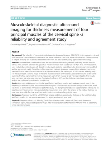
Aorta Normal transverseIVCAortaVertebral body Normal longitudinal
Aorta AAA transverse AAA longitudinal
Aorta AAA
Aorta AAATrue lumenMural thrombus
Rule out DVT Two-site compressionFemoralPopliteal– formal ultrasound– Misses calf DVTs– Follow up in 5-7 days
Normal study
DVT
DVT
Ocular Retinal detachment
Ocular Vitreous hemorrhage
Ocular Nerve sheathdiameter
Ocular Nerve sheathdiameter
Procedural Ultrasound Standard of care forIJ vein catheterization
Procedural Ultrasound Standard of care forIJ vein catheterization
Future Directions Hand-held ultrasound Vscan (GE)– 7000- 8000– One probe
Future Directions MobiUS (Mobisante)– Connects to cell phone– 7000- 8000 Hand-held ultrasound
Conclusion Probe indicator head or rightFree fluid is hypoechoicStandard of care for IJ linesNumerous applications– EM: FAST, Cardiac, Renal, Biliary, Aorta, IUP, DVT, Ocular Hand-held devices coming soon
References Mateer J, Plummer D, et al. Model curriculum for physician training in emergencyultrasonography. Ann Emerg Med 1994; 23(1):95-102.Moore CL, Copel JA. Point-of-Care Ultrasonography. NEJM 2011; 364(8):749-757.Roelandt JRTC. Seeing the invisible. A short history of cardiac ultrasound. Eur JEchocardiography 2000; 1(1):8-11.Sigel B. A brief history of Doppler ultrasound in the diagnosis of peripheral vasculardisease. Ultrasound in Med and Biol 1998; 24(2):169-176.Walcott BM. Mapping the literature of diagnostic medical sonography. Bull Med LibrAssoc 1999; 87(3):287-291.Woo JSK. A short history of the development of ultrasound in obstetrics andgynecology. www.ob-ultrasound.net/history1.html. Accessed Jan. 6, 2012.
Sigel B. A brief history of Doppler ultrasound in the diagnosis of peripheral vascular disease. Ultrasound in Med and Biol 1998; 24(2):169-176. Walcott BM. Mapping the literature of diagnostic medical sonography. Bull Med Libr Assoc 1999; 87(3):287-291. Woo JSK. A short history of the develop