Transcription
Øverås et al. Chiropractic & Manual Therapies (2017) 25:2DOI 10.1186/s12998-016-0132-9RESEARCHOpen AccessMusculoskeletal diagnostic ultrasoundimaging for thickness measurement of fourprincipal muscles of the cervical spine -areliability and agreement studyCecilie Krage Øverås1*†, Birgitte Lawaetz Myhrvold2†, Gro Røsok3 and Eli Magnesen4AbstractBackground: The reliability of musculoskeletal diagnostic ultrasound imaging (MSK-DUSI) for the evaluation of neckmusculature has been sparsely documented in the research literature. Until now, research has featured a limited numberof subjects and only few studies have tested for both inter- and intra-reliability using appropriate methodology.Methods: Four examiners conducted an inter- and intra-rater reliability and agreement study. Fifty females with andwithout neck pain (NP) between the ages of 20–70 were recruited from October 2014 to April 2015. The muscles thatwere evaluated were the longus colli (Lcol), the rectus capitis posterior major (Rcpm), the deep cervical extensors (Dce)and the semispinalis capitis (Sscap). Each of the examiners captured ultrasound images of their allocated muscle andmeasured the thickness of that muscle twice, on separate occasions, for the first part of the intra-rater reliability study.For the second part, a second image of the same muscle was taken on the same subject and measured by the sameexaminer. The four examiners then met to measure on each other’s images, to test inter-rater reliability. Their resultswere compared pair-wise using Interclass Correlation Coefficients (ICC) and Bland-Altman plots. Linear regressionanalysis was performed to evaluate for possible bias.Results: Inter-rater reliability was found to be good for Lcol and Sscap muscles and moderate towards poor for thedeeper Rcpm and Dce muscles. Intra-rater reliability was good for all the muscles, with the exception of the Dce, whichwas found to be moderate in the second part of the study. The B&A plots showed good agreement, few outliers, and nobias. However, the agreement intervals indicated a measurement error within the variance of the method that may nothave been acceptable for these small muscles if the aim is to evaluate change in thickness.Conclusions: This study found that MSK-DUSI had variable reliability in assessing the thickness of the Lcol, Rcpm, Dce,and Sscap muscles. No bias was demonstrated, but agreement intervals were wide.Keywords: Diagnostic ultrasound, Reliability, Cervical spine/neck muscles, Longus colli, Deep cervical extensors,Semispinalis capitis, Rectus capitis posterior major, Intra-class correlation coefficient, Bland-Altman’s limits of agreement,Linear regression analysis* Correspondence: cecilie@nemus.no†Equal contributors1MSc Chiropractic, MSc Ultrasound (Musculoskeletal), NEMUS Trondheim,Fjordgata 80, 7010 Trondheim, NorwayFull list of author information is available at the end of the article The Author(s). 2017 Open Access This article is distributed under the terms of the Creative Commons Attribution 4.0International License (http://creativecommons.org/licenses/by/4.0/), which permits unrestricted use, distribution, andreproduction in any medium, provided you give appropriate credit to the original author(s) and the source, provide a link tothe Creative Commons license, and indicate if changes were made. The Creative Commons Public Domain Dedication o/1.0/) applies to the data made available in this article, unless otherwise stated.
Øverås et al. Chiropractic & Manual Therapies (2017) 25:2BackgroundNeck pain and muscle functionNeck pain (NP) is common and represents a disablingand costly problem to society [1]. It is a global healthproblem ranked as the fourth most common cause ofloss of quality of life and incapacity [2]. The recoveryrate tends to be low and the recurrence of neck painepisodes is common, demonstrating the persistence ofneck pain [3–8]. Altered muscle function and morphological changes in these muscles are recognised featuresof painful neck disorders [9–14]. The underlying mechanisms of these changes and the implications forfunctional and physical impairment are not fully understood. Neck pain may occur in an impaired cervicalmotor system, where functional demands exceed physiological capacity [13]. This is thought to be a factor in thepersistent or recurrent nature of mechanical NP [15]and suggests that clinicians ought to incorporate theevaluation of muscle function into their diagnosticconsiderations. Still, there is a need to establish valid,reliable and useful clinical and biological markers ofneck dysfunction [16].The longus colli (Lcol) function as a stabiliser and flattener of cervical lordosis [10, 17]. The sub-occipitalscontribute to finer movement and stabilisation [18], particularly the rectus capitis posterior major (Rcpm) sinceit crosses the two upper cervical joints [10]. The deepcervical extensors (Dce), gives proprioceptive feedbackand are considered important for intervertebral segmental control [10, 19–23]. The intramuscular function ofthe Semispinalis capitis (Sscap) is not completely clear,but it exerts large extensor movement to the head andneck [15]. Altered muscle activation, atrophy, increasedfatty infiltration and decreased muscle strength has beenreported in these muscles [24–27] and they seem to playa role in cervicocephalic – and tension type headaches,whiplash-mechanism-related complaints, post fusion surgery, work-related and long-lasting NP [25, 26, 28–32].In recent years, MSK-DUSI has emerged as a methodto evaluate morphological changes within neck muscles[33]. The amount of change in thickness at rest andduring isometric contraction is considered an indirectmeasure of muscle function [34]. MSK- DUSI may henceplay a role in the subgrouping process for diagnosis ofNP, but also to evaluate effects of interventions. Fordiagnostic imaging, a practice guideline for spinal disorders state that conventional radiographs are not indicated for non-specific acute, sub-acute or persistent NP,in the absence of red flags [35]. Magnetic resonanceimaging (MRI) is frequently advocated for the evaluationof NP as it can determine the presence of serious underlying disease, and is helpful in confirming the site andlevel of root compression. However, MRI of the cervicalspine gives little attention to muscular tissue, apart fromPage 2 of 13when utilised for research, which again may partly explain why it fails to identify these structural changes possibly related to symptoms [29]. Diagnostic ultrasound issafe, non- invasive, non-ionizing, with the ability to domeasurements in real time. It is both cheaper and morecost-effective than MRI and easy to fit into a clinical settingin primary care dealing with musculoskeletal issues. However, it is known to be operator dependent [28, 36–38].Javanshir et al. [70] reviewed 16 different studies ofultrasonography of the cervical muscles and argued thatthere was not good enough evidence to conclude thatMSK-DUSI was appropriate for assessing neck muscles,even though previous studies had indicated it to be bothreliable and valid [39–41]. For reliability studies, theysuggested using constant landmarks, knowledge ofanatomy and function of target muscles, and proper definition of muscular borders to help obtain a clearerimage. Further, they highlighted the use of standardisedsubject positioning, the correct placement of the transducer, and the use of multiple images for statistical analysis in order to improve results. Thus there is a need forfurther investigation in order to determine the clinicalutility of ultrasound in this area.The main objective in this study was therefore to investigate one aspect of clinical utility by determining theinter-rater reliability and degree of agreement of determining the thickness of the Lcol, Rcpm, Dce and Sscap,by four raters on the same ultrasound image. It was alsodeemed important to ascertain the repeatability betweendays and between scans by investigating the intrareliability of MSK-DUSI in measurement of the thickness of the same muscles.MethodsIntra- and inter-rater reliability and agreement study.RatersFour raters, living in different parts of Norway, wereinvolved in the study. The raters were chiropractors withat least 4 years of clinical experience in MSK-DUSI andhad post-graduate certificates and diplomas in MSKDUSI, from a CASE accredited University. This article isbased on the four raters separate thesis submitted inpartial fulfilment of the requirements leading to thedegree of MSc Ultrasound. Their method was the same,but each rater had separate muscles to concentrate on.Beforehand the raters had agreed on which four musclesthey considered most appropriate to investigate according to the literature describing muscle impairment inneck pain disorders [42], and these were then allocatedby drawing lots to each of them. Hence each rater hadone of the four muscles to scan and evaluate, but all fourraters performed measurements on each other’s
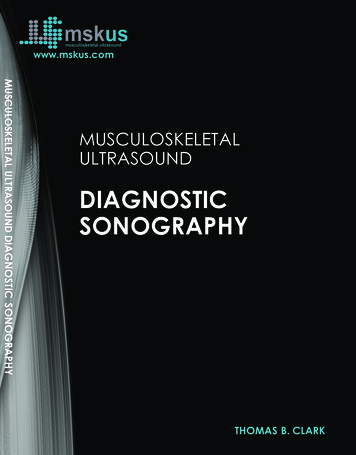
Øverås et al. Chiropractic & Manual Therapies (2017) 25:2Page 3 of 13ultrasound images when they later met up in eachother’s individual clinics.the study group were also a part of preparations prior tocommencement of the study.Study subjectsThickness assessment of the Lcol muscleAn a priori decision was made to include a total of atleast 50 female subjects for each muscle investigated.This was considered an accessible size as the subjectswere invited into the study from the four raters’ privatechiropractic clinics from October 2014 to April 2015.The subjects were enrolled by consecutive invitationover the period of one to three months at each clinic.See details for inclusion/exclusion criteria in Table 1. Allthe participants gave verbal and informed writtenconsent prior to study enrolment. Images and data werecollected and stored anonymously. Application for ethicsapproval was first sent to the Regional Ethics Committee(REC) in Norway. They concluded that; “as this is not acollection of information related to health or illness,apart from NP, but rather a quality assurance of a diagnostic tool for cervical muscles, the project can beaccomplish without approval from REC”.Subjects were supine in a neutral position with a smalltowel placed under the neck to support the cervical lordosis, knees and hips were bent, and arms lying alongthe sides of the body. The thyroid and cricoid cartilageswere palpated and the ultrasound probe was placed inthe sagittal plane in the midline of these cartilages. Thecricoid cartilage corresponded to the C6 level [43], whilethe bottom of the laryngeal prominence of the thyroidcartilage corresponded with the C5 level [40]. With thecricoid cartilage in the middle of the screen, the probewas moved laterally over the thyroid gland until the carotid artery was visible in the longitudinal view. The Lcolwas then visible between the carotid artery and the vertebral bodies (VB). The thickness measurement of theLcol was taken from the midpoint of ventral surface ofthe C6 vertebral body, defined as the posterior border ofthe muscle, to the ventral part of the muscle, at itsborder to the pre-facial tissues, surrounding the carotidartery (Fig. 1).ProceduresUltrasound machine and scanning proceduresThe ultrasound machines used were the one the ratershad available in their clinics. For the Rcpm and Sscap,the Esaote MyLab5 ultrasound machine was used, whilea Medison X 8 was used for Lcol and Dce muscles. Thescanning was performed in B-mode. A linear probeapplied with a 40 mm footprint and high frequency (12–18 MHz chosen individually) was used. The radiologicalprincipal ALARA (as low as reasonably achievable) wasfollowed to obtain the necessary information, withminimal settings and examination time. No standardprotocol (from EFSUMB or BMUS) existed for ultrasoundscanning of the neck muscles. Thus, prior to testing, scanning procedures were based upon extended knowledgeabout anatomy from books and scientific articles. Anexcursion with a professor in anatomy at a dissection lab,including dissection and ultrasound scanning of a femalecadaver, as well as training sessions and discussions withinTable 1 Inclusion and exclusion criteriaInclusion criteria Adults (20–70 years of age) Females With or without neck painExclusion criteria Diagnosis of recent trauma, spinal surgery, fractures or structuralpathologies in the area of interest, to ensure the right anatomicallandmarks on the ultrasound image during the ultrasound procedureof identifying the muscle Not able to comply with the examination procedure If for whatever reason anatomical landmarks and muscle borders couldnot be clearly enough identified on the ultrasound images for analysisThickness assessment of the Rcpm, Dce, and Sscap musclesSubjects were placed in a prone position, as it has previously been shown to give more reliable measurements[61]. Foreheads were placed onto the adjustable headpiece of the bench with slight flexion of the head. Theexaminer was positioned on the left side of the subject.The Rcpm muscle is situated between the posterior tubercle of atlas to the medial region of the inferior nuchalline. It lies deep to the Sscap, splenius capitis and uppertrapezius muscle. The C2 spinous process (SP) was identified by palpation. The transducer was placedtransversely and moved laterally to identify the lamina atthe C2 level. From this position the transducer wasmoved superiorly to identifying the lamina at the C1level, where the examiner tilted the probe upward ordownward to clearly identify the borders of the Rcpmmuscle, in the transverse plane. The anterior-posteriordimension (APD) was used for thickness measurementof the muscle, taken at the largest distance between theinner and outer borders of the Rcpm muscle (Fig. 2).For the Dce, containing the semispinalis cervicis(Sscerv), the cervical multifidus (Cxmult), and therotators, the transverse process on one side of C7 wasidentified in the transverse plane. The probe was movedmedially to visualise the articular pillar and was thenturned longitudinally, to identify the cervical facet jointof C5-6. The articular facet between C5-6 was placed inthe middle of the image and the probe was again turnedtransversely. In this position, the lateral insertion ofCxmult on the articular facet and capsule was identified,
Øverås et al. Chiropractic & Manual Therapies (2017) 25:2Page 4 of 13Fig. 1 Ultrasound image of the longus colli at C6 in transverse view to the left behind the carotid artery, and to the right longitudinal wheremeasurements were taken. The caliper is placed on the midpoint of the ventral surface of the C6 vertebral body and the interface between theLcol and the pre-fascial tissue surrounding the carotid arterywhile the probe was moved medially toward the base ofthe SP. The probe was angled up- or downwards tomake the anterior and posterior borders as sharp aspossible, before the image was captured. Thickness ofthe deep cervical extensors was determined as the measurement between the two echogenic lines of the laminaof C5 and the echogenic line of the hyperechoic fasciabetween Sscap and Sscerv. The calliper was positionedat 90 in relation to the laminae. The measurement wasperformed where the rater considered the muscular unitto be at its thickest (Fig. 3).Sscap, the third muscle in the layer, was recognised byits medial and lateral parts with aponeuroses visualisedinternally. To identify Sscap, the transducer was placedtransversely to the level of C4. The bifurcation of thecarotid artery usually occurs at the level of C4. Thetransducer was placed transversely to the SP and wasFig. 2 Ultrasound image of the Rcpm at rest in transverse view atC1. The caliper is placed around the midpoint above the C1 laminabetwen the inner and outer borders of the muscle where it wasconsidered thickest by the ratersmoved laterally and anteriorly to identify the carotid artery on both sides, to ensure that the C4 level was in theimage. The thickness of the muscle was measured byAPD, at its thickest part over the midline of the laminaof C4, in the longitudinal view. An image in transverseplane was saved to decrease measurement error. Thiswas achieved due to it giving dynamic visualisation ofthe fascia layers, thus clarifying which bright lines wereactually within the muscle or fascial layer dividing themuscles (Fig. 4).One image (image A) was obtained for each musclefrom the left and right sides. Once the first two imageswere taken, the subject stood up from the bench, walkedaround and was re-positioned and the procedure wasrepeated for two more images (image B). In the intrarater reliability part of the study, rater one measured themuscle’s thickness as image A was captured, and randomly again one week after all of the images had beenFig. 3 Ultrasound image of the Dce in transverse view at C5. Thecaliper is placed 90 to the lamina of C5 where the rater consideredthe muscle to be at its thickest and up to the echogenic line of thehyperechoic fascia between the Sscerv and Sscap
Øverås et al. Chiropractic & Manual Therapies (2017) 25:2Page 5 of 13Fig. 4 Ultrasound image of the Sscap at C4 in transverse view to the left for dynamic visualisation of anatomical borders and in longitudinal viewwhere measurements were taken to the right. The caliper is placed from the lower border the Sscap against the Dce above the midpoint of thelamina of C4 to the outer border of Sscap against the Splenius Capitis musclecollected. Thickness measurements performed by raterone on image B of the same subject, captured after re-positioning, were performed one week after all the datawas collected. For the inter-rater reliability part of thestudy, the other raters measured muscle thickness onthe entire A images and these were plotted together withthe first measurements from rater one. Each of theraters’ measurements was compared pairwise to theother raters’ measurements. Thus in total, six ratingswere collected for each muscle. This procedure wasrepeated for all of the muscles. The measurements wereperformed using the calliper software on the machine.The mean of two or more ratings has been recommended to increase reliability [49]. However, it was decided that only one single measurement for each imagewould be recorded, as this was considered more comparable to clinical practice. Still, when in doubt theraters were allowed a couple of measurements withoutrecording them before determining what they consideredto be the correct single measure. The measurementswere recorded manually and not saved on the machine.The method was clarified to the raters prior to themundertaking the measurements. The raters were blindedfor the subjects’ identification, clinical information andeach other results. The results of the measurementswere recorded manually on a list with the correspondingsubject numbers, and transferred to an excel file for laterstatistical analysis. On the sheet of the ratings, commentfields were made available for the raters to comment onpotential difficulties they encountered in performing themeasurements.Data analysisAll of the data were analysed with IBM SPSS version 23software. Descriptive statistics were used to describe thestudy population. The intra- and inter-rater reliabilitywas analysed by calculation of the ICC, known asanalysis of variance (ANOVA), which reflected both thedegree of consistency and agreement among ratings.ICC was determined by a two-way mixed model, typeabsolute agreement, with a 95% CI (confidence interval),for single measures (ICC 3.1). For the estimation of thelevel of agreement and illustration of measurement error,the Bland-Altman plot was considered most relevant[44]. The raters were tested against each other usingseparate pairwise plots. A calculation of the pair-wisedifferences for the six comparisons was made andaveraged together to define the y-axis. The x-axis wasdefined by the average mean of the measurements madeby the four raters. Limits of agreement (LoA) (2 SD)were calculated for each pair and a linear regressionanalysis were performed to evaluate for possible bias.Based on the LoA range we calculated the greatest difference % measured between examiners when applied tothe average thickness of the different muscles.Table 2 Descriptive dataMuscleFemales (N)Mean age (Yrs)Age range (Yrs)SD (Yrs)Neck PainNo Neck PainLcol5446,920–70 13,461%39%Rcpm3144,120–70 10,748%52%Dce56a50,020–70 11,364%36%Sscap5037,120–70 13,564%36%a6 of the subjects were excluded from to the inter- or intra-rater reliability and agreement analysis because of missing measurements due to difficulties defininganatomical borders
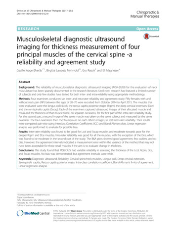
Øverås et al. Chiropractic & Manual Therapies (2017) 25:2Page 6 of 13ResultsDescriptive dataA description of the study subjects is seen in Table 2.50–56 different subjects were recruited for each muscle.Prior to analysis, 19 subjects for the Rcpm and 3 subjects for the Dce were excluded due to difficulties withlandmarks and muscle borders and one for the Sscap asthe splenius capitis muscle could not be identified.Inter-rater reliabilitySeparate analyses were made for the right and left sides.As the results were similar, only the right side isreported. When measurements from the four raters werecompared, the ICC values for the Dce and the Rcpmwere moderate towards poor, and generally lower thanfor the Lcol and Sscap muscles, where inter-rater reliability was good, see Table 3. In addition confidenceintervals for the Dce and Rcpm muscles were wider indicating greater uncertainty around the estimate.The Bland-Altman plots for inter-rater agreement areshown in Figs. 5, 6, 7 and 8 and Table 4. The comparison of measured thickness between raters (Figs. 5, 6, 7and 8) revealed a low mean difference, except for theDce. For Dce the mean difference was approximately20% of the average muscle thickness (2,52 of 12,2 mm),but only 2% (0.13 of 8,3 mm), 4% (0.24 of 6,2 mm) and0.1% (0.003 of 3,9 mm) for Lcol, Rcpm and Sscap,respectively. The greatest difference % was considered asthe maximal possible error of thickness measurement;which for the Dce was 14% (1,67 of 12,2 mm), 13% forLcol (1,11 of 8,3 mm), 25% for Rcpm (1,55 of 6,2 mm)and 10% for Sscap (0,37 of 3,9 mm). Zero did lie withinthe LoA intervals, thus reflecting that there was no fixedbias. A linear regression analysis was performed for allthe plots. The P-values were all 0.05, so there was noproportional bias.Intra-rater reliabilityThere was found to be good intra-rater reliability for allfour muscles when measurements were done on thesame image, see Table 5. However, when measuring ontwo different images of the same muscle the Dce showedpoorer intra-rater reliability. The mean difference wasfound to be small, for both between days and betweenscan repeatability, but the agreement intervals werewider between scans. The greatest difference %measured by the examiner when applied to the averagethickness of the different muscles was the same forSscap and Rcpm, as in the inter-rater reliability part(10% (0,37 of 3,9 mm) and 25% (1,57 of 6,2 mm) respectively), and higher for Lcol (26% (2,14 of 8,3 mm))and Dce (22% (2,66 of 12,2 mm)).DiscussionThe aim of this study was to establish the reliability ofMSK-DUSI in measuring the thickness of the Lcol, Rcpm,Dce, and Sscap by four clinicians. The lower reliabilityfound for the Rcpm and Dce may be because morphological changes, as fat infiltration in the deepest extensormuscles including the Rcpm, may make anatomical landmarks and muscle borders more difficult to define. TheICC values were higher in the intra-reliability part of thestudy, which was expected as the same rater who took theimages also did the measurements, and hence probablyhad a greater understanding of that muscle and its borders. For a test to be useful on a consistent basis in clinicalpractice reproducibility would be considered of moreimportance than repeatability [45]. However, if the interrater reliability is poor, knowledge of the intra-raterreliability might assist in identification of sources of error,as may be the case in this study.Statisticians maintain that one should not seek agreement between different methods or measurers; insteadone should focus on disagreement or bias [46]. Thisstudy therefore focused on this school of thought. However, a priori definition of acceptable limits for the agreement interval based upon clinical necessity andbiological considerations, as proposed by Giavarina [71],had not been made. In general, the mean difference between raters was low, except for the Dce muscle. If wewere to use ultrasound for diagnostic purpose, such asfollow-up measurements, any reported change above themean difference may be associated with actual change ina muscle and not be a result of the reliability of themeasurement method. Still, the agreement interval indicated a measurement error range that appeared to betoo high considering the size of theses muscles andprobable changes seen in relation with NP. LoA has notoften been reported in comparable previous studies, andto our knowledge, no previous literature has yet outlinedacceptable agreement levels for these muscles.Table 3 Inter-rater reliability - ICC values and corresponding confidence intervalsMuscleEx1-2ICC (95% CI)Ex1-3 ICC (95% CI)Ex1-4 ICC (95% CI)Ex2–3 ICC (95% CI)Ex2–4 ICC (95% CI)Ex3–4 ICC (95% CI)Right Lcol.87 (.77–.93).90 (.83–.94).86 (.73–.92).84 (.72–.91).77 (.63–.86).84 (.70–91)Right Rcpm.86 (.71–93).54 (.23–.75).65 (.39–.81).46 (.14–.69).66 (.33–.83).43 (.11–.68)Right Dce.67 (.49–.79).54 (.32–.71).63 (.42–.77).62 (.43–.77).46 (.22–.64).54 (.32–.71)Right Sscap.88 (.72–.95).89 (.75–.95).87 (.77–.93).76 (.33–.89).85 (.75–.91).74 (.47–.86)
Øverås et al. Chiropractic & Manual Therapies (2017) 25:2Page 7 of 13Fig. 5 Bland & Altman Plot with LoA for the right Dce muscle showing agreement between the 4 raters for measurements of thickness on thesame image. (N 53 subjects, 212 ratings). The mean difference was calculated from the 6 pair of comparison of the different examiners and themean thickness were calculated for the four ratersFig. 6 Bland & Altman Plot of LoA for the right Lcol muscle showing agreement between the 4 raters for measurements of thickness on thesame image. (N 54 subjects, 216 ratings). The mean difference was calculated from the 6 pair of comparison of the different examiners and themean thickness were calculated for the four raters
Øverås et al. Chiropractic & Manual Therapies (2017) 25:2Page 8 of 13Fig. 7 Bland & Altman Plot of LoA for the right Rcpm muscle showing agreement between the 4 raters for measurements of thickness on thesame image. (N 31 subjects, 124 ratings). The mean difference was calculated from the 6 pair of comparison of the different examiners and themean thickness were calculated for the four ratersFig. 8 Bland & Altman Plot of LoA for the right Sscap muscle showing agreement between the 4 raters for measurements of thickness on thesame image. (N 50 subjects, 200 ratings). The mean difference was calculated from the 6 pair of comparison of the different examiners and themean thickness were calculated for the four raters
Øverås et al. Chiropractic & Manual Therapies (2017) 25:2Page 9 of 13Table 4 Mean thickness, mean difference, agreement intervals (LoA) & linear regression analysisMuscleMean muscle thickness (mm)Mean difference (mm)Right Lcol8.3 1.60.13( 0.86; 1.11)0.06Right Rcpm6.2 1.2 0.24( 1.55; 1.06)0.9112.2 1.3 2.52( 1.67; 1.16)0.46( 0.37; 0.37)0.58Right DceRight Sscap3.9 0.70.003Methodological considerations - strengths and limitationsTo improve the quality of the study, the methodologyemployed was tailored according to proposed recommendations in the literature [48–50]. It has been highlyrecommended for reliability studies that they reflect thecircumstances in which they would like the results to begeneralised [50]. This study included a representativesample from a typical clinical setting in primary care.The sample size was generally higher than that used inprevious studies. The current study included a wider agerange and was thought to be large enough to represent avariety of different subject types, as well as subjects withand without neck pain. Only females were included,which made the population more homogenous, animportant criterion for reliability studies.Unlike most previous studies both the ICC and theB&A test with LoA were used. An advantage of the B&Aplot is that the graph provides a representation of themagnitude of the degree of agreement. One can easilyidentify bias, outliers, and other relationships betweenthe variance in measure [51].The small thickness of these muscles may have amplified errors, thus influencing the variability of measurements [33]. With lack of variability, measurementsmight have fallen within a restricted range that couldalso have affected the ICC [52]. Four raters were available for the inter-rater part of this study, thus allowingone to yield a more precise reliability estimate. This wereconsidered a strength of this study, even though thenumbers of subjects used was thought to have a greaterimpact on the accuracy of the results than the numberof raters [48]. Owing to the use of more than tworepeated measurements, calculations were more complex. As a result, the sample size needed to be largeenough, preferably greater than 50, to allow the B&A’sLoA range (mm)Linear regression (p 0.05)LoA to be estimated and to avoid the CI becoming toowide [51]. Even though this study included moresubjects than most of the previous studies in this topicarea, the sample may still not have been sufficiently largeenough when 4 raters were included [47]. Reliableresults of small muscles have also reported to be challenging using MRI, despite it being regarded as the goldstandard [40]. However, no validated method existed toquantify atrophic changes and fatty infiltration withMSK-DU
MSK-DUSI was appropriate for assessing neck muscles, . The ultrasound machines used were the one the raters had available in their clinics. For the Rcpm and Sscap, the Esaote MyLab5 ultrasound machine was used, while . about anatom