Transcription
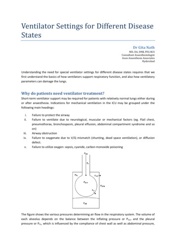
Ventilator Settings for Different DiseaseStatesDr Gita NathMD, DA, DNB, FFA RCSConsultant AnaesthesiologistAxon Anaesthesia AssociatesHyderabadUnderstanding the need for special ventilator settings for different disease states requires that wefirst understand the basics of how ventilators support respiratory function, and also how ventilatoryparameters can damage the lungs.Why do patients need ventilator treatment?Short-term ventilator support may be required for patients with relatively normal lungs either duringor after anaesthesia. Indications for mechanical ventilation in the ICU may be grouped under thefollowing main headings:i.ii.iii.iv.v.Failure to protect the airwayFailure to ventilate due to neurological, muscular or mechanical factors (eg. Flail chest,pneumothorax, bronchospasm, pleural effusion, abdominal compartment syndrome and soon)Airway obstructionFailure to oxygenate due to V/Q mismatch (shunting, dead space ventilation), or diffusiondefect.Failure to utilize oxygen: sepsis, cyanide, carbon monoxide poisoningThe figure shows the various pressures determining air flow in the respiratory system. The volume ofeach alveolus depends on the balance between the inflating pressure or PALV and the pleuralpressure or PPL, which is influenced by the compliance of chest wall as well as abdominal pressure,
PAB. Air flow at any instant during inspiration or expiration is determined by the pressure gradientbetween PALV and PAO as well as resistance of the airways.Adverse effects of mechanical ventilation: Barotrauma: Lung injury associated with high alveolar pressures ( 35-40 cm H2O),presentation ranges from asymptomatic pulmonary interstitial emphysema (PIE), throughsubcutaneous emphysema, pneumomediastinum; to tension pneumothorax.Volutrauma: This term refers to lung injury due to overstretching of alveoli because ofexcessive tidal volumes, which leads to release of inflammatory mediators that have bothpulmonary and systemic effects, increasing morbidity and mortality. When non-homogenouspulmonary pathology is present, tidal volume delivered by the ventilator is preferentiallydelivered to the more compliant, relatively normal areas of the lung. Thus normal alveoli areoverstretched, and the remaining normal lung is damaged, further increasing lung injury anddamage. Repeated closing and re-opening of alveoli (recruitment and collapse) is anotherfactor which causes damage due to shear stress and loss of surfactant. The release ofinflammatory mediators can cause damage to remote organs such as the liver and kidney.Barotrauma is due to high alveolar pressures while volutrauma is related to high transalveolar pressure gradient. Auto-PEEP: This is the positive pressure which develops in the alveoli at the end ofexpiration due to incomplete emptying of alveoli, either due to airway obstruction due tosecretions, bronchospasm or airway closure; or else if expiratory time is too short. Thiscannot be detected at the ventilator end of the tube but causes increased work of breathingand progressive overdistension of the lungs, if not corrected.Oxygen toxicity: High inspired oxygen concentrations (FiO2 0.5) cause cellular damage dueto free radical formation. In addition, high FiO2s are also associated with absorptionatelectasis. Hence it is important to set the lowest FiO2 possible, aiming for SaO2 90%Cardiovascular effects of mechanical ventilation include decreased venous return, RVdysfunction and altered LV compliance.
Initial ventilator settings:Mode of ventilationThe choice depends on whether or not the patient is conscious, sedated or has receivedneuromuscular blockers. SIMV or A/CMV modes are appropriate for initial settings. Patients with agood respiratory drive can be placed on PSV mode.Tidal volume:Currently, lower tidal volumes are recommended than in the past, and 5-8 ml/Kg of ideal bodyweight should be set, adjusting the value so that Pplat is less than 35 cm H2O.Respiratory rate:This should be set at 8-10 breaths per minute, unless hyperventilation is required for intracranialpathology or metabolic acidosis. Higher rates may result in air trapping and intrinsic PEEP.Inspired oxygen level:Though an initial FiO2 of 1 is usually recommended, it is advisable to reduce the FiO2 as rapidly aspossible, aiming for an oxygen saturation 90% and PaO2 60 mmHg.Inspiration/expiration ratio:Normally the I/E ratio is set at 1:2. Expiratory time may be increased if airway obstruction is presentso as to avoid air trapping and auto-PEEP. Inspiratory time may need to be prolonged in the settingof severe ARDS so as to equal or even exceed expiratory time (inverse ratio ventilation).Inspiratory flow rate:Inspiratory peak flow rate may need to be set in some ventilators when volume control ventilation(VCV) is being used. It should be set at 4 times the desired minute volume so that the patient’sinspiratory flow requirement is met. If the flow is too low, the patient needs to make an extra effort,and too high an inspiratory flow causes the peak pressure to be too high. It is usually set at 60 L/minto start with.In newer ventilators, the peak flow is set automatically, depending on the tidal volume, I/E ratio,respiratory rates and inspiratory pause, if present.During pressure modes of ventilation, the flow is determined by the inspiratory pressure and theresistance and compliance of the respiratory system.Positive end-expiratory pressureEven in patients with normal lungs, PEEP of 3-5 cm H2O should be set during mechanical ventilation,in order to prevent decreases in FRC and dynamic airway collapse. Higher levels of PEEP may benecessary in patients with diseased lungs, so that acceptable oxygenation may be achieved withlower inspired oxygen concentrations.Several methods have been described for selecting the optimal PEEP, such as setting the PEEP at 1 or2 cm H2O above the lower inflexion point of a pressure/volume loop. Alternatively, a high PEEP canbe set initially (eg. 20 cm H2O) and then progressively decreased till desaturation occurs. At thispoint, a recruitment manoeuvre is done and the PEEP is set about 2 cm H2O higher.
Sensitivity:If pressure triggering is used, the trigger sensitivity is set at -2 cm H2O initially, keeping in mind thatthe presence of intrinsic PEEP increases the effort required by the patient if an adequate level ofextrinsic PEEP is not set. Modern ventilators provide the option of flow-triggering which needs lesseffort from the patient.Specific disease statesObstructive airway diseaseThe main physiological abnormality in these conditions is an increase in airway resistance, especiallyduring expiration. This has a large reversible component in bronchial asthma, unlike chronicobstructive pulmonary disease (COPD). In COPD, there is an abnormal and amplified inflammatoryresponse to various noxious substances, which affects the airways, parenchyma as well as thepulmonary vasculature. The airway obstruction has only a small reversible component, and is due tothe following factors:i.ii.iii.iv.Oedema, smooth muscle hypertrophy peri-bronchial fibrosisExcessive mucus secretion and pluggingDestruction of alveoli and respiratory bronchioles with reduced recoil. This predisposes tocollapse of conducting airways.Hyper-responsive airway reflexes – bronchospasmThe increased airway resistance leads to expiratory flow limitation (Intra-thoracic airway diameter isalways smaller during expiration, during spontaneous respiratory efforts). This, along withinadequate time for expiration due to tachypnoea, causes dynamic hyperinflation leading torespiratory embarrassment, increased work of breathing and hemodynamic compromise. The goalsof ventilator support are to minimize intrinsic PEEP, rest the respiratory muscles and maintainacceptable blood gases.Initial settings in these patients consist of a low tidal volume (6-8 ml/Kg), high inspiratory peak flow(80-100 ml/min), low PEEP (0-5 cmH2O), low respiratory rate and I:E ratio of 1:4 to 1:5. Traditionally,assist/control mode is chosen, but there is no consensus on which mode of ventilation should be setinitially in obstructive airway disease. Volume ventilation ensures delivery of the set tidal volume,but the distribution of that tidal volume may be patchy, with reduction of flow through constrictedairways causing an increased inspiratory pressure and resultant dilatation of non-constricted airwayselsewhere in the lungs. Changing to pressure controlled ventilation has been shown to improve gasexchange in pediatric patients. A simulation study using an integrative network model of the lungshowed PCV produced a more uniform distribution of tidal volume, but the delivered tidal volumewas highly sensitive to small changes in inspiratory pressure setting. Dual modes such as PCRV orautoflow may be helpful in this situation; these are essentially PCV with the inspiratory pressurecontinually adjusted to deliver the set tidal volume.Dynamic hyperinflation may be estimated by measuring the end-inspiratory volume above FRC, bycollecting the total exhaled volume over 60 sec of apnoea. Other estimates of dynamichyperinflation are the plateau pressure and intrinsic PEEP which can be measured during an
expiratory hold manoeuvre. The ventilator flow-time tracing shows continued expiratory flow at thebeginning of inspiration, showing that the lung has not fully emptied, i.e. air trapping is taking place.These ventilator settings with a low minute ventilation lead to hypercapnia and respiratory acidosis.Permissive hypercapnia is tolerated down to a pH of 7.15, below which sodium bicarbonate or THAMinfusions are recommended. Adequate sedation and sometimes, neuromuscular blockade isrequired in these patients to achieve adequate ventilation.
Acute Respiratory Distress Syndrome / Acute Lung InjuryLung injury due to a variety of pulmonary and extra-pulmonary causes presents with patchyinfiltrates and is included under the heading of ARDS/ALI. There is an oxygenation defect due tointrapulmonary shunting, which can be estimated by the PaO2/FiO2 ratio; with a ratio 200 mmHgdesignated as ARDS and ratio 200-300 mmHg defined as ALI. Depending on the severity, thesepatients may require ventilator support till the lungs recover from the primary insult which causedalveolar damage. It is important to ensure that this ventilatory support does not itself producefurther lung injury by damaging the remaining normal alveoli.The pathophysiology of these conditions involves increased alveolar permeability with patchyatelectatic areas as well as more normal air filled alveoli. On the macro scale, the lung appears stiffwith a low compliance, but there is considerable heterogeneity with different alveoli having a rangeof compliance values and time constants. It is as if there is an atelectatic lung and a normal lungconnected in parallel. Thus, a relatively normal tidal volume is forced into the normal alveoli whichadd up to much less than a normal lung – the much smaller or so-called “baby lung”, overdistendingthe alveoli and causing volutrauma.Thus the ventilator settings for ARDS include a low tidal volume (6 ml/IBW) keeping the Pplat 30cmH2O; and FiO2–PEEP combinations as per the ARDS network protocol, aiming for a PaO2 of 55-80mmHg and SaO2 of 88-95%. Pressure targeted modes of ventilation give better distribution of tidalvolume compared to volume targeted modes but there is no conclusive evidence that these givebetter results as long as the tidal volumes and plateau pressures are kept low. I:E ratio is kept at 1:2to 1:3 to start with, but severe oxygenation defect may benefit from I:E ratio of 1:1 or even higher –inverse ratio ventilation.Ventilator modes which allow spontaneous breathing such as APRV and Bilevel PAP have beenshown to improve oxygenation and hemodynamics, help lung recruitment and also reduce the needfor sedation. Recruitment manoeuvres, which involve the application of sustained positive pressureof 40-50 cm H2O followed by PEEP, have been shown to improve oxygenation. This approach isreferred to as “open the lung and keep it open!”
Abdominal compartment syndromeThe elevated intra-abdominal pressure and cephalad displacement of the diaphragm reduce the FRC.The compliance of the respiratory system is greatly reduced, though lung compliance may benormal. Alveolar volume is determined by transmural pressure and not the absolute alveolarpressure. These factors influence ventilator settings in these patients.Intra-abdominal pressure raises pleural pressure but since the pleural pressure is not routinelymeasured, transmural pressure across the alveolar wall may be estimated by the following formula:Ptm Pplat – IAP/2Volutrauma is mainly related to alveolar over-distension, and hence, the inspiratory pressure may besafely increased as long as the transmural pressure is kept below 35 cm H2O. Moderate PEEP (10 cmH2O) has been shown to increased LV afterload produced by IAH. Recruitment manoeuvres in thesepatients need higher inflation pressures (40 IAP/2 cm H2O).Hemodynamically, filling pressures (CVP and PCWP) are elevated in IAH, hence other techniquesshould be used for preload assessment, such as esophageal Doppler, echocardiography and pulsecontour analysis. Monitoring the extravascular lung water with techniques such as PiCCO is alsohelpful.Heart failure / Pulmonary oedemaMany patients with congestive heart failure respond to noninvasive ventilation, either simple CPAPor BiPAP. These modes help by increasing FRC and opening the alveoli. Fluid is shifted out of thealveoli and ventilation/perfusion balance is improved. Oxygen cost of breathing is reduced bymoving the patient onto a more favourable position on the compliance curve and by unloading therespiratory muscles. PEEP is transmitted to the left ventricle, reducing transmural pressure andafterload, and thus improving performance.For patients who need intubation, ventilator settings are similar to those used for ARDS, namely alow tidal volume (6 ml/IBW) keeping the Pplat 30 cmH2O; and FiO2–PEEP combinations aiming for aPaO2 of 55-80 mmHg and SaO2 of 88-95% and relatively high respiratory rates. Hypotension oftenfollows intubation and ventilation; this should be managed by a combination of fluid therapy andmanipulation of cardiac contractility and peripheral resistance.Intracranial pathologyMechanical ventilation affects intracranial dynamics in many ways. Raised intrathoracic pressure canraise ICP by direct transmission and by lowering CPP due to fall in venous return and cardiac output.However, this effect is relatively modest, and most patients with intracranial pathology tolerate IPPVand PEEP well. In patients with ARDS as well as intracranial hypertension, recruitment manoeuvreswhich would be expected to increase ICP actually produced the opposite effect because of improvedgas exchange and CO2 elimination.CO2 has a powerful effect increasing cerebral blood flow, but the effect is transient and wears offover a period of hours. Hyperventilation is now recommended in the context of acute intracranialhypertension, only for temporary reduction of ICP while other measures become effective. At othertimes, normocapnia should be maintained, since excessive hypocapnia predisposes to cerebralischaemia due to vasoconstriction.
Traditionally, ventilatory strategy in the presence of intracranial pathology focused on airwayprotection, optimizing oxygen delivery to the brain, careful control of CO2 and maintaining thelowest possible intrathoracic pressure. This method used large tidal volumes, high FiO2, low or zeroPEEP, fluids and vasopressors to maintain CPP.However, it is now recognised that volutrauma and atelectrauma caused by this type of settingspredispose to ALI/ARDS as well as extrapulmonary adverse effects on the kidney, liver, gut and otherorgans; especially if they have been “primed” by the initial brain injury. The concern about usinglung-protective ventilation is that it may result in hypercapnia, which is deleterious in the presenceof intracranial hypertension. But it is possible to apply a lung protective strategy without causinghypercapnia, and low tidal volumes (6 ml/IBW), moderate PEEP and relatively high respiratory ratesshould be used in these patients.Neuromuscular Respiratory FailurePatients who need ventilator support for neuromuscular disease have relatively normal lungs tobegin with. Tidal volumes of 8-10 ml may be given initially, with PEEP of 3-5 cm H2O, keeping the Pplatbelow 30 cm H2O. Volume or pressure targeted modes may be used, but pressure targeted modesmay by more comfortable for the patient. Dual modes such as PRVC or autoflow are helpful inensuring adequate ventilation even in pressure targeted modes.Non-invasive ventilationThis term refers to providing ventilatory support without intubation, via a nasal mask, full face maskor a helmet. It requires an alert, cooperative patient with airway protective reflexes. Othercontraindications are GI bleeding, severe hemodynamic instability and airway obstruction (otherthan obstructive sleep apnoea).There are several ways of providing NIV. Simple CPAP or continuous positive airway pressure can begiven with a high flow of oxygen, a well fitting mask and a PEEP valve, without a ventilator.Dedicated NIV ventilators are available which can give CPAP or BiPAP (bi-level positive airwaypressure). BiPAP is essentially pressure support ventilation in which EPAP represents PEEP and IPAPis the level of pressure support. These specialty ventilators have fewer options but are more tolerantof leaks in the circuit.Most of the critical care ventilators nowadays have a NIV option and thus can be used for NIV. Themost common mode employed is CPAP with pressure support. Some of these ventilators can alsoprovide proportional assist ventilation or PAV. Though volume modes can be used for NIV, pressuresupport and PAV are better tolerated by patients and are more leak-tolerant than volume modes.Initial ventilator settings are 5 cm H2O PEEP or EPAP and 10 cm H2O pressure support or IPAP. Theaim is to achieve tidal volumes of5-7 ml/Kg, respiratory rate below 25/min and SaO2 above 90%.Serial blood gas analysis and close clinical monitoring are essential for success with NIV. Thereshould be a low threshold for moving to invasive ventilation, should the patient deteriorate.Exacerbation of COPD and cardiogenic pulmonary edema are the two conditions well suited for NIVand improvement may be seen within 1-2 hours of starting treatment. Reduction in intubation ratesand mortality with NIV has been seen in both these conditions. NIV can also act as a bridge support
after early extubation, since the rates of extubation failure may be as high as 10-20%. This isespecially so in patients with underlying COPD.Other conditions where NIV is beneficial are: Immuno-compromised patients – solid organ transplants, febrile neutropeniaAsthmaPostoperative patientsRib fractures, lung contusionAdvanced malignancy (do not intubate status)ARDS – in less severly ill patientsSARS – able to avoid intubation in 70% patients. Useful in hypoxemic patients with lowAPACHE II scoresWeaning from Ventilatory SupportPlanning for separating the patient from the ventilator should start right from the time of institutionof mechanical ventilation. To this end, the modes used should generally maintain spontaneousrespiratory activity, as well as optimize other factors such as fluid balance, nutrition, electrolyte andmetabolic status. The two major weaning strategies are either using a so-called weaning mode andstep-wise reduction of ventilator support, or alternatively daily assessment of possibility of ventilatorwithdrawal. An evidence based task force has recommended the latter approach.i.ii.iii.The patient is assessed daily for readiness for a spontaneous breathing trial. This alsoinvolves daily interruption of sedation. The primary indication for ventilation should haveresolved and patient should be alert. Pulmonary gas exchange should be adequate, withPaO2/FiO2 ratio 200, FiO2 0.5 and PEEP 5-7 cm H2O. Hemodynamics should be stablewithout high levels of inotropic support. The patient should be able to initiate spontaneousbreaths.The spontaneous breathing trial is performed (using T-piece, CPAP or pressure support of 5cm H2O) for 30 -120 min, monitoring ventilator pattern, gas exchange, hemodynamics,subjective comfort.Patients who pass this trial may be extubated. Those who fail the trial should be put back ona stable, non-fatiguing mode should be instituted till the next SBT. Modes of partial supportshould provide adequate muscle unloading by using sensitive and responsive triggeringsystems and an unrestricted flow pattern which meets the patient’s demands.ConclusionVentilatory settings have profound effects on the outcome; and the interaction of each conditionwith the effects of the different modes of ventilation should be kept in mind so as to avoid furtheriatrogenic damage to the lungs and other organs,
PCVPEPAPFiO2FRCIAHIAPIBWICPI:E EPPLPRVCPSVPtmSARSSaO2SBTSIMVVCVV/Q:Intrinsic pressureAssist control modeAcute lung injuryAcute respiratory distress syndromeBi-level positive airway pressureChronic obstructive pulmonary diseaseContinuous positive airway pressureCerebral perfusion pressureCentral venous pressureExpiratory positive airway pressureInspired oxygen concentrationsFunctional residual capacityIntra-abdominal hypertensionIntra-abdominal pressureIdeal body weightIntracranial pressureInspiration to expiration ratioIntra-abdominal pressureInspiratory positive airway pressureNon-invasive ventilationAbdominal pressureAlveolar pressureAirway pressureArterial partial pressure of oxygenProportional assist ventilationPressure controlled ventilationPulmonary capillary wedge pressurePositive end-expressionPulse induced contour cardiac outputPulmonary interstitial emphysemaPleural pressurePressure regulated volume controlPressure support ventilationTransmural pressureSevere acute respiratory syndromeOxygen saturationSpontaneous breathing trialSynchronised intermittent mandatory ventilationVolume control ventilationVentilation/perfusion
In COPD, there is an abnormal and amplified inflammatory response to various noxious substances, which affects the airways, parenchyma as well as the pulmonary vasculature. The airway obstruction has only a small reversible component, and is due to the following factors: i. Oede